Abstract
Introduction
In order to improve the efficiency of heat moisture exchangers (HMEs), new hybrid humidifiers (active HMEs) that add water and heat to HMEs have been developed. In this study we evaluated the efficiency, both in vitro and in vivo, of a new active HME (the Performer; StarMed, Mirandola, Italy) as compared with that of existing HMEs (Hygroster and Hygrobac; Mallinckrodt, Mirandola, Italy).
Methods
We tested the efficiency by measuring the temperature and absolute humidity (AH) in vitro using a test lung ventilated at three levels of minute ventilation (5, 10 and 15 l/min) and at two tidal volumes (0.5 and 1 l), and in vivo in 42 patients with acute lung injury (arterial oxygen tension/fractional inspired oxygen ratio 283 ± 72 mmHg). We also evaluated the efficiency in vivo after 12 hours.
Results
In vitro, passive Performer and Hygrobac had higher airway temperature and AH (29.2 ± 0.7°C and 29.2 ± 0.5°C, [P < 0.05]; AH: 28.9 ± 1.6 mgH2O/l and 28.1 ± 0.8 mgH2O/l, [P < 0.05]) than did Hygroster (airway temperature: 28.1 ± 0.3°C [P < 0.05]; AH: 27 ± 1.2 mgH2O/l [P < 0.05]). Both devices suffered a loss of efficiency at the highest minute ventilation and tidal volume, and at the lowest minute ventilation. Active Performer had higher airway temperature and AH (31.9 ± 0.3°C and 34.3 ± 0.6 mgH2O/l; [P < 0.05]) than did Hygrobac and Hygroster, and was not influenced by minute ventilation or tidal volume. In vivo, the efficiency of passive Performer was similar to that of Hygrobac but better than Hygroster, whereas Active Performer was better than both. The active Performer exhibited good efficiency when used for up to 12 hours in vivo.
Conclusion
This study showed that active Performer may provide adequate conditioning of inspired gases, both as a passive and as an active device.
Keywords: absolute humidity, airflow resistance, heat moisture exchanger, hot water humidifiers, relative humidity
Introduction
During normal breathing the upper airways condition inspired gases (i.e. with respect to heat and humidity) in order to prevent drying of the mucosal membranes and other structures [1]. However, during invasive mechanical ventilation, when the upper airways are bypassed with an endotracheal tube or tracheostomy, the inspired medical gases – if not conditioned – are heated and humidified by the lower airways with a large loss of heat and moisture from the respiratory mucosa [2]. Conditioning of medical gases by the lower airways causes severe damage to the respiratory epithelium [3], alterations in respiratory function [4] and heat loss [5].
The two most commonly used devices to heat and humidify medical gases are hot water humidifiers and heat moisture exchangers (HMEs) [6]. Hot water humidifiers provide adequate levels of humidity and temperature, but they can increase nursing workload [7,8] and bacterial colonization of the ventilator circuit [9-11]. HMEs are relatively efficient and usually have a microbiological filter [2]. During the expiratory phase, the patient's expired heat and moisture condense on the HME membrane, which then returns the expired heat and moisture during the next inspiration.
To date there is no clear evidence that increasing absolute humidity (AH) to more than 30 mgH2O/l may confer any benefit during invasive mechanical ventilation [12,13]. However, compared with a hot water humidifier, a HME may be inadequate during ventilation with large minute volumes [14], when body temperature is low [15], or when exhaled gas is lost [2]. Furthermore, because of the increase in respiratory workload, HMEs should be used with caution in weak or tired patients with respiratory failure ventilated with pressure support [16].
To overcome these limitations, a new active HME, called the Performer (StarMed, Mirandola, Italy), has been developed. The Performer is similar to a common hygroscopic–hydrophobic HME, but it can also add water and heat to the inspired gas circuit. The water is continuously added from an external source, wetting the hygroscopic–hydrophobic membrane; the membrane is heated, yielding water from evaporation.
In the present study we assessed the efficiency and stability of this new active moisture exchanger in delivering heat and moisture to inspired gases, as compared with widely used heat and moisture exchangers. We conducted the study in a test lung with different ventilatory settings and temperatures, and in a group of patients with acute lung injury.
Methods
Materials
In addition to the Performer, The HMEs evaluated were the Hygroster (Mallinckrodt, Mirandola, Italy) and the Hygrobac (Mallinckrodt). Respectively, the latter two devices weigh 53 and 49 g, with internal volumes 95 and 94 ml; both have microbiological retention greater than 99.99% (as reported by the manufacturer).
The Performer has a single antimicrobial filter, with two cellulose membranes (hygroscopic and hydrophobic) inside a rigid plastic box (Fig. 1). This is a disposable device. Between the membranes there is a thin metal element that has many small holes (diameter 0.3–0.5 cm). The metal plate is heated from the outside by a dedicated heating system, which is not in direct contact with the gas, using mains voltage electrical power (called the Provider) at three plate temperature settings (40, 50 and 60°C). External sterile water is added by a water pump through a port in the upper part of the plastic box to reach the two membranes. As the water is heated by the metal element, it evaporates and increases the amount of water vapor in the inspired gas. The Performer weighs 70 g, with an internal volume of 85 ml and microbiological retention greater than 99.99% (as reported by the manufacturer). Apart from testing the Perfomer in an active mode (active Performer), we also tested it as a passive humidifier, without adding any water or heat (passive Perfomer).
Figure 1.
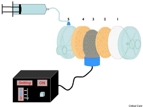
A diagram of the Performer, showing the different parts of the active passive heat moisture exchanger. (1) The single antimicrobial filter; 2 and 4, the two cellulose membranes (hygroscopic and hydrophobic elements); 3, thin metal element with the small holes (diameter 0.3–0.5 cm); and 5, port through which the water is added. The control box (the Provider) for the heating plate has three settings (1, 2 and 3) for temperatures of 40, 50 and 60°C.
The authors did not have any financial interest in any of the devices tested.
Experimental protocol
Humidifiers were tested in random order. Measurements in vitro were taken every 15 min up to 1 hour, and in the in vivo study they were taken after 1 hour of use. The long-term efficiency of the active Performer, with the provider set at level II of heating (50°C), was also evaluated after 12 hours of use in patients.
In vitro
Figure 2 shows the test lung model and measuring devices used to test the HMEs. The lung – a 2-l rubber bag (Mallinckrodt) – was connected with a plastic nonconducting tube to a mechanical ventilator (Servo 300 C; Siemens, Solna, Sweden) that emptied in the hot water humidifier (MR 730; Fisher & Paykel, Auckland, New Zealand). The hot water humidifier, used to condition the gas entering the humidifier, was set to mimic normothermic (i.e. temperature 34°C) and hypothermic (i.e. 28°C) conditions. The temperature and humidity output of the lung model were checked before every measurement.
Figure 2.
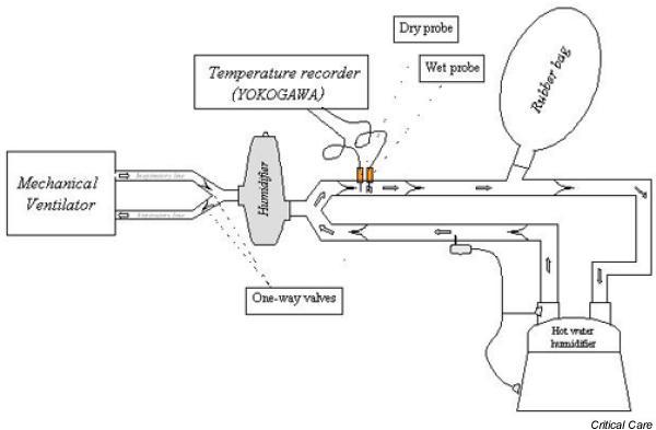
The test lung model and measuring devices used to evaluate heat moisture exchangers in vitro
The ventilator was used to ventilate the test lung with 12 different settings. Combinations of two tidal volumes (0.5 and 1 l), two peak inspiratory flow (0.5 and 1 l/s) and three levels of minute controlled ventilation (5, 10 and 15 l/min), with the ventilator set on volume control, were used. A fractional inspired oxygen of 1 was used throughout. To stabilize the system before taking any measurements, the model lung was ventilated for 2 hours without applying any HME.
The Performer was heated by the Provider set at level III (60°C) and sterile water was added by a pump syringe set to deliver a volume of 6, 7, or 8 ml each hour for minute ventilations of 5, 10 and 15 l/min, respectively (manufacture's recommendation). Two HMEs were tested in each condition.
At the start and after 1 hour, for each humidifier and setting, the airflow resistances were measured by dividing the difference between the inspiratory peak and plateau pressure by the inspiratory flow [17]. The gas flow rate was measured using a heated pneumotachograph (Fleish No 2; Fleish, Lausanne, Switzerland) inserted before the filter in the circuit. The airway pressure was measured using a pressure transducer (MPX 2010 DP; Motorola, Phoenix, AZ, USA).
The room temperature was 24–26°C.
In vivo
Patients with acute lung injury during volume controlled mechanical ventilation were eligible for the study. They were sedated with diazepam (0.03–0.15 mg/kg per hour) and paralyzed with pancuronium (0.05–0.1 mg/kg per hour). Exclusion criteria were body temperature below 34°C or a bronchopleural fistula. The Institutional Review Board of our hospital approved the study, and informed consent was obtained from the patients' next of kin.
The patients were ventilated with a Servo 300 C mechanical ventilator (Siemens) using a standard ventilator circuit. Respiratory rates and tidal volumes were adjusted to maintain arterial carbon dioxide tension at around 40–45 mmHg; oxygen fraction and positive end-expiratory pressure (PEEP) were adjusted to maintain an arterial oxygen tension of at least 80 mmHg.
Unlike in the in vitro study, the Performer was also evaluated at the three Provider levels (levels I, II and III of heating, or 40, 50 and 60°C), and a constant volume of sterile water of 7 ml was added each hour.
Hygrometric measurements
AH and relative humidity were measured during the inspiratory phase in the in vitro and in vivo studies. The Performer, the Hygroster, or the Hygrobac was placed between the Y piece of the ventilator circuit and the test lung or the patient. A device to separate the inspiratory and expiratory gas flows, by four unidirectional valves, was inserted between the humidifiers and the lung model or the patient.
The psychometric method is the one most commonly used by clinicians to measure humidity [18]; it is based on two thermal probes – a dry and a wet one [19]. We used platinum resistance temperature detectors; these exhibited very good accuracy, with an error of 0.3°C and without any variations with time. The two probes were placed on the inspiratory side after the filter in the circuit. Thus, the probe always had to measure the same amount of flow (i.e. same velocity of air), without causing any artefacts in measurements.
Temperatures were electronically measured, displayed and printed on a chart recorder (Yokogawa, Tokyo, Japan). Subsequently, the measurements were analyzed from the chart recorder. The dry probe measures the actual gas temperature. The wet probe is coated with cotton that is wet with sterile water. The evaporation of the sterile water is proportional to the dryness of the gas, and so the difference in temperature between the dry and wet probe is related to the dryness of the gas [19].
At the start of the measurements, we inserted the two probes in a solution of water plus ice to test the offset with respect to a 0°C reading; we checked the two probes (without the wet cotton) in room air; and we verified that the offset was maintained, with no significant variations. We used this offset (in the order of 0.1–0.2°C) to correct the measurements obtained during the study.
In each condition, the average of three or four readings from the wet and dry probe was computed.
Statistical analysis
All data are expressed as mean ± standard deviation. For the in vitro study, we compared the three HMEs using a one-way analysis of variance for repeated measures, followed when appropriate by post hoc multiple comparisons, performed using paired t-test with Bonferroni's correction. Comparisons within the same HME were done using three-way analysis of variance for repeated measures, followed when appropriate by post hoc multiple comparisons, performed using paired t-test with Bonferroni's correction.
Results
In vitro
The temperature and AH of expiratory gases reaching the model lung side of the humidifiers were, respectively, 32.4 ± 0.1°C and 35.6 ± 0.2 mgH2O/l in normothermic conditions, and 26.7 ± 0.2°C and 25.9 ± 0.1 mgH2O/l in hypothermic conditions, with no differences between the settings and the devices tested. This indicates good stability of the test lung model.
Normothermic conditions
The temperature and AH of the inspired gases differed significantly between the devices. In every condition tested, passive Performer and Hygrobac provided a significantly higher temperature and AH in inspired gases than did the Hygroster (Fig. 3). At a minute ventilation of 10 l/min, passive Performer, Hygrobac and Hygroster all had significantly higher temperature and AH than at minute ventilations of 5 and 15 l/min. Increasing the tidal volume decreased the temperature and AH with the passive Performer.
Figure 3.
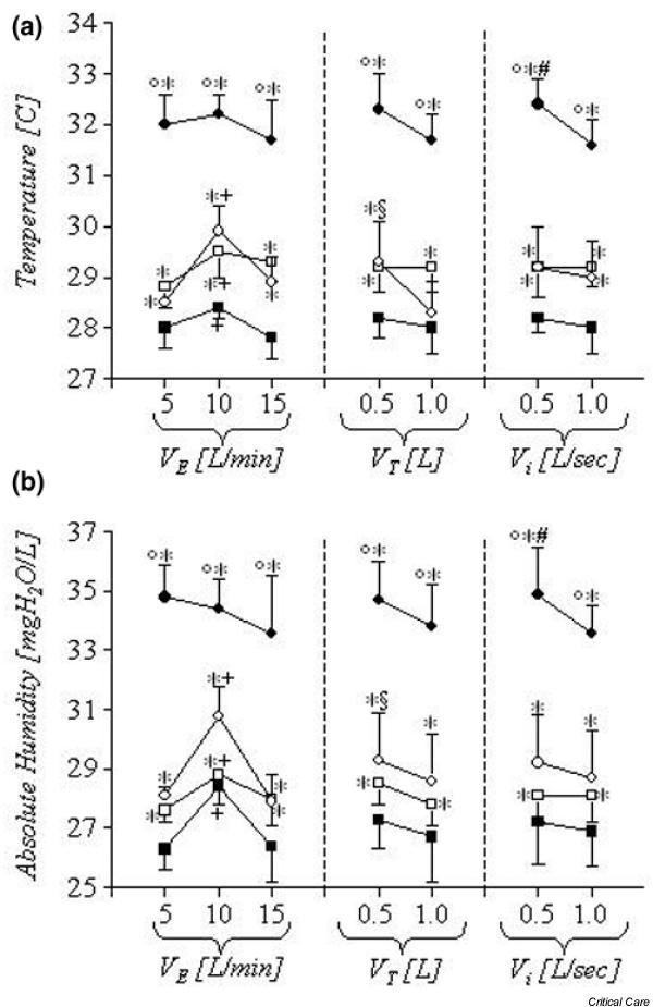
In vitro study in normothermic conditions. (a) Temperatures of the active Performer, and (b) absolute humidity for the Performer (black circle), passive Perfomer (empty circle), Hygrobac (empty square) and Hygroster (black square). Data are presented as means ± standard deviation. Between the devices: °P < 0.05 versus passive Performer and Hygrobac; *P < 0.05 versus Hygroster. Within the same device: +P < 0.05 versus VE 5 and 15 l/min; §P < 0.05 versus VT 1 l; #P < 0.05 versus Vi 1 l/s. VE = minute ventilation; Vi = peak inspiratory flow; VT = tidal volume.
Active Performer had a significantly higher temperature and AH than did passive Performer, Hygrobac and Hygroster, which was unaffected by minute ventilation or tidal volume (Fig. 3). Increasing the peak inspiratory flow rate with active Performer lowered the temperature and AH. The temperature and AH, measured every 15 min in each patient, remained stable and were no different after 1 hour of use in each setting with the different devices.
Hypothermic conditions
Passive Performer, Hygrobac and Hygroster gave similar temperature and AH in the majority of tested conditions (Fig. 4). At a minute ventilation of 10 l/min, passive Performer, Hygrobac and Hygroster had a higher AH than at 5 l/min. Changing the tidal volume or the peak inspiratory flow did not affect the temperature and AH in any humidifier.
Figure 4.
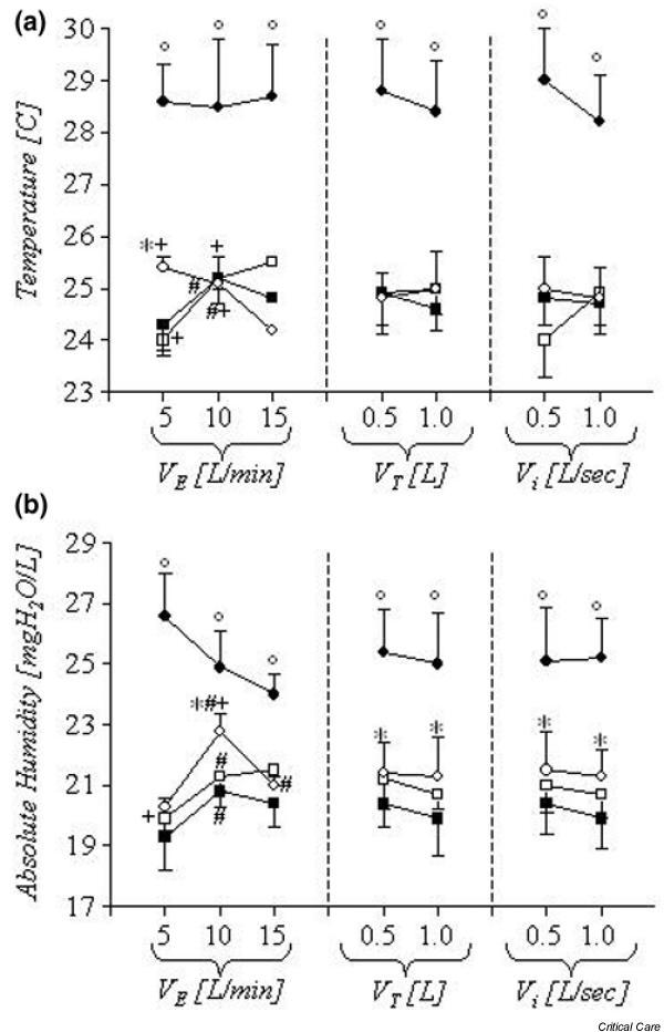
In vitro study conducted in hypothermic conditions. (a) Temperatures of the active Performer and (b) absolute humidity for the Performer (black circle), passive Perfomer (empty circle), Hygrobac (empty square) and Hygroster (black square). Data are presented as means ± standard deviation. Between the devices: °P < 0.05 versus passive Perfomer, Hygrobac and Hygroster. Within the same device: #P < 0.05 versus VE 5 l/min; +P < 0.05 versus VE 15 l/min. VE = minute ventilation; Vi = peak inspiratory flow; VT = tidal volume.
Like under normothermic conditions, the temperature and AH with active Perfomer were significantly higher than with passive Performer, Hygrobac and Hygroster (Fig. 4). Active Performer had a higher AH at a minute ventilation of 5 l/min than at 15 l/min.
Airflow resistance
At the start of the experiment the mean airflow resistances for passive Performer, active Performer, Hygrobac and Hygroster were 1.5 ± 0.8, 1.6 ± 0.5, 1.6 ± 0.3 and 2.1 ± 0.9 cmH2O/l per s. Increasing the peak inspiratory flow from 0.5 to 1.0 l/s significantly increased the airflow resistance for passive Performer from 0.95 ± 0.2 to 2.0 ± 0.8 cmH2O/l per s, for active Perfomer from 1.2 ± 0.1 to 1.9 ± 0.5 cmH2O/l per s, for Hygrobac from 1.4 ± 0.2 to 1.8 ± 0.1 cmH2O/l per s, and for Hygroster from 1.3 ± 0.2 to 2.9 ± 0.3 cmH2O/l per s. The airflow resistances at the start of the experiment and after 1 hour of use were similar.
In vivo
We studied 42 patients (mean age 60.5 ± 16.9 years) who were intubated and mechanically ventilated. The tidal volume was 0.60 ± 0.17 l and a minute ventilation of 8.8 ± 2.4 l/min was applied, with a PEEP of 8 ± 3 cmH2O. This resulted in an arterial oxygen tension/inspired fractional oxygen ratio of 283 ± 72 mmHg. The body temperature was 37.5 ± 0.8°C, with a room temperature of 25.1 ± 1.4°C.
Passive Performer and the Hygrobac had significantly higher airway temperature and AH than did the Hygroster (Fig. 5). Active Performer, regardless of the level of heating, always had a higher temperature and AH than did passive Performer, Hygrobac and Hygroster. Active Performer, with the Provider set at level III of heating (60°C), had the greatest temperature and AH (Fig. 5). There was no difference in the temperature and AH after 12 hours of continuous use.
Figure 5.
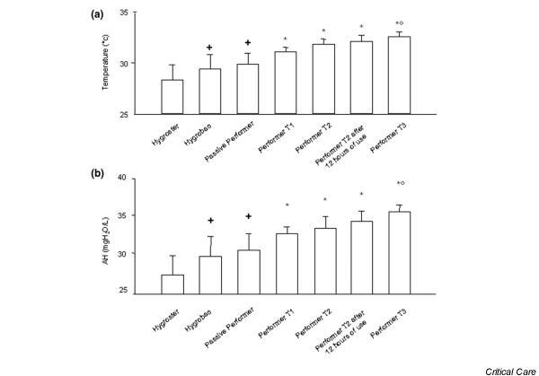
In vivo study. (a) Temperature for the Hygroster, Hygrobac, passive Performer and active Performer with the Provider set at T1 (40°C), active Performer with the provider set at T2 (50°C), active Performer with the Provider set at T2 after 12 hours of use, and active performer with the Provider set at T3 (60°C). (b) Absolute humidity (AH) for the Hygroster, Hygrobac, passive Performer, active Performer with the Provider set at T1, active Performer with the Provider set at T2, active Performer with the Provider set at T2 after 12 hours of use, and active performer with the Provider set at T3. Data are presented as means ± standard deviation. +P < 0.05 versus Hygroster; *P < 0.05 versus Hygroster, Hygrobac and passive Performer; °P < 0.05 versus active Performer set at T1.
Discussion
The Performer, a new type of active HME, when used as a passive device, provided airway conditioning at least comparable to that with other HMEs. When used as an active device, however, the efficiency of the Performer increased beyond that of a purely passive HME.
The optimal level of conditioning remains debatable [1,2]. For example, Williams and coworkers [20] suggested that heating and humidifying the inspired gas to the natural targets of core temperature (i.e. 37°C) and AH of 44 mgH2O/l reduces heat and moisture exchange with the mucosa and maximizes mucociliary clearance. However, Tsuda and coworkers [21] found airway damage after only 3 hours inhalation of gas at 35°C with AH of 39 mgH2O/l.
In normal conditions the temperature of expired gases ranges between 28 and 32°C with an AH of 27–33 mgH2O/l [22,23], and thus a temperature of 29–33°C with an AH of 28–35 mgH2O/l should be adequate for inspired gases [2]. Two previous studies [12,13] showed that a HME that is able to deliver a mean AH of 30 mgH2O/l could safely be used for up to 7 days in mechanically ventilated patients. These data suggest that, in general, it is not necessary to provide an AH greater than 30 mgH2O/l.
The Performer as a 'passive' device
When used as a passive device, the Performer provided an average absolute humidity of 28.9 ± 0.9 mgH2O/l in vitro during normothermic conditions and 30.7 ± 1.6 mgH2O/l in vivo. The passive Performer was consistently more efficient than the Hygroster and was comparable to the Hygrobac. Moreover, in all conditions tested, the Hygroster delivered a temperature and AH significantly lower than that with the Hygrobac – a feature others have noted [18].
Several clinical studies have found a satisfactory AH (i.e. ≥ 30 mgH2O/l) when using HMEs in patients with high minute ventilation (between 10.5 and 16.5 l/min) [19,23,24]. In the present study we found that increasing or decreasing the minute ventilation above or below 10 l/min resulted in a marked reduction in AH, to even below the commonly suggested limits [1,2].
We also investigated the effects of severe hypothermia on HME efficiency. We found that hypothermia markedly reduced the efficiency of HMEs. These findings confirm that HMEs should be used with caution in severely or moderately hypothermic patients.
The Performer as an 'active' device
When the Performer was used as an 'active' humidifier it provided higher levels of humidification (AH range 30–36 mgH2O/l), independently of minute ventilation and expiratory AH, unlike the other HMEs. Active Performer also showed good stability in patients without any loss of efficiency after 12 hours of continuous use, and reached a steady state in terms of temperature and humidity after only 15 min of use.
To improve the efficiency of HMEs, use of two other different devices – the Booster (TomTec, Kapellen, Belgium) and the Humid-Heat (Gibeck, Upplands wasby, Sweden) – has been proposed. The Booster is a small heating element that is placed between the HME and the patient. The heating element, powered electrically, is covered by a Gore-Tex membrane, in which water (added from the outside) vaporizes and thus increases the AH of inspired gases [25]. Patients ventilated with Booster for 96 hours had higher temperature and AH of inspired gases (2–3°C and 2–3 mgH2O/l more than with a standard HME), and there was no bacterial colonization of the ventilatory circuit [25]. Similar in design to the Performer is the Humid-Heat, in which external water and heat are added to the patient side of a HME circuit. The Humid-Heat can boost temperature and AH up to 37°C and 44 mgH2O/l, which are close to the levels achieved with conventional hot water humidifiers [8,26,27]. In addition, if the water supply runs out, all of these devices continue to work as passive HMEs, avoiding the possibility that dry gases will be delivered.
In severe hypothermic conditions, active Perfomer was more efficient than the Hygroster, although the AH was lower than the minimum required levels. In these extreme conditions, hot water humidifiers should be used.
Airflow resistances and dead space
The presence of any HMEs in the ventilatory circuit increases the airflow resistance [28]. We found similar low inspiratory airflow resistances with the Performer, Hygrobac and Hygroster, with no difference between the beginning of the experiment and after 1 hour of use. After increasing the peak inspiratory flow to a very high level (1 l/s) the airflow resistance was still low, with an average value of 2.3 ± 0.6 cmH2O/l per s. This additional resistance, which is lower than that with an endotracheal tube, is not likely to play any significant role during controlled mechanical ventilation [29] and can be considered acceptable during assisted ventilation [28].
Because of the internal volume of HMEs, ranging from 50 to 90 ml, the dead space of the ventilator circuit is increased [16,30], causing an increase in carbon dioxide levels, especially during low tidal volume ventilation [31]. HMEs also cause an increase the inspiratory work of breathing, with an increase in intrinsic PEEP [16,32]. Consequently, because they increase the resistive dead space load, use of HMEs cannot be recommended in patients who are weak or difficult to wean, unless the level of ventilator assistance is increased [16].
Limitations
Potential limitations of the study must be addressed. First, we did not examine the effects on gas exchange, respiratory mechanics, secretions, or microbiological contamination of the ventilator circuit. However, during the study we did not observe any obstruction of the endotracheal tube. Second, we tested the Performer in vivo only at a single minute ventilation and for a relatively brief period of only 12 hours. Third, we did not have any data from a heated humidifier because the heated humidifier, being an active system, can deliver gas at a broad range of temperatures and AHs (i.e. with a relative humidity of 100%), independent from the ventilatory settings.
Possible indications and advantages of the Performer
Although HMEs may be safely used during long-term ventilation [12,13], many centres do not routinely use HMEs for fear of tube obstruction and insufficient humidification [33]. In the presence of thick secretions, the use of HMEs, because of water loss from the airways, may increase the risks for tube occlusion, air trapping and hypoventilation [2]. Because the Performer can deliver higher AH than other HMEs, it may be useful in patients in whom the use of HMEs appears to worsen the clinical characteristics of secretions and in hypothermic patients who would otherwise require the use of heated humidifiers.
Although we did not directly evaluate the cost, in agreement with a previous study that evaluated a similar active HME (Humid-Heat) [8], the Performer should allow a reduction in daily sterile water consumption, avoidance of condensate in the ventilator circuit, a decrease in changes of ventilator circuits, and a reduction in nurses' workload.
Conclusion
The Performer exhibited good stability (up to 12 hours) in maintaining adequate levels of temperature and AH in the inspired gases. These features were less dependent on ventilator settings than with other HMEs. Because an AH greater than 30 mgH2O/l is not necessary in the majority of mechanically ventilated patients, we believe that active HMEs are useful only in those patients with variable minute ventilation or with thickened secretions when passive HMEs have failed or when moderate hypothermia is present.
Key messages
• A new active heat moisture exchanger is able to increase the temperature and absolute humidity of the inspired gases compared to the commonly used heat moisture exchangers. However, the use of active heat moisture exchangers should be restricted in patients with variable minute ventilation or in presence of thickened secretions or hypothermia.
Competing interests
None declared.
Abbreviations
AH = absolute humidity; HME = heat moisture exchanger; PEEP = positive end-expiratory pressure.
References
- Shelly MP, Lloyd GM, Park GR. A review of the mechanism and methods of humidification of inspired gases. Intensive Care Med. 1988;14:1–9. doi: 10.1007/BF00254114. [DOI] [PubMed] [Google Scholar]
- Branson RD. Humidification for patients with artificial airways. Respir Care. 1999;44:630–642. [Google Scholar]
- Marfatia S, Donhaoe PK, Hendren WH. Effects of dry and humidified gases on the respiratory epithelium in rabbits. J Pediatr Surg. 1975;10:583–592. doi: 10.1016/0022-3468(75)90360-7. [DOI] [PubMed] [Google Scholar]
- Noguchi H, Tkumi Y, Aochi O. A study of humidification in tracheostomised dogs. Br J Anaesth. 1973;45:844–848. doi: 10.1093/bja/45.8.844. [DOI] [PubMed] [Google Scholar]
- Chalon J, Loew DAY, Malebranche J. Effect of dry anesthetic gases on tracheobronchial epithelium. Anesthesiology. 1972;37:338–345. doi: 10.1097/00000542-197209000-00010. [DOI] [PubMed] [Google Scholar]
- Cook D, Ricard JD, Reeve B, Randall J, Wigg M, Brochard L, Dreyfuss D. Ventilator circuit and secretion management strategies: a Franco-Canadian survey. Crit Care Med. 2000;28:3547–3554. doi: 10.1097/00003246-200010000-00034. [DOI] [PubMed] [Google Scholar]
- Kirton OC, DeHaven B, Morgan J, Morejon O, Civetta J. A prospective, randomized comparison of an in-line heat and moisture exchanger filter and heated wire humidifiers. Chest. 1997;112:1055–1059. doi: 10.1378/chest.112.4.1055. [DOI] [PubMed] [Google Scholar]
- Larsson A, Gustafsson A, Svanborg L. A new device for 100 per cent humidification of inspired air. Crit Care. 2000;4:54–60. doi: 10.1186/cc651. [DOI] [PMC free article] [PubMed] [Google Scholar]
- Branson RD, Davis K, Brown R, Rashkin M. Comparison of three humidification techniques during mechanical ventilation: patient selection, cost, and infection considerations. Respir Care. 1996;41:809–816. [Google Scholar]
- Craven DE, Goularte TA, Make BJ. Contaminated condensate in mechanical ventilator circuits: A risk factor for nosocomial pneumonia? Am Rev Respir Dis. 1984;29:625–628. [PubMed] [Google Scholar]
- Dreyfuss D, Djedaini K, Gros I, Mier L, Le Bourdelles G, Cohen Y, Estagnasie P, Coste F, Boussougant Y. Mechanical ventilation with heat and moisture exchangers: effects on patient colonization and incidence of nosocomial pneumonia. Am J Respir Crit Care Med. 1995;151:986–992. doi: 10.1164/ajrccm/151.4.986. [DOI] [PubMed] [Google Scholar]
- Ricard JD, Le Miere E, Markowicz P, Lasry S, Saumon G, Djedaini K, Coste F, Dreyfuss D. Efficiency and safety of mechanical ventilation with a heat and moisture exchanger changed only once a week. Am J Respir Crit Care Med. 2000;161:104–109. doi: 10.1164/ajrccm.161.1.9902062. [DOI] [PubMed] [Google Scholar]
- Djedaini K, Billiard M, Mier L, Le Bourdelles G, Brun P, Markowicz P, Estagnasie P, Coste F, Boussougant Y, Dreyfuss D. Changing heat and moisture exchangers every 48 hours rather than 24 hour does not affect their efficacy and the incidence of nosocomial pneumonia. Am J Respir Crit Care Med. 1995;152:1562–1569. doi: 10.1164/ajrccm.152.5.7582295. [DOI] [PubMed] [Google Scholar]
- Unal N, Kanhai JK, Buijk SL, Pompe JC, Holland WP, Gultuna I, Ince C, Saygin B, Bruining HA. A novel method evaluation of three heat-moisture exchangers in six different ventilator settings. Intensive Care Med. 1998;24:138–146. doi: 10.1007/s001340050535. [DOI] [PubMed] [Google Scholar]
- Roustan JP, Kienlen J, Aubas P, Aubas S, du Cailar J. Comparison of hydrophobic heat and moisture exchangers with heated humidifier during prolonged mechanical ventilation. Intensive Care Med. 1992;18:97–100. doi: 10.1007/BF01705040. [DOI] [PubMed] [Google Scholar]
- Girault C, Breton L, Richard JC, Tamion F, Vandelet P, Aboab J, Leroy J, Bonmarchand G. Mechanical effects of airway humidification devices in difficult to wean patients. Crit Care Med. 2003;31:1306–1311. doi: 10.1097/01.CCM.0000063284.92122.0E. [DOI] [PubMed] [Google Scholar]
- D'Angelo E, Calderini E, Torri G, Ribatto FM, Bono D, Milic-Emili J. Respiratory mechanics in anesthetized paralyzed humans: effect of flow, volume and time. J Appl Physiol. 1989;67:2556–2664. doi: 10.1152/jappl.1989.67.6.2556. [DOI] [PubMed] [Google Scholar]
- Thiery G, Boyer A, Pigne E, Salah A, De Lassence A, Dreyfuss D, Ricard JD. Heat and moisture exchangers in mechanically ventilated intensive care patients: a plea for an independent assessment of their performance. Crit Care Med. 2003;31:699–704. doi: 10.1097/01.CCM.0000050443.45863.F5. [DOI] [PubMed] [Google Scholar]
- Martin C, Thomachot L, Quinio B, Viviand X, Albanese J. Comparing two heat and moisture exchangers with one vaporizing humidifier in patients with minute ventilation greater than 10 L/min. Chest. 1995;107:1411–1415. doi: 10.1378/chest.107.5.1411. [DOI] [PubMed] [Google Scholar]
- Williams R, Rankin N, Smith T, Galler D, Seakins P. Relationship between the humidity and temperature of inspired gas and the function of the airway mucosa. Crit Care Med. 1996;24:1920–1929. doi: 10.1097/00003246-199611000-00025. [DOI] [PubMed] [Google Scholar]
- Tsuda T, Noguchi H, Takimag Y, Aochi O. Optimum humidification of air administered to a tracheostomy in dogs. Br J Anaesth. 1977;49:965–977. doi: 10.1093/bja/49.10.965. [DOI] [PubMed] [Google Scholar]
- Chiumello D, Gattinoni L, Pelosi P. In Yearbook of Intensive Care and Emergency Medicine. Heidelberg: Springer-Verlag; 2002. Conditioning of inspired gases in mechanically ventilated patients. [Google Scholar]
- Martin C, Papazian L, Perrin G. Performance evaluation of three vaporizing humidifiers and two heat and moisture exchangers in patient with minute ventilation >10 l/min. Chest. 1992;102:1347–1350. doi: 10.1378/chest.102.5.1347. [DOI] [PubMed] [Google Scholar]
- Martin C, Papazian L, Perrin G, Saux P, Gouin F. Preservation of humidity and heat of respiratory gases in patients with a minute ventilation greater than 10 L/min. Crit Care Med. 1994;22:1871–1876. [PubMed] [Google Scholar]
- Thomachot L, Viviand X, Boyadjiev I, Vialet R, Martin C. The combination of a heat and moisture exchanger and a BoosterTM: a clinical and bacteriological evaluation over 96 h. Intensive Care Med. 2002;28:147–153. doi: 10.1007/s00134-001-1193-2. [DOI] [PubMed] [Google Scholar]
- Branson R, Campbell RS, Johannigman JA, Ottaway M, Davis K, Luchette FA, Frame S. Comparison of conventional heated humidification with a new active hygroscopic heat and moisture exchanger in mechanically ventilated patients. Respir Care. 1999;44:912–917. [Google Scholar]
- Kapadia F, Shelly MP, Anthony JM, Park GR. An active heat and moisture exchanger. Br J Anaesth. 1992;69:640–642. doi: 10.1093/bja/69.6.640. [DOI] [PubMed] [Google Scholar]
- Chiaranda M, Verona L, Pinamonti O, Dominioni L, Minoja G, Conti G. Use of heat and moisture exchanging (HME) filters in mechanically ventilated ICU patients: influence on airway flow-resistance. Intensive Care Med. 1993;19:462–466. doi: 10.1007/BF01711088. [DOI] [PubMed] [Google Scholar]
- Conti G, De Blasi RA, Rocco M, Pelaia P, Antonelli M, Bufi M, Mattia C, Gasparetto A. Effects of the heat moisture exchangers on dynamic hyperinflation of mechanically ventilated COPD patients. Intensive Care Med. 1990;16:441–443. doi: 10.1007/BF01711222. [DOI] [PubMed] [Google Scholar]
- Pelosi P, Solca M, Ravagnan I, Tubiolo D, Ferrario L, Gattinoni L. Effects of heat and moisture exchangers on minute ventilation, ventilatory drive, and work of breathing during pressure support ventilation in acute respiratory failure. Crit Care Med. 1996;24:1184–1188. doi: 10.1097/00003246-199607000-00020. [DOI] [PubMed] [Google Scholar]
- Prat G, Renault A, Tonnelier JM, Goetghebeur D, Oger E, Boles JM, L'Her E. Influence of the humidification device during acute respiratory distress syndrome. Intensive Care Med. 2003;29:2211–2215. doi: 10.1007/s00134-003-1926-5. [DOI] [PubMed] [Google Scholar]
- Le Bourdelles G, Mier L, Fiquet B, Djedaini K, Saumon G, Coste F, Dreyfuss D. Comparison of the effects of heat and moisture exchangers and heated humidifiers during mechanical ventilation and gas exchange during weaning trials from mechanical ventilation. Chest. 1996;110:1294–1298. doi: 10.1378/chest.110.5.1294. [DOI] [PubMed] [Google Scholar]
- Ricard JD, Cook D, Griffith L, Brochard L, Dreyfuss D. Physician' attitude to use heat and moisture exchangers or heated humidifiers: a Franco-Canadian survey. Intensive Care Med. 2002;28:719–725. doi: 10.1007/s00134-002-1285-7. [DOI] [PubMed] [Google Scholar]


