Abstract
An immobilization scheme for bacterial cells is described, in which the antimicrobial peptide cecropin P1 was used to trap Escherichia coli K-12 and O157:H7 cells on microtiter plate well surfaces. Cecropin P1 was covalently attached to the well surfaces, and E. coli cells were allowed to bind to the peptide-coated surface. The immobilized cells were detected colorimetrically with an anti-E. coli antibody-horseradish peroxidase conjugate. Binding curves were obtained in which the signal intensities were dependent upon the cell concentration and upon the amount of peptide attached to the well surface. After normalization for the amount of peptide coupled to the surface and the relative binding affinity of the antibody for each strain, the binding data were compared, which indicated that there was a strong preference for E. coli O157:H7 over E. coli K-12. The cells could be immobilized reproducibly at pH values ranging from 5 to 10 and at ionic strengths up to 0.50 M.
Cecropins are a class of antimicrobial peptides which exhibit selectivity for prokaryotic cells over eukaryotic cells (24). They are largely alpha-helical and consist of an amphipathic N-terminal helix and a hydrophobic C-terminal helix (7, 21). The mechanism by which these peptides kill is not fully understood, however. It has been demonstrated that insect cecropins can aggregate at high peptide/lipid ratios (13). Positively charged residues on the N-terminal helix interact preferentially with acidic phospholipid membranes (7, 8). The hydrophobic C-terminal helix can interact with hydrophobic lipids, allowing the penetration of the lipid bilayer in a manner similar to that proposed for magainins and dermaseptins (18, 26). Cecropins have also been shown to form ion channels in planar lipid bilayers (5, 6). However, conflicting data have been reported; attenuated total reflectance Fourier transform infrared spectroscopy studies have shown that cecropin P1 orients itself parallel to planar lipid bilayers, suggesting that the C-terminal helix does not penetrate into the membrane to form a channel (9). This supports a carpet mechanism, in which the peptide binds to negatively charged elements and orients itself on the membrane surface in such a way as to destabilize it, leading to cell lysis. Finally, cecropin P1 has been shown to kill only when sufficient peptide is present, enabling the formation of a monolayer over the cell (25). Here we propose that the inherent cell binding properties of cecropin P1 can be used to immobilize bacterial cells on solid surfaces.
Immobilized cells have been used in a wide spectrum of applications, such as degradation of phenols (14), production of ethanol (15), and biosensors (19). The methods of cell immobilization include nonspecific adsorption, covalent attachment, and entrapment. Immobilization by nonspecific adsorption may require a long incubation time (up to 12 h) and is sensitive to changes in pH, temperature, and ionic strength. All of these factors can result in removal of cells from the surface (29). Covalent attachment is often achieved by activating some surface with a cross-linker, such as glutaraldehyde, and then attaching the cell through surface amine groups (10). Cells can also be immobilized by the formation of coordinate bonds with metal-activated supports (3). Although effective, covalent attachment can result in a loss of cell activity and viability. Entrapment can be accomplished by forming gels around the cells. Gels have been made from alginates (2), carrageenans (4), and polyacrylamide (22). Materials such as cellulose acetate have been used to form membranes around cells. None of these methods utilize any specific affinity binding mechanisms that may produce cell immobilization strategies with the ability to effortlessly regenerate active surfaces.
We aim to demonstrate that the affinity of cecropin P1 for bacterial cells may provide an alternative way of immobilizing such cells on surfaces such as microplate wells. This affinity-based approach would alleviate the need for covalent attachment, trapping, and adsorption, as well as the problems associated with each method. The fact that a large dose of the peptide is required to lyse cells suggests that, if immobilized in smaller quantities, the peptide can capture cells on a surface without disrupting the cell membrane. Here, a method for immobilizing Escherichia coli cells on microtiter plate well surfaces with the antimicrobial peptide cecropin P1 is described. The peptide was immobilized through amine residues in maleic anhydride-functionalized microtiter plate wells. E. coli K-12 and a nonpathogenic strain of E. coli O157:H7 were immobilized on the peptide-coated surface and subsequently detected by using a rabbit anti-E. coli antibody-horseradish peroxidase conjugate. Binding curves were obtained with signal intensities that were dependent upon the concentration of cells and the amount of peptide coupled to the surface. These curves indicated that under mild pH conditions, the peptide bound to E. coli O157:H7 more strongly than it bound to E. coli K-12. Cells could be immobilized in a pH range from 5 to 10, and E. coli O157:H7 was more efficiently immobilized at pH 5 to 8. E. coli K-12 could be immobilized over a pH range from 5 to 10. The peptide was not able to bind to cells at ionic strengths greater than 0.5 M.
MATERIALS AND METHODS
Attachment of cecropin P1 to microplate well surfaces.
Cecropin P1(Sigma, St. Louis, Mo.) was dissolved in phosphate-buffered saline (PBS) (100 mM sodium phosphate, 0.1 M NaCl; pH 7.2) at concentrations of 100, 50, and 25 μg/ml. Aliquots (100 μl) of the cecropin P1 solutions were added to the wells of a Reacti-bind maleic anhydride-functionalized microtiter plate (Pierce, Rockford, Ill.). A total of 32 wells were used for each concentration of peptide. The plate was covered and shaken overnight at room temperature, after which the plate wells were rinsed three times with PBS. To cap any unreacted maleic anhydride groups, 10% (vol/vol) ethanolamine in PBS was added to the wells, and the plate was shaken for 1 h at room temperature; this was followed by three more rinses with PBS.
To determine the amount of cecropin P1 bound to the plate well surfaces, sulfosuccinimidyl-4-O-(4,4′-dimethoxytrityl)butyrate (Pierce) was dissolved in 100 mM sodium bicarbonate (pH 8.5) to a final concentration of 0.1 mM. Portions (100 μl) of this solution were added to the first four wells of each block of 32 wells. The same volume of bicarbonate buffer was added to four more wells of each block for use as a blank. The plate was shaken for 1 h at room temperature. The wells were then rinsed three times with PBS, and color development was initiated by addition of 250 μl of 37% perchloric acid. The absorbance at 498 nm was determined. The absorbance data were used to calculate the number of moles of free amine groups.
Immobilization and detection of E. coli K-12 and O157:H7.
In the following procedures, all microplate incubations were carried out at room temperature with mild shaking, unless otherwise specified. The concentrations of viable cells in the cultures were determined by diluting the cultures, plating the dilutions on Luria-Bertani agar plates, and counting the number of resultant colonies. A blocking solution consisting of 50 mM Tris (pH 7.8) supplemented with 1% (wt/vol) bovine serum albumin (BSA) was added in 100-μl aliquots to each well. The plate was shaken at room temperature for 1 h, and the wells were rinsed three times with the Tris buffer. Overnight cultures of E. coli K-12 (Pierce) and nonpathogenic E. coli O157:H7 strain ATCC 43888 (American Type Culture Collection, Rockville, Md.) were grown in Luria-Bertani medium at 37°C with shaking. Serial dilutions of the cell cultures were prepared in Tris buffer. The dilutions were added to the plate wells in 200-μl aliquots. Four replicates of each dilution were used. A blank was made with Tris buffer and no cells. The plate was then incubated for 2 h, and this was followed by three rinses with Tris buffer. One hundred microliters of rabbit anti-E. coli antibody-horseradish peroxidase conjugate (Virostat Inc., Portland, Maine) diluted 1:500 in Tris buffer supplemented with 6% (wt/vol) BSA was added to each well, and the plate was incubated for 1 h. The wells were rinsed six times with 10 mM Tris (pH 7.8)-0.15 M NaCl-0.05% (vol/vol) Tween 20. Immunopure 3,3′,5,5′-tetramethylbenzidine substrate (Pierce) was prepared according to the manufacturer's instructions. The substrate solution was added in 100-μl aliquots to the wells. The color was allowed to develop for 25 min, and the reaction was stopped by addition of an equal volume of 2 M sulfuric acid. The absorbance at 450 nm was then determined by using a reference wavelength of 550 nm.
Comparison of the relative binding affinities of rabbit anti-E. coli for E. coli O157:H7 and K-12.
Overnight cultures of E. coli O157 (nonpathogenic) and E. coli K-12 were grown with shaking at 37°C in Luria-Bertani broth until an optical density at 600 nm of approximately 2.0 was reached. The cells were diluted 1:10 in 50 mM sodium bicarbonate (pH 9.6) and added to the wells of a Nunc Maxisorp microtiter plate (Fisher, Hanover Park, Ill.). The amount added corresponded to a saturating amount of cells. The plate was then covered and incubated for 3 h at 37°C. The wells were washed three times with PBS and subsequently blocked at room temperature with 200 μl of 1% BSA for 1 h. The wells were rinsed again with PBS. Rabbit anti-E. coli antibody-horseradish peroxidase conjugate was then diluted 1:50, 1:100, 1:200, 1:400, 1:800, 1:1,600, 1:3,200, and 1:6,400 in 0.75% BSA. Three 100-μl aliquots of each dilution were added to the wells, and the plate was incubated at room temperature for 1 h. The wells were rinsed four times with PBS. Color development and measurement of absorbance were carried out as described above.
The data were plotted as absorbance versus the logarithm of the dilution factor of the antibody. The resulting binding curves were then fitted to a four-parameter logistic equation with ABI Prism 3.0. The slope at the center of each curve was determined and used as a measure of the relative binding affinity of the antibody for the two E. coli strains.
pH study.
Overnight cultures of E. coli K-12 and O157:H7 were diluted in the following buffers: 50 mM sodium acetate (pH 5.0), 50 mM morpholineethanesulfonic acid (MES) (pH 6.0), 50 mM sodium phosphate (pH 7.0), 50 mM Tris (pH 8.0), 50 mM 2-(cyclohexylamino)ethanesulfonic acid (pH 9.0), and 50 mM 3-(cyclohexylamino)-1-propanesulfonic acid (pH 10.0). Immobilization and detection of the cells were carried out as described above.
Ionic strength study.
Overnight cultures of E. coli K-12 and O157:H7 were diluted in 50 mM Tris (pH 7.8) supplemented with 0.05, 0.1, 0.25, 0.5, and 1.0 M NaCl. The ionic strength of each solution was calculated based on the buffer composition and the concentration of NaCl. Immobilization and detection of the cells were carried out as described above.
RESULTS
In order to demonstrate that the interaction between cecropin P1 and bacterial cells could be used to develop an affinity-based immobilization scheme for the cells, binding curves were obtained for experiments in which increasing concentrations of bacterial cells were immobilized in microtiter plate wells coated with different amounts of cecropin P1. Figure 1 shows the binding curves obtained for the immobilization of E. coli O157:H7. As expected, both the steepness of the curve and the maximum signal decreased as the amount of peptide on the surface decreased, indicating that the signal was dependent on the binding of the peptide to the cells and not due to nonspecific adsorption of the cells on the plate well surfaces. Binding curves obtained with very small amounts of cecropin P1 or no cecropin P1 gave signals that were not significantly different from the background signals (data not shown). The binding curves for E. coli K-12 are shown in Fig. 2. The same trend was observed, but the curve steepness and maximum signals suggest that cecropin P1 did not bind to the K-12 strain as tightly as it bound to the O157:H7 strain. An interesting feature of these curves is that no saturation occurred with smaller amounts of peptide. Assuming a standard Scatchard model of binding, one would expect to see saturation at lower concentrations of cells with smaller amounts of peptide on the surface. However, this is not the case. In these experiments and in the development and optimization of the methods used, this phenomenon always occurred. The reasons for this remain unclear.
FIG. 1.
Immobilization of E. coli O157:H7 on cecropin P1-coated microplate wells. Cells were immobilized in wells containing 33 ± 1 pmol (○), 26 ± 3 pmol (•), and 20 ± 1 pmol (▪) of cecropin P1.
FIG. 2.
Immobilization of E. coli K-12 on cecropin P1. Cells were immobilized in microplate wells containing 40 ± 1 pmol (○), 31 ± 1 pmol (•), and 25 ± 1 pmol (▪) of cecropin P1.
In order to compare the relative binding affinities of E. coli O157:H7 and K-12 for the rabbit anti-E. coli antibody, binding curves for the two strains were determined (data not shown). The slopes of the centers of the two curves were determined and used as a measure of the relative binding affinities of the two strains. The slope of the E. coli O157:H7 curve was approximately twice that of the E. coli K-12 curve. The slopes were then used to adjust the absorbance data for a direct comparison of the binding of the two strains to cecropin P1.
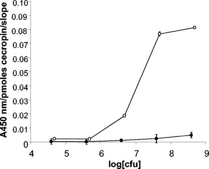
A direct comparison of the E. coli O157 and K-12 binding curves is shown in Fig. 3. For this figure, the signals were normalized with respect to the amount of peptide bound in the wells, as well as the relative binding affinity of the antibody for the strain. The data are plotted as absorbance at 450 nm per picomoles of cecropin per slope, where the slope is the slope calculated from the binding data for each strain with the anti-E. coli antibody. This comparison suggests that cecropin P1, when coupled to a surface, preferentially binds to E. coli O157:H7. Clearly, the fact that the relative binding affinities of the two strains for the antibody differ by a factor of approximately two makes little difference.
FIG. 3.
Comparison of binding curves for E. coli O157:H7 (○) and K-12 (•) with cecropin P1. Data were plotted as absorbance at 450 nm per picomole of cecropin per slope versus log CFU. The number of picomoles of cecropin reflects the amount of peptide coupled to the plate well surface, whereas the slope is the slope of the center of the binding curve for each strain binding to anti-E. coli antibody-horseradish peroxidase conjugate.
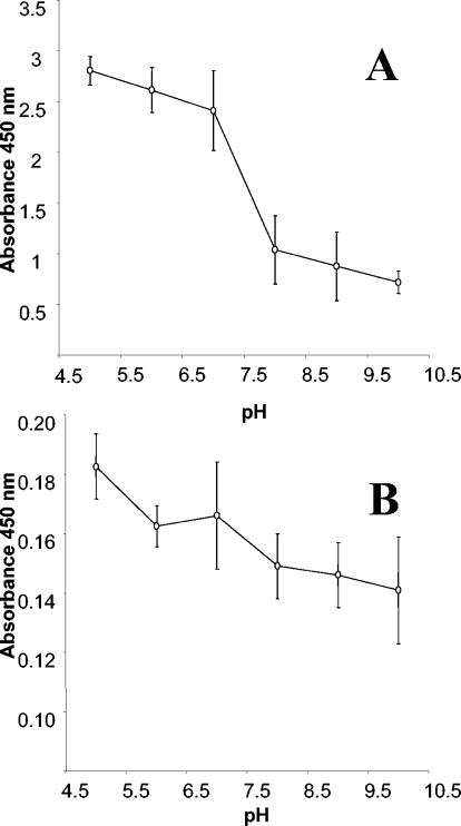
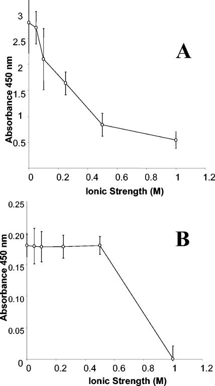
In order to determine the effect of pH on the interaction between cecropin P1 and the cells, both E. coli strains were diluted in buffers with pH values ranging from 5 to 10. The cells were incubated in the plate wells and were detected as described above. Figure 4 shows that cecropin P1 captured cells at pH values from 5 to 10, although E. coli O157:H7 was bound more efficiently at lower pH values (pH 5 to 7). Figure 5 shows the effect of ionic strength on the interaction between the peptide and the cells. E. coli O157:H7 could be immobilized effectively at an ionic strength of only 50 mM, but the signal dropped to less than 80% of the maximum signal at an ionic strength of 0.1 M or more. E. coli K-12 was able to withstand ionic strengths up to 0.5 M.
FIG. 4.
pH profiles for E. coli O157:H7 (A) and K-12 (B) immobilized on cecropin P1.
FIG. 5.
Effect of ionic strength on the binding of E. coli O157:H7 (A) and K-12 (B) to cecropin P1.
DISCUSSION
We demonstrated the feasibility of using the antimicrobial peptide cecropin P1 to immobilize bacterial cells on solid surfaces. Cells could be immobilized at a pH range from 5 to 10, and E. coli O157:H7 was more efficiently immobilized at pH 5 to 7. Since the positively charged residues of cecropin P1 are involved in the initial binding to cell surfaces, it could be supposed that the binding interaction might be stronger at lower pH values, because the population of amine-containing residues would be more highly protonated. The peptide was not able to bind to cells at ionic strengths greater than 0.5 M. The sharp reduction in binding at ionic strengths greater than 0.5 M may have been due to interference with the initial charge-charge interaction between the N-terminal helix of cecropin P1 and cell surface groups or to release of the cell periplasm by an osmotic shock effect. To our surprise, cecropin P1 bound to E. coli O157:H7 cells more strongly than it bound to E. coli K-12 cells. The maximum signal obtained for K-12 was approximately 23-fold lower than that obtained for O157:H7.
Many excellent reports that provide insight into the importance of peptide sequences and structures in antimicrobial activity have been published (for a review see reference 23). In several cases, it has been demonstrated that antimicrobial activity does not correlate with binding affinity (25, 27). When binding affinity and antimicrobial activity are not interrelated, the peptide often binds to the membrane with high affinity but lacks the ability to exhibit antimicrobial activity. Several factors have been implicated in cell binding, including peptide sequence, chirality, hydrophobicity, and helicity (1, 12, 16, 28). Understanding the biophysical properties that drive these peptide-cell interactions is essential for rationally designing peptides with better specificity and selectivity.
In the present study, however, peptide structure remained constant and therefore could not explain the preferential binding observed. More likely, differences in cell surface chemistry between the two strains of E. coli were responsible for the specific binding. Studies of the surface localization of outer membrane constituents indicate that lipopolysaccharide is the most likely site of antimicrobial peptide binding (17, 20, 27). Lipopolysaccharide is an amphipathic molecule with a hydrophobic, well-conserved lipid A region and a highly variable hydrophilic region. The hydrophilic portion consists of an oligosaccharide core, which usually is replaced by the O antigen. Even within a single species like E. coli, the O antigen shows extreme diversity and can therefore contribute considerably to the net charge and chemical composition of the bacterial cell surface (11). The preferential binding observed in these studies may be due to the fact that the K-12 strain lacks both the O and H antigens that O157:H7 possesses.
It is important to explore the selective binding of antimicrobial peptides to cell surfaces in greater detail, in particular with regard to ionic strength, temperature, pH, and peptide sequence. Elucidation of cell type-specific binding domains and essential amino acid motifs could create an opportunity to tailor peptides and peptide hybrids for enhanced selectivity.
Finally, the affinity-based cell capture approach may provide an alternative method of cell immobilization, which does not depend upon covalent attachment, trapping, or adsorption. The potential applications for this approach include immobilization of cells for whole-cell sensing, cell-based detoxification of toxic organic compounds, and study of the binding characteristics of various antimicrobial peptides and cells. The fact that certain antimicrobial peptides bind various microorganisms with different affinities might have applications in sensing and identifying cells.
Acknowledgments
We are grateful to the National Science Foundation IGERT Fellowship program and the Natick Research Development and Engineering Center AH52 program for financial support of this work.
Special thanks are extended to Leonidas Bachas, Steven Arcidiacono, Jason Soares, and Jean Herbert for helpful discussions and suggestions.
Footnotes
This document reports research undertaken at the University of Kentucky and the U.S. Army Research, Development and Engineering Command, Natick Soldier Center, Natick, Mass., and is no. NATICK/TP-03/058 in a series of papers approved for publication by the U.S. Army Research, Development and Engineering Command, Natick Soldier Center.
REFERENCES
- 1.Andreu, D., and R. B. Merrifield. 1985. N-terminal analogues of cecropin A: synthesis, antibacterial activity, and conformational properties. Biochemistry 24:1683-1688. [DOI] [PubMed] [Google Scholar]
- 2.Bucke, C. 1987. Cell immobilization in calcium alginate. Methods Enzymol. 135:175-189. [Google Scholar]
- 3.Cabral, J. M. S., and J. F. Kennedy. 1987. Immobilization of microbial cells on transition metal-activated supports. Methods Enzymol. 135:357-372. [DOI] [PubMed] [Google Scholar]
- 4.Chibata, I., T. Tosa, T. Sata, and I. Takata. 1987. Immobilization of cells in carrageenan. Methods Enzymol. 135:189-198. [DOI] [PubMed] [Google Scholar]
- 5.Christensen, B., J. Fink, R. B. Merrifield, and D. Mauzerall. 1988. Channel-forming properties of cecropins and related model compounds incorporated into planar lipid membranes. Proc. Natl. Acad. Sci. USA 85:5072-5076. [DOI] [PMC free article] [PubMed] [Google Scholar]
- 6.Durell, S. R., G. Raghunathan, and H. R. Guy. 1992. Modeling the ion channel structure of cecropin. Biophys. J. 63:1623-1631. [DOI] [PMC free article] [PubMed] [Google Scholar]
- 7.Gazit, E., W. J. Lee, P. T. Brey, and Y. Shai. 1994. Mode of action of the antibacterial cecropin B2: a spectrofluorometric study. Biochemistry 33:10681-10692. [DOI] [PubMed] [Google Scholar]
- 8.Gazit, E., A. Boman, H. G. Boman, and Y. Shai. 1995. Interaction of the mammalian antimicrobial peptide cecropin P1 with phospholipids vesicles. Biochemistry 34:11479-11488. [DOI] [PubMed] [Google Scholar]
- 9.Gazit, E., I. Miller, P. C. Biggin, M. S. P. Sansom, and Y. Shai. 1996. Structure and orientation of the mammalian antibacterial peptide cecropin P1 within phospholipids membranes. J. Mol. Biol. 258:860-870. [DOI] [PubMed] [Google Scholar]
- 10.Jirku, V., and J. Turkova. 1987. Cell immobilization by covalent linkage. Methods Enzymol. 135:341-357. [Google Scholar]
- 11.Lugtenberg, B., and L. van Alphen. 1983. Molecular architecture and functioning of the outer membrane of Escherichia coli and other gram-negative bacteria. Biochim. Biophys. Acta 737:51-115. [DOI] [PubMed] [Google Scholar]
- 12.Maloy, W. L., and U. P. Kari. 1995. Structure-activity studies on magainins and other host defense peptides. Biopolymers 37:105-122. [DOI] [PubMed] [Google Scholar]
- 13.Mchaourab, H. S., J. S. Hyde, and J. B. Feix. 1994. Binding and state of aggregation of spin-labeled cecropin AD in phospholipids bilayers: effects of surface charge and fatty acyl chain length. Biochemistry 33:6691-6699. [DOI] [PubMed] [Google Scholar]
- 14.Mordocco, A., C. Kuek, and R. Jenkins. 1999. Continuous degradation of phenol at low concentration using immobilized Pseudomonas putida. Enzyme Microb. Technol. 25:530-536. [Google Scholar]
- 15.Nigam, J. N. 2000. Continuous ethanol production from pineapple cannery waste using immobilized yeast cells. J. Biotechnol. 8:189-193. [DOI] [PubMed] [Google Scholar]
- 16.Park, C. B., K.-S. Yi, K. Matsuzaki, M. S. Kim, and S. C. Kim. 2000. Structure-activity analysis of buforin II, a histone H2A-derived antimicrobial peptide: the proline hinge is responsible for the cell-penetrating ability of buforin II. Proc. Natl. Acad. Sci. USA 97:8245-8250. [DOI] [PMC free article] [PubMed] [Google Scholar]
- 17.Piers, K. L., M. H. Brown, and R. E. W. Hancock. 1994. Improvement of outer-membrane-permeabilizing and lipopolysaccharide-binding activities of an antimicrobial cationic peptide by C-terminal modification. Antimircrob. Agents Chemother. 38:2311-2316. [DOI] [PMC free article] [PubMed] [Google Scholar]
- 18.Pouny, Y., D. Rapaport, A. Mor, P. Nicolas, and Y. Shai. 1992. Interaction of antimicrobial dermaseptin and its fluorescently labeled analogues with phospholipid membranes. Biochemistry 31:12416-12423. [DOI] [PubMed] [Google Scholar]
- 19.Rainina, E. I., E. N. Efremenco, S. D. Varfolomeyer, A. L. Simonian, and J. R. Wild. 1996. The development of a new biosensor based on recombinant E. coli for the direct detection of organophosphorus neurotoxins. Biosens. Bioelectron. 11:991-1000. [DOI] [PubMed] [Google Scholar]
- 20.Rana, F. R., and J. Blazyk. 1991. Interactions between antimicrobial peptide, magainin 2, and Salmonella typhimurium lipopolysaccharides. FEBS Lett. 293:11-15. [DOI] [PubMed] [Google Scholar]
- 21.Sipos, D., M. Andersson, and A. Ehrenberg. 1992. The structure of the mammalian antibacterial peptide cecropin in solution, determined by proton-NMR. Eur. J. Biochem. 209:163-169. [DOI] [PubMed] [Google Scholar]
- 22.Skryabin, G. K., and K. A. Koscheenko. 1987. Immobilization of living microbial cells in polyacrylamide gel. Methods Enzymol. 135:198-216. [DOI] [PubMed] [Google Scholar]
- 23.Soares, J. W., and C. M. Mello. 2003. Antimicrobial peptides: a review of how peptide structure impacts antimicrobial activity, p. 20-27. In B. S. Bennedsen, Y.-R. Chen, G. E. Meyer, A. G. Senecal, and S.-I. Tu (ed.), Proceedings of SPIE—The International Society for Optical Engineering. SPIE, Providence, R.I.
- 24.Steiner, H., D. Hultmark, Å. Engstrøm, H. Bennich, and H. G. Boman. 1981. Sequence and specificity of two antibacterial proteins involved in insect immunity. Nature 292:246-248.7019715 [Google Scholar]
- 25.Steiner, H., D. Andreu, and R. B. Merrifield. 1988. Binding and action of cecropin and cecropin analogues: antibacterial peptides from insects. Biochim. Biophys. Acta 939:260-266. [DOI] [PubMed] [Google Scholar]
- 26.Strahilevitz, J., A. Mor, P. Nicolas, and Y. Shai. 1994. Spectrum of antimicrobial activity and assembly of dermaseptin and its precursor form in phospholipid membranes. Biochemistry 33:10951-10960. [DOI] [PubMed] [Google Scholar]
- 27.Tsubery, H., I. Ofek, S. Cohen, M. Eisenstein, and M. Fridkin. 2002. Modulation of the hydrophobic domain of polymyxin B nonapeptide: effect on outer-membrane permeabilization and lipopolysaccharide neutralization. Mol. Pharmacol. 62:1036-1042. [DOI] [PubMed] [Google Scholar]
- 28.Vunnam, S., P. Juvvadi, and R. B. Merrifield. 1997. Synthesis and antibacterial action of cecropin and proline-arginine-rich peptides from pig intestine. J. Pept. Res. 49:59-66. [DOI] [PubMed] [Google Scholar]
- 29.Wang, A. W., A. Mulchandani, and W. Chen. 2001. Whole-cell immobilization using cell surface-exposed cellulose-binding domain. Biotechnol. Prog. 17:407-411. [DOI] [PubMed] [Google Scholar]


