Key Points
-
•
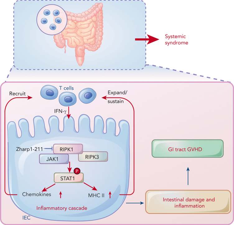
RIPK1/RIPK3 in IECs activates JAK1/STAT1-mediated chemokines and MHC II molecules and triggers an inflammatory cascade.
-
•
Nonimmunosuppressive RIPK1 kinase inhibitor Zharp1-211 restores intestinal homeostasis and reduces GVHD without compromising the GVL effect.
Visual Abstract
Abstract
Intestinal epithelial cells (IECs) are implicated in the propagation of T-cell–mediated inflammatory diseases, including graft-versus-host disease (GVHD), but the underlying mechanism remains poorly defined. Here, we report that IECs require receptor-interacting protein kinase-3 (RIPK3) to drive both gastrointestinal (GI) tract and systemic GVHD after allogeneic hematopoietic stem cell transplantation. Selectively inhibiting RIPK3 in IECs markedly reduces GVHD in murine intestine and liver. IEC RIPK3 cooperates with RIPK1 to trigger mixed lineage kinase domain-like protein-independent production of T-cell–recruiting chemokines and major histocompatibility complex (MHC) class II molecules, which amplify and sustain alloreactive T-cell responses. Alloreactive T-cell–produced interferon gamma enhances this RIPK1/RIPK3 action in IECs through a JAK/STAT1-dependent mechanism, creating a feed-forward inflammatory cascade. RIPK1/RIPK3 forms a complex with JAK1 to promote STAT1 activation in IECs. The RIPK1/RIPK3-mediated inflammatory cascade of alloreactive T-cell responses results in intestinal tissue damage, converting the local inflammation into a systemic syndrome. Human patients with severe GVHD showed highly activated RIPK1 in the colon epithelium. Finally, we discover a selective and potent RIPK1 inhibitor (Zharp1-211) that significantly reduces JAK/STAT1-mediated expression of chemokines and MHC class II molecules in IECs, restores intestinal homeostasis, and arrests GVHD without compromising the graft-versus-leukemia (GVL) effect. Thus, targeting RIPK1/RIPK3 in IECs represents an effective nonimmunosuppressive strategy for GVHD treatment and potentially for other diseases involving GI tract inflammation.
Yu and colleagues delineate the role of receptor-interacting protein kinase (RIPK) 3 in driving gastrointestinal (GI) graft-versus-host disease (GVHD). The authors report that RIPK3 cooperates with RIPK1 to drive GI tract GVHD through the triggering release of T-cell-recruiting chemokines and major histocompatibility complex class II molecules that sustain an alloreactive T-cell response. They describe a novel RIPK1 inhibitor that arrests murine GI tract GVHD without impairing graft-versus-leukemia reactivity, suggesting a novel nonimmunosuppressive therapeutic option.
Introduction
Nonhematopoietic cells, including epithelial cells and stromal cells, are also important for enhancing or dampening inflammatory immune responses, especially at “barrier sites” such as the gastrointestinal (GI) tract and skin.1, 2, 3, 4, 5, 6 The GI tract is crucial for inducing many T-cell–mediated inflammatory disorders, including graft-versus-host disease (GVHD) in individuals who receive allogeneic hematopoietic cell transplantation (allo-HCT). Alloreactive T-cell–mediated damage to the GI tract is a determinant of GVHD severity.7, 8, 9 Importantly, intestinal epithelial cells (IECs) are known as the site of amplifying GVHD.4,5 Some studies suggested that IEC presentation of major histocompatibility complex (MHC) class II initiates a GVHD inflammatory cascade,5 whereas others showed that inflammation-mediated cell death in the intestinal epithelium contributes to severe GVHD.10, 11, 12, 13, 14 A better understanding of the molecular basis through which IECs contribute to GVHD may enable innovative therapeutic strategies for managing inflammatory disorders in a broad context.
Receptor-interacting protein kinase (RIPK) 1 and RIPK3 are activators of necroptotic cell death.15, 16, 17 Upon inflammatory stimulation, RIPK1 and RIPK3 are activated, followed by the phosphorylation of mixed lineage kinase domain-like protein (MLKL).18, 19, 20, 21, 22, 23 Activated MLKL oligomerizes, translocates to the cell membrane, and induces necroptosis.24, 25, 26, 27 Necroptosis is a form of inflammatory cell death that causes the release of damage-associated molecular patterns to amplify the inflammatory responses.28 Necroptosis in IECs causes intestinal inflammation.3,14,29, 30, 31, 32 Many pathogenic factors, such as tumor-necrosis factor (TNF), toll-like receptor agonists, type I interferons, and viral infection, have been shown to activate RIPK3.15, 16, 17 These immune stimuli induce IEC necroptosis upon dampening of caspase-8 activity.28,29 Deletion of the autophagic protein ATG16L1 enhances this RIPK3-mediated effect in IECs.13,14 However, whether targeting RIPK1/RIPK3 may lead to inhibition of both intestinal inflammation and cell death in ATG16L1-sufficient hosts (ie, wild-type [WT] hosts) remains elusive.
Addressing these possibilities is important to establish the efficacy of pharmacologically inhibiting RIPK1/RIPK3 to reduce intestinal GVHD. Several RIPK1/RIPK3 inhibitors have been developed for the treatment of inflammatory diseases. For example, necrostatin-1 was the first RIPK1 kinase inhibitor identified.33,34 However, necrostatin-1 showed limited clinical application due to its moderate activity and poor metabolic stability.35 Clinical studies of another RIPK1 inhibitor, GSK2982772, did not demonstrate meaningful improvement in patients with rheumatoid arthritis.36 The selective effect of GSK2982772 on human cells rather than rodent cells prevents the assessment of its action in preclinical models.37 Therefore, RIPK1/RIPK3 inhibitors that are active both in humans and animals remain an unmet need.
Here, we identify that RIPK1/RIPK3 is required for IECs to drive both GI tract and systemic GVHD in mice. Besides mediating necroptosis, RIPK1/RIPK3 forms a complex with JAK1 to promote STAT1 activation in IECs, resulting in a feed-forward inflammatory cascade of alloreactive T-cell responses. Furthermore, we discover a selective and potent RIPK1 inhibitor (Zharp1-211) that significantly reduces JAK/STAT1-mediated IEC inflammation, restores intestinal homeostasis, and arrests GVHD without compromising the graft-versus-leukemia (GVL) effect.
Materials and methods
Human participants
Colon biopsies samples were collected after diagnosis or surgery, in accordance with research ethics board approval from The First Affiliated Hospital of Soochow University and The Children’s Hospital of Soochow University (No. 2020CS080). The inflammation status of tissue was confirmed by pathological examination. The characteristics in 10 pediatric patients with GVHD are shown in supplemental Table 1, available on the Blood website. GVHD grading was performed based on the Glucksberg grade standard.38
Mice
C57BL/6J (B6) and BALB/c mice were purchased from Beijing Vital River. Ripk3−/− and Mlkl−/− B6 mice were kindly provided by Dr. Xiaodong Wang (National Institute of Biological Sciences, Beijing). Ripk1K45A/K45A B6 mice were generated as described.39 Villin-cre (Vil-cre) Ripk3fl/fl B6 mice were generated by crossing Vil-cre B6 with Ripk3fl/fl B6 mice. Ripk3−/−caspase-8−/− were generated by crossing Ripk3−/− B6 with Caspase-8+/− B6 mice. Stat1−/− mice were from Jackson Lab. Zbp1−/− mice were generated using the CRISPR/Cas9 system by Bioray Laboratories Inc (Shanghai, China). All mice were housed in the specific pathogen-free animal facility at the Suzhou Institutes of Systems Medicine. All animal experiments were performed in accordance with protocols approved by the Suzhou Institutes of Systems Medicine Institutional Animal Care and Use Committee.
Methods
GVHD mouse models, cell lines, reagents, cell viability assay, western blot analysis, immunoprecipitation, measurement of cytokines, drug treatment in vivo, histology and immunohistochemistry, immunofluorescence confocal, culture of murine and human intestinal organoids, proximity ligation assay, chromatin immunoprecipitation assays, cell migration assays, T-cell analysis, intestinal crypt cells isolation, treatment of human intestinal explants, molecular docking study, quantitative polymerase chain reaction (qPCR), flow cytometry, RNA sequencing (RNA-seq) and analysis, quantification, statistical analysis, and detailed synthetic procedure for Zharp1-211 are detailed in supplemental Material and methods.
Results
Loss of RIPK3 in IECs reduces both local and systemic GVHD
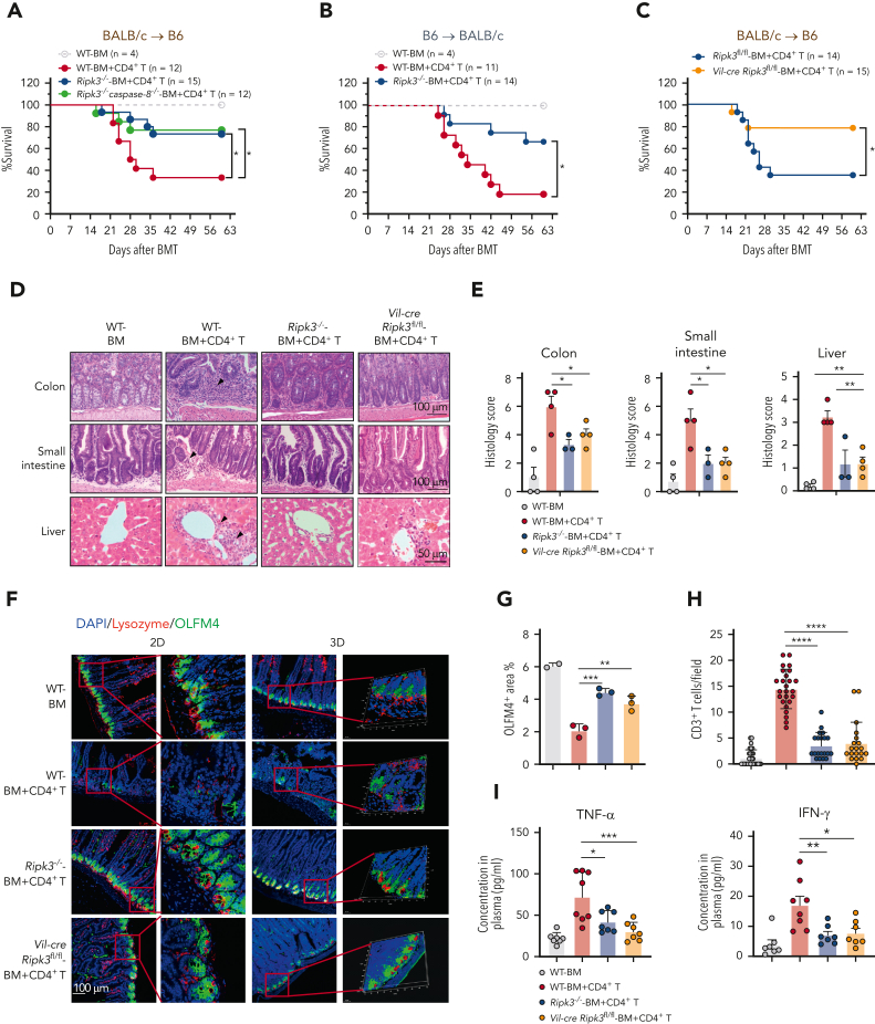
To assess the impact of RIPK3 on GVHD development in ATG16L1-sufficient hosts, we transferred BALB/c mouse CD4+ T cells plus T-cell–depleted bone marrow (TCD-BM) into lethally irradiated WT and Ripk3−/− B6 mice. Ripk3−/− B6 mice showed significantly reduced GVHD and improved survival rates compared with WT recipients (Figure 1A; supplemental Figure 1A). The requirement of host RIPK3 for inducing GVHD was confirmed in a second GVHD model using Ripk3−/− BALB/c host mice (Figure 1B; supplemental Figure 1B-C). Ripk3−/− B6 mice receiving both CD4+ T and CD8+ T cells showed significantly improved survival compared with WT recipients (supplemental Figure 1D). Intriguingly, although deletion of caspase-8 in Ripk3−/− mice eliminates both caspase-8–mediated apoptosis and RIPK3-mediated necroptosis,40 deletion of both caspase-8 and Ripk3 in B6 recipient mice resulted in similar reduction of GVHD as did Ripk3−/− mice undergoing allo-HCT (Figure 1A; supplemental Figure 1A,E-G). Thus, deletion of RIPK3 in the host protects against GVHD, whereas inhibition of caspase-8–mediated apoptosis provides no further protection against GVHD in Ripk3−/− mice.
Figure 1.
Loss of RIPK3 in IECs reduces both local and systemic GVHD. (A) Survival of the lethally irradiated WT, Ripk3−/−, and Ripk3−/−caspase-8−/− B6 recipients (BALB/c→B6) that received BALB/c TCD BM cells (BM) with or without CD4+ T cells (BM+CD4+ T). (B) Survival of the lethally irradiated WT and Ripk3−/− BALB/c recipients (B6→BALB/c) that received B6 TCD BM cells with CD4+ T cells. (C) Survival of the lethally irradiated Ripk3fl/fl and Vil-cre Ripk3fl/fl B6 recipients (BALB/c→B6) that received BALB/c TCD BM cells with CD4+ T cells. (D-E) Histology analysis of small intestine, colon, and liver obtained from WT, Ripk3−/− and Vil-cre Ripk3fl/fl B6 recipients (BALB/c→B6) on day 17 after allo-HCT. Representative hematoxylin and eosin (H&E) images (D) and quantification of pathology scores (E). Bars in colon and small intestine represent 100 μm. Bar in liver represents 50 μm. Black arrows indicate areas of inflammatory cell infiltration or cell damage. (F) Representative 2D and 3D confocal images of OLFM4+ ISCs and lysozyme+ Paneth cells in the small intestine from the indicated B6 recipients on day 17 after allo-HCT. Bar represents 100 μm. (G) Quantification of IHC staining of OLFM4 in the small intestine from the indicated B6 recipients on day 17 after allo-HCT. (H) Quantification of infiltrated CD3+ T cells in the small intestine from the indicated B6 recipients (BALB/c→B6) on day 17 after allo-HCT. Three mice were examined in each group. (I) TNF-α and IFN-γ levels in serum from the indicated B6 recipients on day 17 after allo-HCT. Data shown are representative of 2 or 3 independent experiments. Data are shown as the mean ± SD (G-H), histology scores, and concentrations of cytokines are shown as the mean ± SEM (E,I). ∗P < .05; ∗∗P < .01; ∗∗∗P < .001; ∗∗∗∗P < .0001. Survival comparisons were evaluated by log-rank test (A-C). Multiple comparisons were evaluated by one-way ANOVA (E,G-I). ANOVA, analysis of variance; IHC, immunohistochemistry; SD, standard deviation; SEM, standard error of the mean.
To determine the effect of RIPK3 in IECs on GVHD, we generated Vil-cre Ripk3fl/fl B6 mice to selectively delete Ripk3 in the intestine (supplemental Figure 1H-I). Vil-cre Ripk3fl/fl B6 mice had significantly decreased GVHD when given BALB/c mouse T cells compared with Ripk3fl/fl (Ripk3-sufficient) B6 mice (Figure 1C; supplemental Figure 1J). Like Ripk3−/− mice, Vil-cre Ripk3fl/fl B6 mice showed decreased inflammation in the intestine (Figure 1D-E). GVHD is associated with the loss of intestinal stem cells (ISCs, marked by OLFM4) and their niche Paneth cells (marked by lysozyme).10,41,42 Three-dimensional imaging showed that both Ripk3−/− and Vil-cre Ripk3fl/fl recipients had markedly increased number of OLFM4+ ISCs and lysozyme+ Paneth cells in the small intestine compared with WT recipients, indicating that inhibiting IEC RIPK3 significantly protects the crypt base compartment against GVHD (Figure 1F-G; supplemental Figure 1K). Inhibition of GI tract GVHD in Vil-cre Ripk3fl/fl mice was associated with significantly decreased infiltration of T cells in the GI tract and reduced serum TNF-α and interferon gamma (IFN-γ) (Figure 1H-I). Furthermore, loss of RIPK3 in the GI tract led to protection against GVHD in the liver (Figure 1D-E). Our findings indicate that activation of IEC RIPK3 signaling causes both GI tract GVHD and systemic syndrome in mice.
IEC RIPK3 promotes MLKL-independent alloreactive T-cell responses
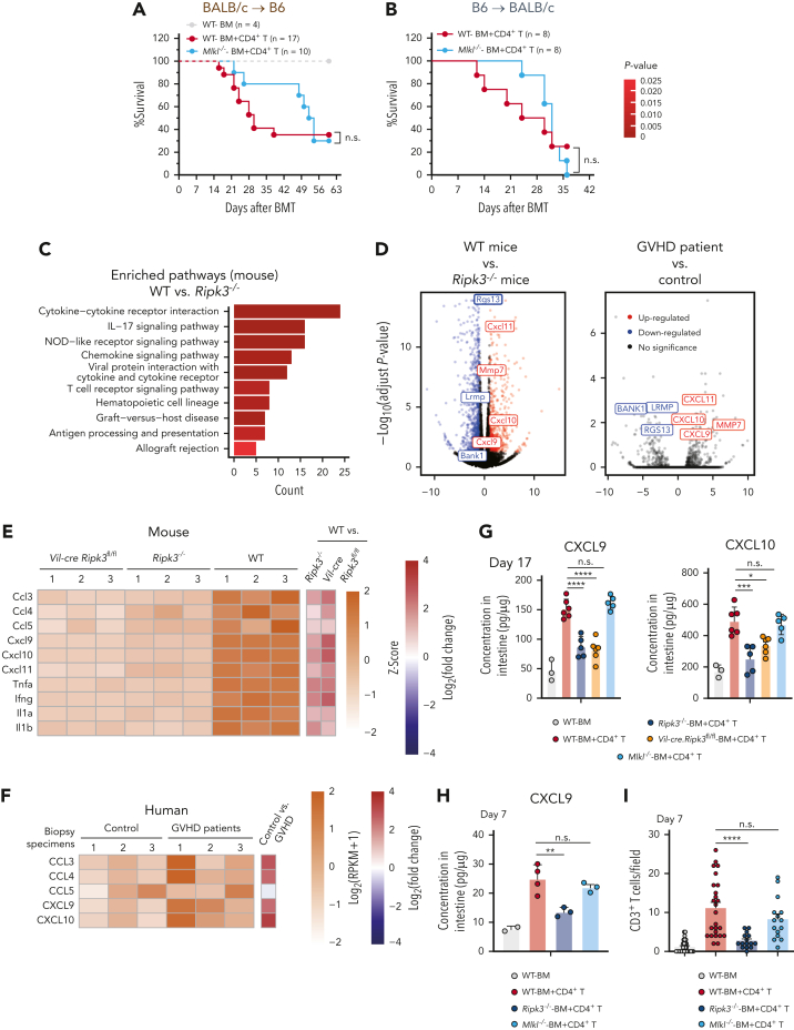
It is believed that IEC death is sufficient to cause severe GVHD.13,14 We examined whether the observed RIPK3-mediated GVHD in IECs resulted from MLKL-dependent necroptosis. Deleting Mlkl in B6 hosts delayed GVHD progression but did not prevent recipients from lethal GVHD (Figure 2A). Similarly, the deletion of both MLKL and caspase-8 did not improve GVHD-associated overall survival rate (supplemental Figure 2A). Using Mlkl−/− BALB/c recipient mice (supplemental Figure 2B), we confirmed that deletion of host Mlkl delayed GVHD onset without improving the overall survival rates or GVH inflammation in the GI tract when examined 30 days after transplantation (Figure 2B; supplemental Figure 2C-D). Moreover, the crypt cells of colon and small intestine from WT but not Ripk3−/− B6 mice displayed activated MLKL 7 days after transplantation (supplemental Figure 2E-F). Thus, although MLKL-mediated necroptosis occurs in the intestine during GVHD and contributes to the severity of the disease, it is not the determinant factor responsible for IEC RIPK3 induction of lethal GVHD.
Figure 2.
IEC RIPK3 promotes MLKL-independent alloreactive T-cell responses. (A) Survival of the lethally irradiated WT and Mlkl−/− B6 recipients (BALB/c→B6) received BALB/c TCD BM alone (BM) or TCD BM plus CD4+ T cells (BM+CD4+ T). (B) Survival of the lethally irradiated WT and Mlkl−/− BALB/c recipients (B6→BALB/c) received B6 TCD BM alone (BM) or TCD BM plus CD4+ T cells (BM+CD4+ T). (C) KEGG pathway enrichment analyses of the DEGs in small intestine samples from WT and Ripk3−/− mice collected on day 17 after allo-HCT (P < .05, the P value is calculated by hypergeometric distribution). (D) Volcano plot showing fold changes of genes in small intestine samples collected from WT vs Ripk3−/− mice on day 17 after allo-HCT (left), or fold changes of genes in colonic biopsy specimens from patients with GVHD vs control colonic biopsy specimens from children without signs of intestinal injury (right). Significantly upregulated (red) and downregulated (blue) mouse or human homologous genes are shown. (E) Gene expression of chemokines and cytokines in the small intestine collected from WT, Ripk3−/−, and Vil-cre Ripk3fl/fl mice on day 17 after allo-HCT. (F) Gene expression of human chemokines in control colonic biopsy specimens and colonic biopsy specimens from patients with GVHD. (G) ELISA analysis of CXCL9 and CXCL10 proteins in the small intestine from the indicated B6 recipients (BALB/c→B6) 17 days after allo-HCT. (H) ELISA analysis of CXCL9 proteins in the small intestine from the indicated B6 recipients (BALB/c→B6) 7 days after allo-HCT. (I) Quantification of infiltrated CD3+ T cells in the small intestine from the indicated B6 recipients (BALB/c→B6) on day 7 after allo-HCT. Data shown are representative of 2 or 3 independent experiments. Data are shown as the mean ± SD (I), histology scores and chemokine concentrations are shown as the mean ± SEM (G-H). ∗P < .05; ∗∗P < .01; ∗∗∗P < .001; ∗∗∗∗P < .0001. Survival comparisons were evaluated by log-rank test (A-B). Multiple comparisons were evaluated by one-way ANOVA (G-I). ANOVA, analysis of variance; DEGs, differentially expressed genes; ELISA, enzyme-linked immunosorbent assay analysis; n.s., not significant; SD, standard deviation; SEM, standard error of the mean.
These findings are unexpected and suggest that neither caspase-8–mediated apoptosis nor MLKL-mediated necroptosis is sufficient for inducing lethal GVHD. We reasoned that there exist additional mechanisms responsible for the IEC’s RIPK3-induced lethal GVHD. To test it, we performed RNA-seq analysis of the intestine samples that were isolated from WT, Ripk3−/−, and Vil-cre Ripk3fl/fl B6 mice on day 17 after allo-HCT. KEGG pathway analysis of the WT vs Ripk3−/− samples and WT vs Vil-cre Ripk3fl/fl samples identified enrichment for differentially expressed genes with annotations for GVHD, signaling pathways for cytokines and chemokines, as well as antigen processing and presentation (Figure 2C; supplemental Figure 3A). Ripk3 deletion in IECs led to drastically decreased expression of proinflammatory cytokines (eg, Tnfa, Ifnγ, Il1a, and Il1b) and chemokines (eg, Cxcl9, Cxcl10, Cxcl11, and Ccl5) (Figure 2D-E). Significant reductions of Cxcl9 and Cxcl10 expression levels were further confirmed in the colon derived from Ripk3−/− recipient mice compared with WT mice on day 17 after allo-HCT (supplemental Figure 3B). RNA-seq analysis using colon biopsy specimens from pediatric patients with GI tract GVHD (grade III-IV) also revealed upregulation of genes associated with cytokine and chemokine signaling pathways, inflammatory responses, and immune cell activation compared with control specimens derived from patients who did not receive transplant (Figure 2D,F; supplemental Figure 3C-D).
Chemokines, including CXCL9, CXCL10, and CCL5, are known to promote the recruitment of T cells and other inflammatory cells to inflamed intestinal tissues.43, 44, 45 We observed higher levels of Cxcl9, Cxcl10, and Cxcl11 in the small intestines of WT mice compared with those of Ripk3−/− mice and in colonic biopsy specimens from patients with GVHD relative to control biopsy specimens (Figure 2D). Enzyme-linked immunosorbent assay analysis confirmed a significant reduction of intestinal CXCL9 and CXCL10 proteins in both Ripk3−/− and Vil-cre Ripk3fl/fl B6 recipients compared with WT and Mlkl−/− B6 recipients (Figure 2G). Moreover, Ripk3−/− B6, but not Mlkl−/− B6 recipients, showed significant decreases in CXCL9 production and donor T-cell infiltration in the intestine on day 7 after allo-HCT than in WT recipients (Figure 2H-I). These findings demonstrate a previously unrecognized RIPK3 function in inducing necroptosis-independent inflammatory responses in IECs.
RIPK1 activates RIPK3 signaling in IECs during GVHD
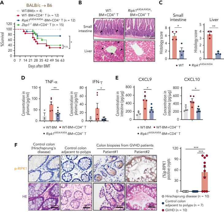
RIPK3 can be activated by RIPK1 kinase19, 20, 21 and Z-nucleic acid–binding protein 1 (ZBP1).46,47 We evaluated the impact of RIPK1 on GVHD in Ripk1 K45A/K45A mice carrying a RIPK1 K45A kinase-dead knockin mutation.39 Inactivation of RIPK1 in Ripk1K45A/K45A B6 recipients significantly reduced GVHD-associated lethality, whereas Zbp1−/− B6 recipients displayed no effect on GVHD severity (Figure 3A; supplemental Figure 4A-B). Furthermore, Ripk1 K45A/K45A recipient mice showed significantly reduced GVH inflammation in the intestine and liver (Figure 3B-C) and led to marked decreases in serum TNF-α and IFN-γ (Figure 3D).
Figure 3.
RIPK1 activates RIPK3 signaling in IECs during GVHD. (A-E) The lethally irradiated WT, Ripk1K45A/K45A, and Zbp1−/− B6 recipients (BALB/c→B6) received BALB/c TCD BM cells with or without CD4+ T cells. (A) Survival of mice after allo-HCT. (B-C) Histology analysis of small intestine and liver samples obtained from WT and Ripk1K45A/K45A B6 recipients on day 17 after allo-HCT. Representative H&E images (B) and quantification of pathology scores (C). Bar in small intestine represents 100 μm. Bar in liver represents 50 μm. Black arrows indicate areas of inflammatory cell infiltration or cell damage. (D) Levels of TNF-α and IFN-γ in serum from WT and Ripk1K45A/K45A B6 recipients on day 17 after allo-HCT. (E) ELISA analysis of CXCL9 and CXCL10 proteins in the small intestine from WT and Ripk1K45A/K45A B6 recipients on day 17 after allo-HCT. (F) Sections of 10 colon biopsies from patients with GVHD involved in the GI tract after allo-HCT, 10 control colon samples (from patients with Hirschsprung's disease) and 7 control colon samples adjacent to polyps were stained with an anti–p-RIPK1 antibody. Shown are representative images of IHC staining with anti–p-RIPK1 antibody and H&E staining from the control colon sample and 2 biopsies of patients with GVHD (left panel), and quantification of p-RIPK1 positive crypts in each sample (right panel). Bars represent 50 μm. Data shown are representative of 2 or 3 independent experiments. Data are shown as the mean ± SD (D-F), histology scores are shown as the mean ± SEM (C). ∗P < .05; ∗∗P < .01; ∗∗∗P < .001. Survival comparisons were evaluated by log-rank test (A). Multiple comparisons were evaluated by one-way ANOVA (D-F), two-group comparisons used unpaired t tests (two-tailed) (C). ANOVA, analysis of variance; ELISA, enzyme-linked immunosorbent assay analysis; IHC, immunohistochemistry; SD, standard deviation; SEM, standard error of the mean.
Consistent with these observations for Ripk3−/− and Vil-cre Ripk3fl/fl recipients, intestines of Ripk1K45A/K45A recipient mice showed significantly decreased levels of CXCL9 and CXCL10 after allo-HCT (Figure 3E). Phosphorylation of RIPK1 at S166 has been established as a marker for RIPK1 activation.16 Active p-RIPK1 was also seen in 7 out of 10 colonic samples from patients with GVHD with GI tract inflammation (grade III-IV), but not in 10 control colon specimens obtained from patients with Hirschsprung disease and 7 adjacent normal colon samples from patients with polyps (Figure 3F; supplemental Table 1). These results indicate the importance of RIPK1 signaling in the pathogenesis of both murine and human GVHD.
JAK/STAT1 signaling mediates RIPK1/RIPK3 induction of chemokines and MHC class II molecules in IECs
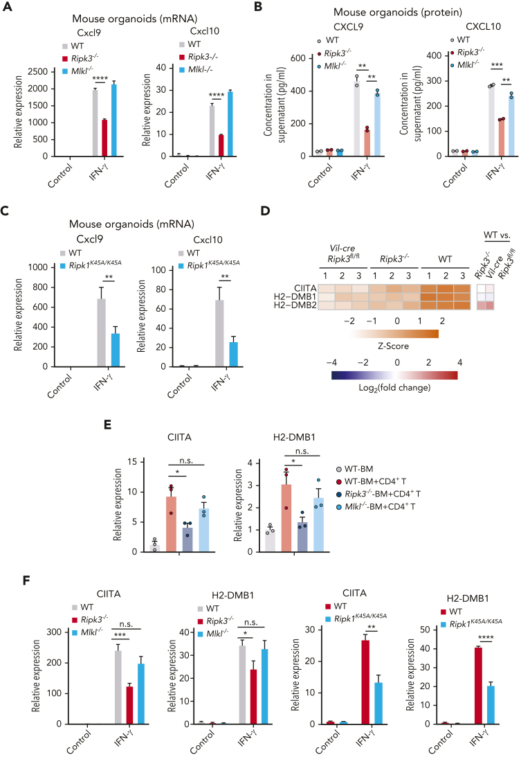
To determine how RIPK1/RIPK3 triggers IEC inflammation during GVHD, we used an ex vivo intestinal organoid culture.12 Intestinal organoids generated from Ripk3−/−, Mlkl−/−, and Ripk1K45A/K45A mice showed similar morphology and growth rates (supplemental Figure 5A-B). WT organoids treated with IFN-γ, but not TNF-α, led to dramatically elevated expression of Cxcl9 and Cxcl10 transcripts at 12 hours after treatment, a time point when cell death was not evident (Figure 4A; supplemental Figure 5C). Deletion of Ripk3 rather than Mlkl from intestinal organoids led to marked reductions in the expression of Cxcl9 and Cxcl10 and their proteins (Figure 4A-B). To exclude the effect of immune cells, we highly purified crypt cells from the colon after the depletion of immune cells. Likewise, RIPK3 deficiency significantly decreased IFN-γ-induced expression of Cxcl9 and Cxcl10 in these crypt cells (supplemental Figure 5D-E). Moreover, RIPK1 inactivation also reduced Cxcl9 and Cxcl10 expression in intestinal organoids treated with IFN-γ (Figure 4C). Neither treatment with the pan-caspase inhibitor z-VAD nor genetic deletion of Mlkl caused any reduction in the expression of these chemokines in the IFN-γ–treated organoids (supplemental Figure 5F), thereby excluding any requirement for apoptosis or necroptosis in IFN-γ–induced, RIPK1/RIPK3-controlled chemokine expression.
Figure 4.
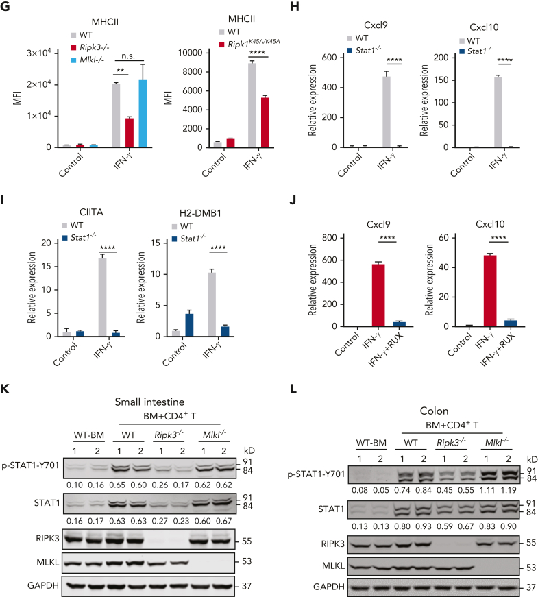
JAK/STAT1 signaling mediates RIPK1/RIPK3 induction of chemokines and MHC class II molecules in IECs. (A-B) Small intestinal organoids prepared from WT, Ripk3−/−, and Mlkl−/− B6 mice were treated with control (PBS) or IFN-γ (10 ng/mL) for 12 hours, with qPCR measurement of Cxcl9 and Cxcl10 expression (A); ELISA measurement of CXCL9 and CXCL10 protein levels (B). Identical concentrations for IFN-γ were used in later experiments unless otherwise stated. (C) qPCR analysis for the expression of Cxcl9 and Cxcl10 in small intestinal organoids from WT and Ripk1K45A/K45A B6 mice that were treated with control, IFN-γ for 12 hours. (D) Gene expression of antigen presentation-related genes in small intestine samples collected from WT, Ripk3−/−, and Vil-cre Ripk3fl/fl mice on day 17 after allo-HCT. (E) qPCR analysis of CIITA and H2-DMB1 mRNA levels in the small intestine from the indicated B6 recipients (BALB/c→B6) 17 days after allo-HCT. (F) Small intestinal organoids prepared from indicated B6 mice were treated with IFN-γ or control for 12 hours. CIITA and H2-DMB1 expression levels were analyzed using qPCR. (G) Small intestinal organoids prepared from indicated B6 mice were treated with IFN-γ or control for 24 hours. The MFI of MHC II was determined by flow cytometer analysis. (H-I) Small intestinal organoids prepared from WT and Stat1−/− B6 mice were treated with control or IFN-γ for 12 hours. qPCR for expression of Cxcl9, Cxcl10 (H), CIITA, and H2-DMB1 (I). (J) Small intestinal organoids from WT B6 mice were treated with 300 nM ruxolitinib (RUX) for 2 hours before IFN-γ treatment for 12 hours; gene expression was analyzed by qPCR. (K-L) Immunoblotting analysis of p-STAT1, STAT1, RIPK3, MLKL, and GAPDH in small intestine and colon from the indicated B6 recipients on day 7 after allo-HCT. Quantification of p-STAT1 or STAT1 normalized to GAPDH was shown under the band. Data shown are representative of 3 independent experiments. Data are shown as the mean ± SD (A-C,F-J), qPCR analysis in the small intestine are shown as the mean ± SEM (E). ∗P < .05; ∗∗P < .01; ∗∗∗P < .001; ∗∗∗∗P < .0001. Multiple comparisons were evaluated by one-way ANOVA (A-B,E-G,J), two-group comparisons used unpaired t tests (two-tailed) (C,F-I). ANOVA, analysis of variance; ELISA, enzyme-linked immunosorbent assay analysis; GAPDH, glyceraldehyde-3-phosphate dehydrogenase; MFI, mean fluorescence intensity; n.s., not significant; PBS, phosphate-buffered saline; SD, standard deviation; SEM, standard error of the mean.
MHC class II expressed on the surface of IECs plays important roles in activating donor CD4 T cells to mediate GVHD in mice.5 Our RNA-seq analysis also showed that the gene encoding class II transactivator (CIITA), a master regulator of MHC class II transcription, was significantly reduced in intestines of both Vil-cre Ripk3fl/fl and Ripk3−/− mice compared with those of WT mice (Figure 4D). Allo-HCT resulted in increased levels of CIITA and H2-DMB1 expression in the intestine, which were significantly attenuated by the deletion of Ripk3, but not by the deletion of Mlkl in recipients (Figure 4E). IFN-γ–stimulated Ripk3−/− organoids and Ripk1 K45A/K45A organoids had significantly reduced expression of CIITA and H2-DMB1 mRNA as well as MHC class II protein compared with stimulated WT organoids, confirming the function of RIPK1/RIPK3 in regulating MHC class II molecules in IECs (Figure 4F-G; supplemental Figure 5G-H). Thus, RIPK1/RIPK3 in IECs promotes the expression of MHC class II molecules known to enhance the expansion of alloreactive CD4+ T cells in the GI tract.
Upon IFN-γ ligation, JAK1/JAK2 and STAT1 are activated to induce the transcription of IFN-stimulated genes.48 We next tested whether RIPK3 induction of chemokines and MHC class II molecules may require IFN-γ activation of JAK1/2-STAT1 signaling in IECs. The absence of Stat1 in intestinal organoids led to greatly reduced expression of chemokines and MHC class II molecules upon IFN-γ induction (Figure 4H-I; supplemental Figure 5I). Moreover, ruxolitinib, a JAK1/JAK2 inhibitor used for GVHD treatment,49 abolished IFN-γ–induced expression of Cxcl9 and Cxcl10 in intestinal organoids (Figure 4J). Activation of STAT1 (phosphorylated STAT1) was observed in the intestine of WT mice with GVHD (Figure 4K-L). Notably, Ripk3−/− recipient mice, but not Mlkl−/− recipient mice, showed reduced phosphorylation of STAT1 and total protein level of STAT1, which can be induced by IFNs,50 in both the small intestine and colon after allo-HCT (Figure 4K-L). Thus, IEC RIPK1/RIPK3 engages the JAK/STAT1 pathway to promote the expression of chemokines and MHC class II molecules.
IFN-γ enhances the binding of RIPK1 to JAK1 to activate STAT1 in IECs
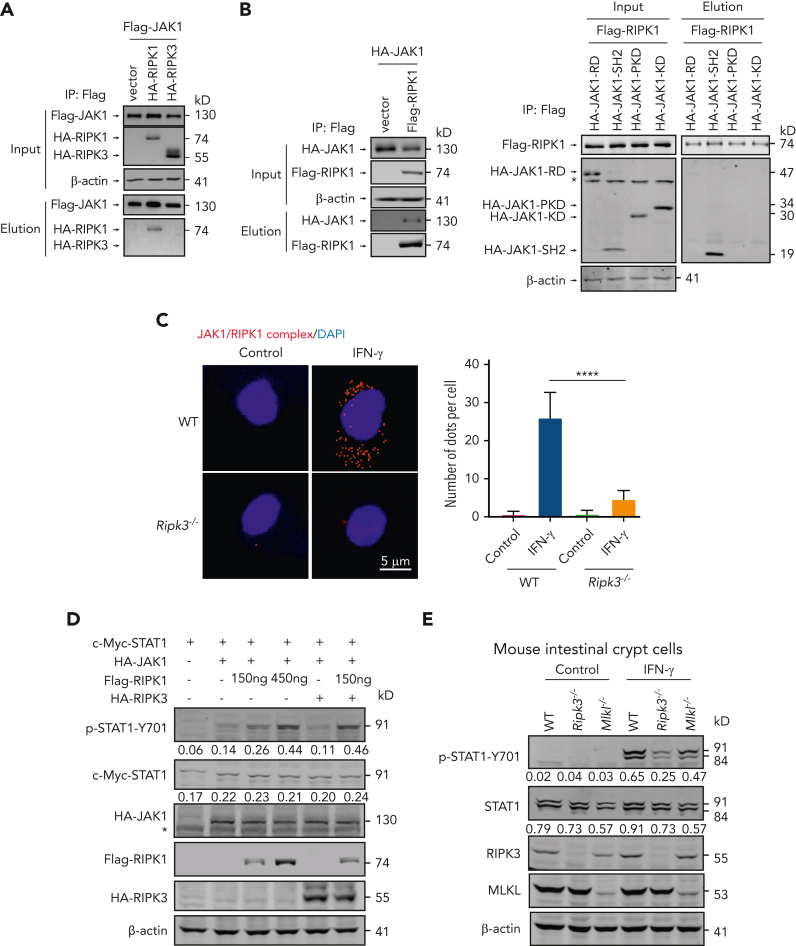
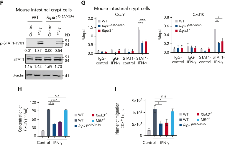
To explore how RIPK1/RIPK3 engages the JAK/STAT1 pathway, we sought to examine the association between RIPK1/RIPK3 and JAK1. We observed that Flag-tagged JAK1 could directly bind to RIPK1 rather than RIPK3 (Figure 5A). The binding of RIPK1 to JAK1 was further confirmed by precipitating a Flag-tagged RIPK1 immunocomplex (Figure 5B). The SH2 domain of JAK1 was found to interact with RIPK1 (Figure 5B). Furthermore, in situ proximity ligation assay showed that IFN-γ stimulation significantly enhanced the formation of a RIPK1/JAK1 complex in crypt cells from mouse intestine, which was attenuated by RIPK3 deficiency (Figure 5C). Of note, overexpression of RIPK1 resulted in increased JAK1-mediated STAT1 activation, which was further enhanced in the presence of RIPK3 (Figure 5D). This suggests that the RIPK1/RIPK3 complex promotes JAK1/ STAT1 activation. Of note, IFN-γ–induced activation of STAT1 was attenuated by RIPK3 loss or RIPK1 inactivation in crypt cells from mouse intestine, whereas MLKL deficiency had no apparent effect on the activation of STAT1 (Figure 5E-F). Moreover, chromatin immunoprecipitation analysis showed that IFN-γ–induced binding of STAT1 to the promoter regions of Cxcl9 and Cxcl10 was significantly inhibited by RIPK3 loss or RIPKI inactivation in intestinal crypt cells (Figure 5G). Compared with WT intestinal crypt cells, Ripk3−/− and Ripk1K45A/K45A intestinal crypt cells produced significantly lower levels of CXCL9 in culture medium upon IFN-γ stimulation (Figure 5H), which was associated with significantly reduced recruitment of T cells (Figure 5I). Collectively, these findings indicate that IEC RIPK1/RIPK3 forms a complex with JAK1 to promote STAT1-mediated expression of T-cell–recruiting chemokines.
Figure 5.
IFN-γ enhances the binding of RIPK1 to JAK1 to activate STAT1 in IECs. (A) HEK293T cells were cotransfected with a DNA plasmid expressing Flag-tagged JAK1 plus the plasmid expressing HA-tagged RIPK1 or RIPK3. Cell lysates were collected and immunoprecipitated with anti-Flag agarose. The Flag-JAK1 immunocomplex was analyzed by immunoblotting analysis. (B) HEK293T cells were cotransfected with a DNA plasmid expressing Flag-tagged RIPK1 plus the plasmid expressing HA-tagged JAK1 or the truncated form of JAK1 as indicated. Cell lysates were collected and immunoprecipitated with anti-Flag agarose. The Flag-RIPK1 immunocomplex was analyzed by immunoblotting analysis. (C) Intestinal crypt cells isolated from WT and Ripk3−/− B6 mice were treated with IFN-γ for 1 hour. Representative images of in situ PLA between JAK1 and RIPK1 (red). 4′,6-diamidino-2-phenylindole was shown (blue). Bar represents 5 μm. (D) Immunoblotting analysis of p-STAT1, c-myc-tagged STAT1, Flag tagged-RIPK1, HA tagged-RIPK3, and β-actin in HEK293T cells transfected with the indicated DNA plasmid(s). Quantification of p-STAT1 or STAT1 normalized to β-actin was shown under the band. (E) Intestinal crypt cells isolated from WT, Ripk3−/−, and Mlkl−/− B6 mice were treated with IFN-γ for 0.5 hour. Immunoblotting analysis of p-STAT1, STAT1, RIPK3, MLKL, and β-actin. Quantification of p-STAT1 or STAT1 normalized to β-actin was shown under the band. (F) Intestinal crypt cells isolated from WT and Ripk1K45A/K45A B6 mice were treated with IFN-γ for 0.5 hour. Immunoblotting analysis of p-STAT1, STAT1, and β-actin. Quantification of p-STAT1 or STAT1 normalized to β-actin was shown under the band. (G) Intestinal crypt cells isolated from WT, Ripk1K45A/K45A, and Ripk3−/− B6 mice were treated with IFN-γ for 0.5 hour. STAT1 binding to selected regions of the Cxcl9 and Cxcl10 promoters was determined by ChIP-qPCR. The amount of precipitated DNA was calculated as percent input. (H-I) Intestinal crypt cells isolated from WT, Ripk3−/−, Mlkl−/−, and Ripk1K45A/K45A B6 mice were treated with IFN-γ for 24 hours, and the culture medium was collected for ELISA measurement of CXCL9 protein (H), and for further experiment (I). The culture medium was then added into the bottom compartment of a transwell chamber and T cells isolated from WT B6 mice were plated into the upper compartment. After 6 hours, migration of T cells was assessed by transwell assay (I). Data shown are representative of 3 independent experiments. Data are shown as the mean ± SD. ∗P < .05; ∗∗∗P < .001; ∗∗∗∗P < .0001. Multiple comparisons were evaluated by one-way ANOVA. ANOVA, analysis of variance; ChIP, chromatin immunoprecipitation; ELISA, enzyme-linked immunosorbent assay analysis; n.s., not significant; PLA, proximity ligation assay. SD, standard deviation.
Development of a novel RIPK1 kinase inhibitor
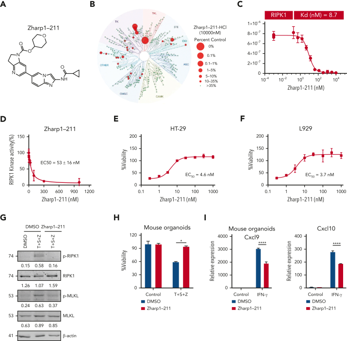
We previously discovered a selective RIPK1 kinase inhibitor, PK68.51 To optimize the efficacy, selectivity, and bioavailability, we synthesized a group of PK68 analogs and identified Zharp1-211 as the most potent RIPK1 inhibitor (Figure 6A; supplemental Figure 6A-B). Selectivity analysis against a panel of 468 kinases revealed that Zharp1-211 (10 μM) was selective for RIPK1 kinase (Figure 6B; supplemental Table 2). Zharp1-211 displayed potent binding affinity for RIPK1 and efficiently inhibited RIPK1’s kinase activity without affecting that of RIPK3 (Figure 6C-D; supplemental Figure 6C). Molecular docking indicated that Zharp1-211 works as a type II inhibitor of RIPK1 kinase, targeting the adenosine triphosphate-binding pocket in RIPK1’s inactive DLG (Asp156(D)–Leu157(L)–Gly158(G))-out kinase conformation (supplemental Figure 6D). Collectively, these data support the hypothesis that Zharp1-211 is a potent and selective type II RIPK1 inhibitor.
Figure 6.
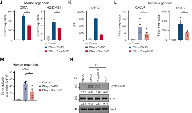
Development of a novel RIPK1 kinase inhibitor. (A) Chemical structure of Zharp1-211. (B) Assessment of Zharp1-211 (10 μM) binding against 468 human kinases (DiscoverX kinase panel). TREEspot kinase interaction map of Zharp1-211 with 468 kinases. Red circles indicate kinases that were inhibited by AC002 >65%. Full data are presented in extended Data supplemental Table 2. (C) The binding constant (Kd) of Zharp1-211 with human recombinant RIPK1. (D) In vitro kinase activity assays using recombinant RIPK1. (E-F) Dose response curve and EC50 for Zharp1-211 in TNF-α–induced necroptosis in human HT-29 cells and mouse L929 cells. HT-29 cells were pretreated with Zharp1-211 at the indicated concentration for 2 hours before the treatment with necroptotic stimuli, TNF-α (40 ng/mL), the inhibitor of apoptosis protein (IAP) antagonist Smac mimetic (100 nM), and the pan-caspase inhibitor z-VAD (20 μM), for 48 hours (E). L929 cells were pretreated with Zharp1-211 at the indicated concentration for 2 hours before treatment with TNF-α (40 ng/mL) and z-VAD (20 μM) for 24 hours (F). Cell viability was assessed by measuring ATP levels. (G-H) Small intestinal organoids from WT B6 mice were treated with Zharp1-211 (100 nM) for 2 hours and then treated with TNF-α (40 ng/mL), Smac mimetic (100 nM), and z-VAD (20 μM). Immunoblotting of cell lysates from organoids with antibodies against RIPK1, p-RIPK1, MLKL, p-MLKL, and β-actin was performed at 8 hours after treatment with TNF-α, Smac mimetic, and z-VAD (G). Quantification of p-RIPK1, RIPK1, p-MLKL, or MLKL normalized to β-actin was shown under the band. Cell viability in organoids at 24 hours aftertreatment with TNF-α, Smac mimetic, and z-VAD, assessed by measuring ATP levels (H). (I-J) Small intestinal organoids prepared from B6 mice were pretreated with Zharp1-211 (100 nM) for 2 hours before the treatment with IFN-γ for 12 hours. qPCR measurement of Cxcl9, Cxcl10 expression (I), CIITA, and H2-DMB1 expression (J). (K) Small intestinal organoids prepared from B6 mice were pretreated with Zharp1-211 (100 nM) for 2 hours before the treatment with IFN-γ for 24 hours. The MFI of MHC II was determined by flow cytometer analysis. (L-M) Human intestinal organoids from 5 individual donors (n = 5) were treated with Zharp1-211 (100 nM) for 2 hours and then stimulated with IFN-γ for 12 hours. Expression of the indicated genes was analyzed by qPCR (L) and protein levels were measured by ELISA (M). (N) Intestinal crypt cells isolated from WT B6 mice were treated with DMSO, 100 nM Zharp1-211, or ruxolitinib (RUX) for 2 hours before stimulation with IFN-γ (100 ng/mL) for 1 hour. Cell lysates was collected for immunoblotting with antibodies against p-STAT1, STAT1, and β-actin. Quantification of p-STAT1 or STAT1 normalized to β-actin was shown under the band. Data shown are representative of 3 independent experiments. Data are shown as the mean ± SD. ∗P < .05; ∗∗P < .01; ∗∗∗∗P < .0001. Multiple comparisons were evaluated by one-way ANOVA (J-M), two-group comparisons used unpaired t test (two-tailed) (H-I). ANOVA, analysis of variance; ATP, adenosine triphosphate; ELISA, enzyme-linked immunosorbent assay analysis; MFI, mean fluorescence intensity; SD, standard deviation.
Zharp1-211 was highly potent for blocking TNF-induced necroptosis in human colon cancer HT-29 cells (EC50 of ∼4.6 nM) and mouse fibroblast L929 cells (EC50 of ∼3.7 nM) (Figure 6E-F). Zharp1-211 efficiently inhibited p-RIPK1 and p-MLKL and necroptosis in intestinal organoids exposed to necroptotic stimuli (Figure 6G-H). Zharp1-211 also significantly reduced the transcription of Cxcl9, Cxcl10, CIITA, and H2-DMB1 as well as the expression of MHCII protein in IFN-γ–stimulated mouse intestinal organoids (Figure 6I-K; supplemental Figure 6E). Zharp1-211’s inhibitory effect on IFN-γ–induced chemokine production was also evident in cultured human intestinal organoids (Figure 6L-M).
Zharp1-211 treatment reduced IFN-γ–induced STAT1 activation in mouse intestinal crypt cells (Figure 6N). The JAK/STAT pathway is required for normal immunological function.48 We noted that inhibiting JAK1/2 with ruxolitinib suppressed the proliferation, survival, and effector differentiation of activated CD4+ T cells upon T-cell receptor (TCR) ligation, whereas Zharp1-211 inhibition of RIPK1 resulted in no significant effects on TCR-activated T-cell responses (supplemental Figure 6F-G). This difference was also supported by the finding that deletion of RIPK3 did not affect TCR-activated T-cell responses (supplemental Figure 6F,H). Our results indicate that RIPK1/RIPK3 engages JAK/STAT1 signaling to regulate IFN-γ–induced inflammatory responses in IECs rather than in T cells.
Pharmacological inhibition of RIPK1 reduces ongoing GVHD while preserving GVL activity
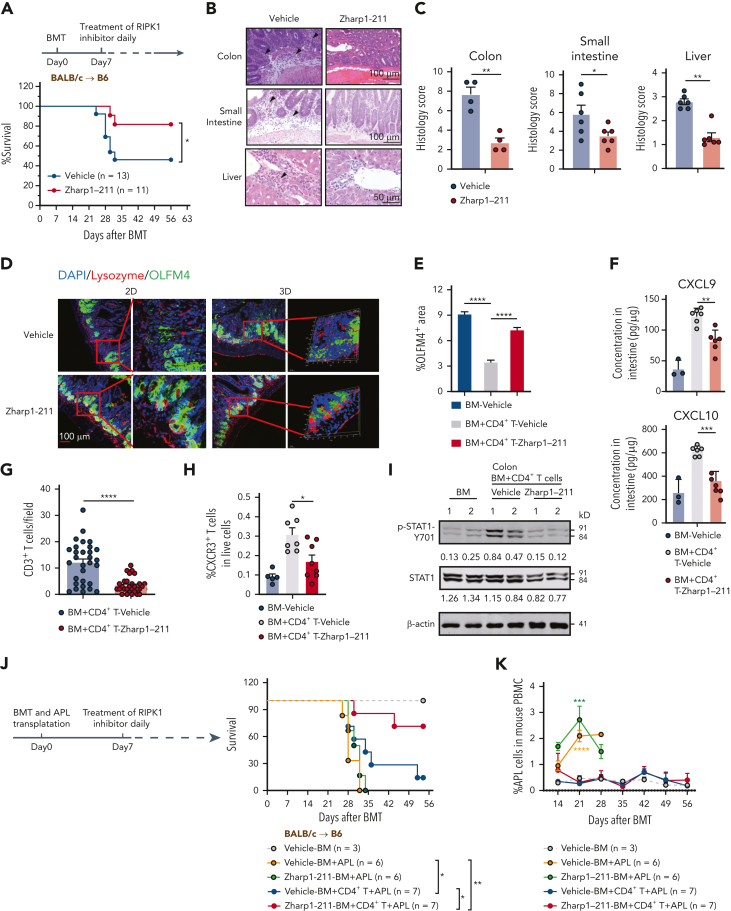
Zharp1-211 possessed favorable physicochemical properties and in vitro safety profiles, displaying minimal inhibition effects against CYP isozymes and hERG potassium channels (supplemental Figure 7). Moreover, Zharp1-211 demonstrated excellent pharmacokinetic profiles in mice and rats (supplemental Figure 8). To test the impact of RIPK1 inhibition on GVHD, we administered Zharp1-211 to C57BL/6 mice 7 days after receiving BALB/c T cells, a time point when IEC RIPK1/RIPK3 was already activated and GVHD occurred. Zharp1-211 treatment significantly reduced the severity of GVHD, leading to improved overall survival rates (Figure 7A; supplemental Figure 9A). This beneficial effect of Zharp1-211 treatment on GVHD inhibition was observed using the secondary B6 anti-BALB/c mouse model (supplemental Figure 9B). Moreover, Zharp1-211 treatment ameliorated the haploidentical B6 anti-BDF1 mouse model of GVHD (supplemental Figure 9C-D).
Figure 7.
Pharmacological inhibition of RIPK1 reduces ongoing GVHD while preserving GVL activity. (A-I) The lethally irradiated B6 recipients (BALB/c→B6) received BALB/c TCD BM cells with CD4+ T cells. Starting at 7 days after allo-HCT, mice were treated daily (intraperitoneally) with vehicle or Zharp1-211 (5 mg/kg). (A) Survival of mice after allo-HCT. (B-C) Histological analysis of small intestine and liver, assessed on day 17 after allo-HCT. Representative H&E images (B) and quantification of pathology scores (C). Bars in colon and small intestine represent 100 μm. Bar in liver represents 50 μm. Black arrows indicate areas of inflammatory cell infiltration or cell damage. (D) Representative 2D and 3D confocal images of OLFM4+ ISCs and lysozyme+ Paneth cells in small intestines from the indicated mice at 17 days after allo-HCT. Bar represents 100 μm. (E) Quantitation of IHC staining of OLFM4 in small intestines on day 17 after allo-HCT. (F) ELISA analysis of CXCL9 and CXCL10 protein levels in the small intestine from the indicated mice at 17 days after allo-HCT. (G) Quantification of infiltrated CD3+ T cells in the small intestines from the indicated B6 recipients at day 17 after allo-HCT. (H) Quantification of infiltrated CXCR3+ CD3+ T cells in the small intestine from the indicated B6 recipients at day 17 after allo-HCT. (I) Immunoblotting analysis of p-STAT1, STAT1, and β-actin in the intestine from the indicated B6 recipients after allo-HCT. Quantification of p-STAT1 or STAT1 normalized to β-actin was shown under the band. (J-K) The lethally irradiated B6 recipients received TCD BM cells (BM) alone, or in combination with CD4+ T cells from BALB/c mice. Subsequently, these mice were challenged with 1 × 10⁶ APL at the time of the BM transplant. Starting at 7 days after allo-HCT, recipients were treated daily (intraperitoneally) with vehicle or Zharp1-211 (5 mg/kg). The survival rates (J) and percentage of APL cells in peripheral blood from day 14(K) were measured. Vehicle-BM+APL group and Zharp1-211-BM+APL group were compared with Vehicle-BM group on day 21. Data shown are representative of 2 or 3s independent experiments. Data are shown as the mean ± SD (E), histology scores and concentrations of cytokines are shown as the mean ± SEM (C,F-H,K). ∗P < .05; ∗∗P < .01; ∗∗∗P < .001; ∗∗∗∗P < .0001. Survival comparisons were evaluated by log-rank test (A,J). Multiple comparisons were evaluated by one-way ANOVA (E-F,H); two-group comparisons used unpaired t tests (two-tailed) (C,G); APL cell measurements were evaluated by two-way ANOVA (K). ANOVA, analysis of variance; ELISA, enzyme-linked immunosorbent assay analysis; IHC, immunohistochemistry; SD, standard deviation; SEM, standard error of the mean.
Zharp1-211 treatment markedly reduced GVH inflammation in the colon, small intestine, and liver (Figure 7B-C) and attenuated skin inflammation (supplemental Figure 9E-F). Zharp1-211 also prevented the loss of ISCs and Paneth cells (Figure 7D-E; supplemental Figure 9G). Zharp1-211–treated mice displayed significant decreases in intestinal chemokines and reduced numbers of alloreactive T cells and CXCR3+T cells (Figure 7F-H). Of note, the Zharp1-211 treatment inhibited STAT1 activation in the intestine (Figure 7I). Moreover, Zharp1-211 treatment reduced MLKL phosphorylation and caspase-3 cleavage in the intestine (supplemental Figure 9H), as well as MLKL phosphorylation in intestinal crypt cells (supplemental Figure 9I), supporting Zharp1-211–mediated suppression of necroptosis and apoptosis in the GI tract. Our results indicate that Zharp1-211 inhibition of RIPK1 strongly reduces an inflammatory cascade in the GI tract and inhibits of GVHD.
After allo-HCT, donor immune cells have the ability to eliminate host leukemic cells, which is known as the GVL effect and is critical for preventing cancer relapse.52 We finally evaluated the impact of Zharp1-211 on the GVL effect in B6 recipients receiving acute promyelocytic leukemia (APL) cells, which are driven by the PML-RARA fusion gene and recapitulate the human disease.53 B6 mice receiving BALB/c TCD-BM and APL cells died from leukemia (Figure 7J-K), whereas B6 mice receiving BALB/c TCD-BM plus T cells showed reduced leukemia but died from GVHD (Figure 7J-K). In contrast, Zharp1-211 treatment preserved potent antileukemia activity without causing severe GVHD in mice receiving BALB/c T cells and APL cells, leading to significantly improved overall survival of leukemia mice undergoing allo-HCT (Figure 7J-K). Zharp1-211–mediated inhibition of GVHD without affecting the GVL effect was further confirmed in BALB/c recipients receiving allo-HCT and A20 murine B lymphoma cells (supplemental Figure 10A-B). Thus, Zharp1-211 is effective for inhibiting GVHD without compromising the GVL effect.
Discussion
Our study has demonstrated that RIPK1/RIPK3 in IECs is required to trigger local and systemic GVHD via a mechanism involving both cell death and activation of a necroptosis-independent inflammatory cascade. RIPK1/RIPK3 promotes JAK/STAT1-induced production of chemokines and MHC class II molecules in IECs, which amplify the recruitment and expansion of alloreactive T cells. We show that alloantigen-primed T-cell–derived IFN-γ activates the RIPK1/RIPK3/JAK1/STAT1 axis in IECs to trigger a feed-forward inflammatory cascade, thereby amplifying alloreactive T-cell responses in the GI tract and inducing subsequent conversion to systemic GVHD syndrome. We also discovered the RIPK1 inhibitor Zharp1-211, which blocks the RIPK1/RIPK3-mediated inflammatory cascade and inhibits both local and systemic GVHD without compromising the GVL effect. Our study therefore demonstrates proof of concept for targeting RIPK1/RIPK3-regulated inflammatory responses in IECs as nonimmunosuppressive therapies to prevent and treat GVHD.
Nonhematopoietic cells in the GI tract have been implicated in GVHD propagation,4,5,54 but the underlying molecular mechanisms remain poorly understood. The MLKL-independent role for RIPK3 identified in this study regulates the pathogenesis of GVHD and does so by generating an inflammatory cascade between IECs and alloreactive T cells. The activation and recruitment of alloreactive T cells to target organs are viewed as determinants of GVHD pathogenesis.7 Blockade of chemokine receptors CXCR3 or CCR5 inhibited GVHD and significantly improved the survival of hosts undergoing allo-HCT,43, 44, 45,55 although the regulatory mechanism(s) of this chemokine production in the GI tract during GVHD remains unknown. We found that RIPK3 regulates IEC production of alloreactive T-cell–recruiting chemokines. Loss of RIPK3 in IECs led to markedly reduced T-cell numbers in the intestine after allo-HCT. Thus, RIPK3-mediated chemokine production in IECs functionally affects the recruitment of alloreactive T cells and other inflammatory cells into the intestine. Besides this regulation of chemokines in IECs, we found that loss of RIPK3 significantly reduced the expression of MHC class II molecules in IECs. This finding supports a previous notion that MHC class II molecules in IECs can promote alloreactive T-cell activation and GVHD pathogenesis.5 Thus, our study reveals a previously unrecognized role of RIPK3 in regulating the IEC-localized production of chemokines and MHC class II molecules, which amplify and sustain alloreactive T-cell responses in the GVHD microenvironment, thereby creating an inflammatory cascade.
RIPK3 is known to be activated by interacting with RIPK1,18, 19, 20 ZBP1,46,47 or TRIF56,57 via their RHIM domains.58,59 Recent studies suggest that ZBP1 activates RIPK3-mediated necroptosis to drive intestinal pathology.32,60 However, we found that deletion of ZBP1 did not protect against GVHD after allo-HCT. Ripk1 K45A/K45A recipient mice exerted effective protection against intestinal and systemic GVHD as seen in Ripk3−/− mice. Notably, we detected RIPK1 phosphorylation in most colon biopsies from human patients with GVHD involved in GI. Thus, our work indicates RIPK1 phosphorylation as a biomarker for RIPK1 activation–mediated pathology in GI tract GVHD. However, the observation that some colon biopsies examined showed no signal of p-RIPK1 could be the result of heterogeneity among different regions of the samples or heterogeneity across patients. Limited by clinical practice and rules, we cannot perform biopsies to examine RIPK1 phosphorylation in the colon from patients without GVHD after allo-HCT. Future studies using the noninvasive method are needed to examine the correlation between active p-RIPK1 and GVHD severity in patients who did not receive allo-HCT.
IFN-γ signaling through nonhematopoietic cells has been implicated in the pathogenesis of GI tract GVHD.61 We delineated cell death–independent function of RIPK1/RIPK3 activating JAK/STAT1 signaling in IECs upon IFN-γ stimulation, inducing high expression of chemokines and MHC class II molecules to promote T-cell recruitment and maintenance. RIPK1 and RIPK3 have been implicated in the regulation of inflammatory responses independent of cell death.62,63 Macrophage RIPK1 and RIPK3 act as regulators of proinflammatory genes in response to LPS.62 Neuronal RIPK3 promotes chemokine expression and the nervous system’s recruitment of T cells in viral encephalitis mice downstream of toll-like receptors.63 We show that RIPK1 can form a complex with JAK1 through the binding of RIPK1 to the SH2 domain of JAK1, facilitating STAT1 activation. RIPK3 deficiency reduces the formation of RIPK1/JAK1 complex upon INF-γ stimulation, suggesting an important role of RIPK3 in promoting the RIPK1/JAK1 interaction. Importantly, RIPK1/RIPK3-controlled activation of STAT1 occurs in the intestine during GVHD. Thus, our study demonstrates that IFN-γ derived from alloreactive T cells activates a RIPK1/RIPK3/JAK/STAT1 axis to generate a feed-forward inflammatory cascade during life-threatening alloreactive T-cell responses.
The existing methods to treat the GVHD using immunosuppressive drugs, including glucocorticoids and the JAK1/2 inhibitor ruxolitinib, cause immunosuppression and adverse effects.49 For example, ruxolitinib therapy causes an increased incidence of thrombocytopenia, anemia, and infection.49 Our study illustrates an innovative therapeutic intervention based on RIPK1 inhibition that does not cause immunosuppression. Distinct from ruxolitinib, inhibition of RIPK1 by Zharp1-211 causes no significant effect on JAK-STAT–mediated TCR-activated T-cell proliferation or effector differentiation. Thus, targeting RIPK1 kinase activity decreases JAK1/STAT1 activity in IECs rather than T cells. Treatment with Zharp1-211 led to the suppression of inflammatory responses and less activation of apoptosis and necroptosis in GI epithelial cells in experimental mice undergoing allo-HCT. Moreover, Zharp1-211 does not affect the beneficial GVL effect. Although RIPK1 is required for IEC homeostasis and bone marrow development,56,57,64 selective inhibition of RIPK1 kinase activity by Zharp1-211 has no obvious side effects on IECs (supplemental Figure 11A-B) and bone marrow reconstruction (supplemental Figure 11C-D). Therefore, inhibition of RIPK1 kinase activity by Zharp1-211 presents a nonimmunosuppressive approach with promising therapeutic potential to treat GVHD.
In summary, our study has revealed that RIPK1/RIPK3 signaling in IECs activates JAK1/STAT1-mediated chemokines and MHC II molecules and triggers an inflammatory cascade of alloreactive T-cell responses. In addition, our discovery of Zharp1-211, which selectively inhibits RIPK1 activation of the JAK1/STAT1 signaling in IECs, illustrates a unique strategy for treating GVHD and potentially for other T-cell–mediated inflammatory diseases (eg, inflammatory bowel disease). Finally, our findings open new perspectives to better understand the molecular mechanisms that govern the activation of epithelial cell RIPK1/RIPK3 signaling, as well as its cell death–dependent and cell death–independent functions that drive inflammatory and immune responses.
Conflict-of-interest disclosure: X. Zhang and S. He are co-founders, consultants, and shareholders of Accro Bioscience Inc, which supports research in their labs. The remaining authors declare no competing financial interests.
Acknowledgments
The authors thank Dr. Xiaodong Wang (National Institute of Biological Sciences, Beijing) for kindly providing Ripk3−/−, Mlkl−/− B6 mice, and Smac mimetic. The authors appreciate the technical support from the RNA technology platform of the Suzhou Institute of Systems Medicine.
This work was supported by the National Natural Science Foundation of China (31830051, 82170218, 31671436, 81973161, 81773561, 31900526, 31771533, and 31600133), the National Key Research and Development Program of China (2022YFC2502702, 2022YFC2502701 and 2018YFA0900803), the CAMS Innovation Fund for Medical Sciences (2021-I2M-1-041, 2021-I2M-1-047, 2021-I2M-1-061, and 2022-I2M-2-004), Nonprofit Central Research Institute Fund of Chinese Academy of Medical Sciences (2021-PT180-001, 2019PT310028, 2017NL31004, 2017NL31002), the Special Research Fund for Central Universities, Peking Union Medical College (3332022077), the Priority Academic Program Development of the Jiangsu Higher Education Institutes (PAPD), the Jiangsu Key Laboratory of Neuropsychiatric Diseases (BM2013003), China Postdoctoral Science Foundation funded project (2019M650563), Natural Science Foundation of Jiangsu Province Grant (BK20160314, BE2021654), and Fok Ying Tung Education Foundation for Young Teachers (151020).
Authorship
Contribution: S. He, S. Hu, X. Zhang, and Y. Zhang designed this study and wrote the manuscript; X.Y., Y.J., Y.D., C.Z., Q.D., Y.T., F.Z., Y. Zheng, and J.W. performed experiments involving mouse GVHD model and organoids, and X.Y. carried out most of the experiments and analyzed the data; H.M., Z.L., and Y.H. performed the chemical synthesis and related analysis; B.L., Z.J., X. Zhu, X.C., and X.S. collected human samples and performed related experiments; S.T. performed molecular docking study; X.Y., C.Y., J.J., W. Zhang, T.Y., S.D., and H.L. performed cell culture, immunoblotting, enzyme-linked immunosorbent assay analysis, flow cytometry, and immunostaining; S.L. and F.M. performed RNA-seq analysis and provided expertise; X.W., J.L., and X.X. performed pathology analysis; H.Z. provided reagents and expertise.
Footnotes
RNA-seq data are available at GEO under No. GSE165605, GSE165606. The crystal structure of Zharp1-211 has been deposited in the CCDC under No.2050039.
Data are available on request from the corresponding author, Sudan He (hesd@ism.pumc.edu.cn).
The online version of this article contains a data supplement.
There is a Blood Commentary on this article in this issue.
The publication costs of this article were defrayed in part by page charge payment. Therefore, and solely to indicate this fact, this article is hereby marked “advertisement” in accordance with 18 USC section 1734.
Contributor Information
Yi Zhang, Email: yi.zhang@hmh-cdi.org.
Xiaohu Zhang, Email: xiaohuzhang@suda.edu.cn.
Shaoyan Hu, Email: hsy139@126.com.
Sudan He, Email: hesd@ism.pumc.edu.cn.
Supplementary Material
References
- 1.Peterson LW, Artis D. Intestinal epithelial cells: regulators of barrier function and immune homeostasis. Nat Rev Immunol. 2014;14(3):141–153. doi: 10.1038/nri3608. [DOI] [PubMed] [Google Scholar]
- 2.Lane RS, Lund AW. Non-hematopoietic control of peripheral tissue T cell responses: implications for solid tumors. Front Immunol. 2018;9:2662–2677. doi: 10.3389/fimmu.2018.02662. [DOI] [PMC free article] [PubMed] [Google Scholar]
- 3.Patankar JV, Becker C. Cell death in the gut epithelium and implications for chronic inflammation. Nat Rev Gastroenterol Hepatol. 2020;17(9):543–556. doi: 10.1038/s41575-020-0326-4. [DOI] [PubMed] [Google Scholar]
- 4.Koyama M, Kuns RD, Olver SD, et al. Recipient nonhematopoietic antigen-presenting cells are sufficient to induce lethal acute graft-versus-host disease. Nat Med. 2011;18(1):135–142. doi: 10.1038/nm.2597. [DOI] [PubMed] [Google Scholar]
- 5.Koyama M, Mukhopadhyay P, Schuster IS, et al. MHC class II antigen presentation by the intestinal epithelium initiates graft-versus-host disease and is influenced by the microbiota. Immunity. 2019;51(5):885–898.e7. doi: 10.1016/j.immuni.2019.08.011. [DOI] [PMC free article] [PubMed] [Google Scholar]
- 6.Chung J, Ebens CL, Perkey E, et al. Fibroblastic niches prime T cell alloimmunity through Delta-like Notch ligands. J Clin Invest. 2017;127(4):1574–1588. doi: 10.1172/JCI89535. [DOI] [PMC free article] [PubMed] [Google Scholar]
- 7.Zeiser R, Blazar BR. Acute graft-versus-host disease - biologic process, prevention, and therapy. N Engl J Med. 2017;377(22):f2167–2179. doi: 10.1056/NEJMra1609337. [DOI] [PMC free article] [PubMed] [Google Scholar]
- 8.Naymagon S, Naymagon L, Wong SY, et al. Acute graft-versus-host disease of the gut: considerations for the gastroenterologist. Nat Rev Gastroenterol Hepatol. 2017;14(12):711–726. doi: 10.1038/nrgastro.2017.126. [DOI] [PMC free article] [PubMed] [Google Scholar]
- 9.Ferrara JL, Levine JE, Reddy P, Holler E. Graft-versus-host disease. Lancet. 2009;373(9674):1550–1561. doi: 10.1016/S0140-6736(09)60237-3. [DOI] [PMC free article] [PubMed] [Google Scholar]
- 10.Hanash AM, Dudakov JA, Hua G, et al. Interleukin-22 protects intestinal stem cells from immune-mediated tissue damage and regulates sensitivity to graft versus host disease. Immunity. 2012;37(2):339–350. doi: 10.1016/j.immuni.2012.05.028. [DOI] [PMC free article] [PubMed] [Google Scholar]
- 11.Fu YY, Egorova A, Sobieski C, et al. T cell recruitment to the intestinal stem cell compartment drives immune-mediated intestinal damage after allogeneic transplantation. Immunity. 2019;51(1):90–103.e3. doi: 10.1016/j.immuni.2019.06.003. [DOI] [PMC free article] [PubMed] [Google Scholar]
- 12.Takashima S, Martin ML, Jansen SA, et al. T cell-derived interferon-gamma programs stem cell death in immune-mediated intestinal damage. Sci Immunol. 2019;4(42):eaay8556–8569. doi: 10.1126/sciimmunol.aay8556. [DOI] [PMC free article] [PubMed] [Google Scholar]
- 13.Matsuzawa-Ishimoto Y, Hine A, Shono Y, et al. An intestinal organoid-based platform that recreates susceptibility to T-cell-mediated tissue injury. Blood. 2020;135(26):2388–2401. doi: 10.1182/blood.2019004116. [DOI] [PMC free article] [PubMed] [Google Scholar]
- 14.Matsuzawa-Ishimoto Y, Shono Y, Gomez LE, et al. Autophagy protein ATG16L1 prevents necroptosis in the intestinal epithelium. J Exp Med. 2017;214(12):3687–3705. doi: 10.1084/jem.20170558. [DOI] [PMC free article] [PubMed] [Google Scholar]
- 15.He S, Wang X. RIP kinases as modulators of inflammation and immunity. Nat Immunol. 2018;19(9):912–922. doi: 10.1038/s41590-018-0188-x. [DOI] [PubMed] [Google Scholar]
- 16.Mifflin L, Ofengeim D, Yuan J. Receptor-interacting protein kinase 1 (RIPK1) as a therapeutic target. Nat Rev Drug Discov. 2020;19(8):553–571. doi: 10.1038/s41573-020-0071-y. [DOI] [PMC free article] [PubMed] [Google Scholar]
- 17.Linkermann A, Green DR. Necroptosis. N Engl J Med. 2014;370(5):455–465. doi: 10.1056/NEJMra1310050. [DOI] [PMC free article] [PubMed] [Google Scholar]
- 18.Holler N, Zaru R, Micheau O, et al. Fas triggers an alternative, caspase-8-independent cell death pathway using the kinase RIP as effector molecule. Nat Immunol. 2000;1(6):489–495. doi: 10.1038/82732. [DOI] [PubMed] [Google Scholar]
- 19.He S, Wang L, Miao L, et al. Receptor interacting protein kinase-3 determines cellular necrotic response to TNF-alpha. Cell. 2009;137(6):1100–1111. doi: 10.1016/j.cell.2009.05.021. [DOI] [PubMed] [Google Scholar]
- 20.Cho YS, Challa S, Moquin D, et al. Phosphorylation-driven assembly of the RIP1-RIP3 complex regulates programmed necrosis and virus-induced inflammation. Cell. 2009;137(6):1112–1123. doi: 10.1016/j.cell.2009.05.037. [DOI] [PMC free article] [PubMed] [Google Scholar]
- 21.Zhang DW, Shao J, Lin J, et al. RIP3, an energy metabolism regulator that switches TNF-induced cell death from apoptosis to necrosis. Science. 2009;325(5938):332–336. doi: 10.1126/science.1172308. [DOI] [PubMed] [Google Scholar]
- 22.Sun L, Wang H, Wang Z, et al. Mixed lineage kinase domain-like protein mediates necrosis signaling downstream of RIP3 kinase. Cell. 2012;148(1-2):213–227. doi: 10.1016/j.cell.2011.11.031. [DOI] [PubMed] [Google Scholar]
- 23.Zhao J, Jitkaew S, Cai Z, et al. Mixed lineage kinase domain-like is a key receptor interacting protein 3 downstream component of TNF-induced necrosis. Proc Natl Acad Sci U S A. 2012;109(14):5322–5327. doi: 10.1073/pnas.1200012109. [DOI] [PMC free article] [PubMed] [Google Scholar]
- 24.Cai Z, Jitkaew S, Zhao J, et al. Plasma membrane translocation of trimerized MLKL protein is required for TNF-induced necroptosis. Nat Cell Biol. 2014;16(1):55–65. doi: 10.1038/ncb2883. [DOI] [PMC free article] [PubMed] [Google Scholar]
- 25.Wang H, Sun L, Su L, et al. Mixed lineage kinase domain-like protein MLKL causes necrotic membrane disruption upon phosphorylation by RIP3. Mol Cell. 2014;54(1):133–146. doi: 10.1016/j.molcel.2014.03.003. [DOI] [PubMed] [Google Scholar]
- 26.Chen X, Li W, Ren J, et al. Translocation of mixed lineage kinase domain-like protein to plasma membrane leads to necrotic cell death. Cell Res. 2014;24(1):105–121. doi: 10.1038/cr.2013.171. [DOI] [PMC free article] [PubMed] [Google Scholar]
- 27.Murphy JM, Czabotar PE, Hildebrand JM, et al. The pseudokinase MLKL mediates necroptosis via a molecular switch mechanism. Immunity. 2013;39(3):443–453. doi: 10.1016/j.immuni.2013.06.018. [DOI] [PubMed] [Google Scholar]
- 28.Pasparakis M, Vandenabeele P. Necroptosis and its role in inflammation. Nature. 2015;517(7534):311–320. doi: 10.1038/nature14191. [DOI] [PubMed] [Google Scholar]
- 29.Welz PS, Wullaert A, Vlantis K, et al. FADD prevents RIP3-mediated epithelial cell necrosis and chronic intestinal inflammation. Nature. 2011;477(7364):330–334. doi: 10.1038/nature10273. [DOI] [PubMed] [Google Scholar]
- 30.Gunther C, Martini E, Wittkopf N, et al. Caspase-8 regulates TNF-alpha-induced epithelial necroptosis and terminal ileitis. Nature. 2011;477(7364):335–339. doi: 10.1038/nature10400. [DOI] [PMC free article] [PubMed] [Google Scholar]
- 31.Schwarzer R, Jiao H, Wachsmuth L, Tresch A, Pasparakis M. FADD and caspase-8 regulate gut homeostasis and inflammation by controlling MLKL- and GSDMD-mediated death of intestinal epithelial cells. Immunity. 2020;52(6):978–993.e6. doi: 10.1016/j.immuni.2020.04.002. [DOI] [PubMed] [Google Scholar]
- 32.Wang R, Li H, Wu J, et al. Gut stem cell necroptosis by genome instability triggers bowel inflammation. Nature. 2020;580(7803):386–390. doi: 10.1038/s41586-020-2127-x. [DOI] [PubMed] [Google Scholar]
- 33.Degterev A, Huang Z, Boyce M, et al. Chemical inhibitor of nonapoptotic cell death with therapeutic potential for ischemic brain injury. Nat Chem Biol. 2005;1(2):112–119. doi: 10.1038/nchembio711. [DOI] [PubMed] [Google Scholar]
- 34.Degterev A, Hitomi J, Germscheid M, et al. Identification of RIP1 kinase as a specific cellular target of necrostatins. Nat Chem Biol. 2008;4(5):313–321. doi: 10.1038/nchembio.83. [DOI] [PMC free article] [PubMed] [Google Scholar]
- 35.Takahashi N, Duprez L, Grootjans S, et al. Necrostatin-1 analogues: critical issues on the specificity, activity and in vivo use in experimental disease models. Cell Death Dis. 2012;3(11):e437–446. doi: 10.1038/cddis.2012.176. [DOI] [PMC free article] [PubMed] [Google Scholar]
- 36.Weisel K, Berger S, Thorn K, et al. A randomized, placebo-controlled experimental medicine study of RIPK1 inhibitor GSK2982772 in patients with moderate to severe rheumatoid arthritis. Arthritis Res Ther. 2021;23(1):85–96. doi: 10.1186/s13075-021-02468-0. [DOI] [PMC free article] [PubMed] [Google Scholar]
- 37.Harris PA, Berger SB, Jeong JU, et al. Discovery of a first-in-class receptor interacting protein 1 (RIP1) kinase specific clinical candidate (GSK2982772) for the treatment of inflammatory diseases. J Med Chem. 2017;60(4):1247–1261. doi: 10.1021/acs.jmedchem.6b01751. [DOI] [PubMed] [Google Scholar]
- 38.Glucksberg H, Storb R, Fefer A, et al. Clinical manifestations of graft-versus-host disease in human recipients of marrow from HL-A-matched sibling donors. Transplantation. 1974;18(4):295–304. doi: 10.1097/00007890-197410000-00001. [DOI] [PubMed] [Google Scholar]
- 39.Liu Y, Fan C, Zhang Y, et al. RIP1 kinase activity-dependent roles in embryonic development of Fadd-deficient mice. Cell Death Differ. 2017;24(8):1459–1469. doi: 10.1038/cdd.2017.78. [DOI] [PMC free article] [PubMed] [Google Scholar]
- 40.Kaiser WJ, Upton JW, Long AB, et al. RIP3 mediates the embryonic lethality of caspase-8-deficient mice. Nature. 2011;471(7338):368–372. doi: 10.1038/nature09857. [DOI] [PMC free article] [PubMed] [Google Scholar]
- 41.Lindemans CA, Calafiore M, Mertelsmann AM, et al. Interleukin-22 promotes intestinal-stem-cell-mediated epithelial regeneration. Nature. 2015;528(7583):560–564. doi: 10.1038/nature16460. [DOI] [PMC free article] [PubMed] [Google Scholar]
- 42.Takashima S, Kadowaki M, Aoyama K, et al. The Wnt agonist R-spondin1 regulates systemic graft-versus-host disease by protecting intestinal stem cells. J Exp Med. 2011;208(2):285–294. doi: 10.1084/jem.20101559. [DOI] [PMC free article] [PubMed] [Google Scholar]
- 43.Reshef R, Luger SM, Hexner EO, et al. Blockade of lymphocyte chemotaxis in visceral graft-versus-host disease. N Engl J Med. 2012;367(2):135–145. doi: 10.1056/NEJMoa1201248. [DOI] [PMC free article] [PubMed] [Google Scholar]
- 44.He S, Cao Q, Qiu Y, et al. A new approach to the blocking of alloreactive T cell-mediated graft-versus-host disease by in vivo administration of anti-CXCR3 neutralizing antibody. J Immunol. 2008;181(11):7581–7592. doi: 10.4049/jimmunol.181.11.7581. [DOI] [PubMed] [Google Scholar]
- 45.Murai M, Yoneyama H, Ezaki T, et al. Peyer's patch is the essential site in initiating murine acute and lethal graft-versus-host reaction. Nat Immunol. 2003;4(2):154–160. doi: 10.1038/ni879. [DOI] [PubMed] [Google Scholar]
- 46.Upton JW, Kaiser WJ, Mocarski ES. DAI/ZBP1/DLM-1 complexes with RIP3 to mediate virus-induced programmed necrosis that is targeted by murine cytomegalovirus vIRA. Cell Host Microbe. 2012;11(3):290–297. doi: 10.1016/j.chom.2012.01.016. [DOI] [PMC free article] [PubMed] [Google Scholar]
- 47.Balachandran S, Mocarski ES. Viral Z-RNA triggers ZBP1-dependent cell death. Curr Opin Virol. 2021;51:134–140. doi: 10.1016/j.coviro.2021.10.004. [DOI] [PMC free article] [PubMed] [Google Scholar]
- 48.Stark GR, Darnell JE., Jr. The JAK-STAT pathway at twenty. Immunity. 2012;36(4):503–514. doi: 10.1016/j.immuni.2012.03.013. [DOI] [PMC free article] [PubMed] [Google Scholar]
- 49.Zeiser R, von Bubnoff N, Butler J, et al. Ruxolitinib for glucocorticoid-refractory acute graft-versus-host disease. N Engl J Med. 2020;382(19):1800–1810. doi: 10.1056/NEJMoa1917635. [DOI] [PubMed] [Google Scholar]
- 50.Liu SY, Sanchez DJ, Aliyari R, Lu S, Cheng G. Systematic identification of type I and type II interferon-induced antiviral factors. Proc Natl Acad Sci U S A. 2012;109(11):4239–4244. doi: 10.1073/pnas.1114981109. [DOI] [PMC free article] [PubMed] [Google Scholar]
- 51.Hou J, Ju J, Zhang Z, et al. Discovery of potent necroptosis inhibitors targeting RIPK1 kinase activity for the treatment of inflammatory disorder and cancer metastasis. Cell Death Dis. 2019;10(7):493–505. doi: 10.1038/s41419-019-1735-6. [DOI] [PMC free article] [PubMed] [Google Scholar]
- 52.Dickinson AM, Norden J, Li S, et al. Graft-versus-leukemia effect following hematopoietic stem cell transplantation for leukemia. Front Immunol. 2017;8:496–511. doi: 10.3389/fimmu.2017.00496. [DOI] [PMC free article] [PubMed] [Google Scholar]
- 53.Brown D, Kogan S, Lagasse E, et al. A PMLRARalpha transgene initiates murine acute promyelocytic leukemia. Proc Natl Acad Sci U S A. 1997;94(6):2551–2556. doi: 10.1073/pnas.94.6.2551. [DOI] [PMC free article] [PubMed] [Google Scholar]
- 54.Ferrara JL, Smith CM, Sheets J, Reddy P, Serody JS. Altered homeostatic regulation of innate and adaptive immunity in lower gastrointestinal tract GVHD pathogenesis. J Clin Invest. 2017;127(7):2441–2451. doi: 10.1172/JCI90592. [DOI] [PMC free article] [PubMed] [Google Scholar]
- 55.Moy RH, Huffman AP, Richman LP, et al. Clinical and immunologic impact of CCR5 blockade in graft-versus-host disease prophylaxis. Blood. 2017;129(7):906–916. doi: 10.1182/blood-2016-08-735076. [DOI] [PMC free article] [PubMed] [Google Scholar]
- 56.Dannappel M, Vlantis K, Kumari S, et al. RIPK1 maintains epithelial homeostasis by inhibiting apoptosis and necroptosis. Nature. 2014;513(7516):90–94. doi: 10.1038/nature13608. [DOI] [PMC free article] [PubMed] [Google Scholar]
- 57.Takahashi N, Vereecke L, Bertrand MJ, et al. RIPK1 ensures intestinal homeostasis by protecting the epithelium against apoptosis. Nature. 2014;513(7516):95–99. doi: 10.1038/nature13706. [DOI] [PubMed] [Google Scholar]
- 58.He S, Liang Y, Shao F, Wang X. Toll-like receptors activate programmed necrosis in macrophages through a receptor-interacting kinase-3-mediated pathway. Proc Natl Acad Sci U S A. 2011;108(50):20054–20059. doi: 10.1073/pnas.1116302108. [DOI] [PMC free article] [PubMed] [Google Scholar]
- 59.Kaiser WJ, Sridharan H, Huang C, et al. Toll-like receptor 3-mediated necrosis via TRIF, RIP3, and MLKL. J Biol Chem. 2013;288(43):31268–31279. doi: 10.1074/jbc.M113.462341. [DOI] [PMC free article] [PubMed] [Google Scholar]
- 60.Jiao H, Wachsmuth L, Kumari S, et al. Z-nucleic-acid sensing triggers ZBP1-dependent necroptosis and inflammation. Nature. 2020;580(7803):391–395. doi: 10.1038/s41586-020-2129-8. [DOI] [PMC free article] [PubMed] [Google Scholar]
- 61.Burman AC, Banovic T, Kuns RD, et al. IFNgamma differentially controls the development of idiopathic pneumonia syndrome and GVHD of the gastrointestinal tract. Blood. 2007;110(3):1064–1072. doi: 10.1182/blood-2006-12-063982. [DOI] [PubMed] [Google Scholar]
- 62.Najjar M, Saleh D, Zelic M, et al. RIPK1 and RIPK3 kinases promote cell-death-independent inflammation by toll-like receptor 4. Immunity. 2016;45(1):46–59. doi: 10.1016/j.immuni.2016.06.007. [DOI] [PMC free article] [PubMed] [Google Scholar]
- 63.Daniels BP, Snyder AG, Olsen TM, et al. RIPK3 restricts viral pathogenesis via cell death-independent neuroinflammation. Cell. 2017;169(2):301–313.e311. doi: 10.1016/j.cell.2017.03.011. [DOI] [PMC free article] [PubMed] [Google Scholar]
- 64.Roderick JE, Hermance N, Zelic M, et al. Hematopoietic RIPK1 deficiency results in bone marrow failure caused by apoptosis and RIPK3-mediated necroptosis. Proc Natl Acad Sci U S A. 2014;111(40):14436–14441. doi: 10.1073/pnas.1409389111. [DOI] [PMC free article] [PubMed] [Google Scholar]
Associated Data
This section collects any data citations, data availability statements, or supplementary materials included in this article.


