Case Presentation
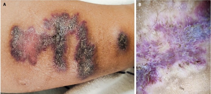
A 10-year-old boy presented with two distinct reddish-purple plaques on his left calf, which had been present since birth and were associated with hyperhidrosis (Figure 1A). On examination, there were two depressed erythematous to violaceous firm plaques, measuring approximately 5 cm × 3 cm and 1 cm × 1 cm. Dermoscopy revealed whitish striations and round lacunae on a prominent pinkish-blue background, surrounded by an area of hypopigmentation (Figure 1B). An ultrasound showed diffuse soft tissue thickening in the subcutaneous plane. Histopathological examination showed epidermal hyperkeratosis and psoriasiform acanthosis, in addition to increased numbers of normal eccrine glands and small blood vessels in the mid and deep dermis. Based on these findings, a diagnosis of verrucous hemangioma like eccrine angiomatous nevus was made, and sclerotherapy was attempted with partial improvement.
Figure 1.
(A) Depressed erythematous to violaceous firm plaques on left calf. (B) Whitish striations and round lacunae on a prominent pinkish-blue background, surrounded by hypopigmentation (DermLite DL3N, dry, contact, polarized, ×10)
Teaching Point
Eccrine angiomatous hamartoma is an uncommon, benign proliferation of eccrine glands and thin-walled vascular channels on the dermis [1]. The commonest sites of involvement are the extremities, especially the palms and soles. Characteristic papules, plaques, and nodules are single, flesh-colored, blue-brown, or reddish in color. Pain and excessive sweating are two signs of eccrine angiomatous hamartoma. Intriguingly, changes in the epidermis were observed in the current case, is an unusual finding in eccrine angiomatous hamartoma. The lesions seen in our patient were more indicative of verrucous hemangiomas, both clinically and dermoscopically. Verrucous hemangiomas are characterized by their multiple, grouped plaques or nodules. Spitzoid or popcorn patterns are the dermoscopic features of eccrine angiomatous hamartomas, whereas reddish-blue or bluish lacunae or dermal features with a bluish-white color are those of verrucous hemangiomas. There is a single case report in literature about dermoscopic characteristics of a verrucous hemangiomas like eccrine angiomatous hamartoma, which was similar to our case [2].
Footnotes
Competing Interests: None.
Authorship: All authors have contributed significantly to this publication.
Funding: None.
References
- 1.Sanusi T, Li Y, Sun L, Wang C, Zhou Y, Huang C. Eccrine angiomatous hamartoma: A clinicopathological study of 26 cases. Dermatology. 2015;231(1):63–69. doi: 10.1159/000381421. [DOI] [PubMed] [Google Scholar]
- 2.Liu L, Zhou L, Zhao Q, Wei D, Jiang X. Eccrine angiomatous hamartoma with verrucous hemangioma-like features–an unusual combination. Indian J Dermatol Venereol Leprol. 2021;87(6):842–844. doi: 10.25259/IJDVL_195_2021. [DOI] [PubMed] [Google Scholar]


