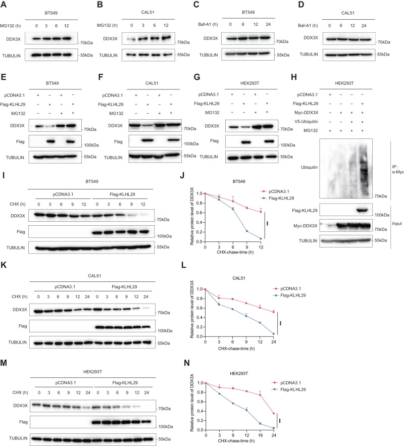
Fig. 5. KLHL29 promotes the ubiquitin-proteasome degradation of DDX3X.
A, B Protein levels of DDX3X in BT549 (A) and CAL51 (B) cells are elevated upon the treatment with 20 μM MG132 at the indicated time points. C, D Protein levels of DDX3X in BT549 (C) and CAL51 (D) cells are not affected by 200 nM Baf-A1 treatment at the indicated time points. E–G MG132 prevents KLHL29-mediated DDX3X degradation in BT549 (E), CAL51 (F) and HEK293T (G) cells. Cells expressing the empty vector or Flag-KLHL29 plasmid were treated with DMSO or 20 μM MG132 for 6 h before harvested for western blotting. H KLHL29 enhances the ubiquitination level of DDX3X. HEK293T cells were transfected with the indicated plasmids and treated with 20 μM MG132 for 6 h before harvested for ubiquitination assays. I–N The half-life of DDX3X is shortened upon KLHL29 overexpression in BT549 (I), CAL51 (K) and HEK293T (M) cells. Cells expressing the empty vector or Flag-KLHL29 plasmid were treated with 100 μg/mL of CHX and harvested for western blotting at the indicated time points. Quantification of relative KLHL29 protein levels (KLHL29/TUBULIN) by ImageJ are shown in J, L, and N. ***p < 0.001 by two-tailed Student’s t test.

