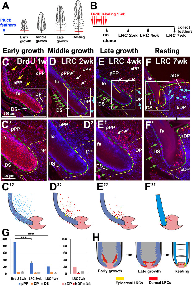
Fig. 3.
The behavior of long-term label-retaining dermal cells during feather cycling. (A) Contour feathers are plucked and allowed to regenerate. (B) Strategy to identify LRDCs. BrdU labeling begins 1 week after contour feather regeneration. Chickens are labeled with BrdU for 1 week. Feathers are collected at time points representing different cycle stages. (C) A feather follicle labeled with BrdU for 1 week without chasing. (D-F) Feather follicles are chased for 2 (D), 4 (E) and 7 (F) weeks. Collected feathers are in the middle growth, late growth and resting phase, respectively. Green dashed line outlines the feather epidermis. Yellow dashed line surrounds the DP. Yellow arrows (C) indicate the BrdU-positive cells in PP after 1-week labeling. White arrows (D,E) indicate the LRDCs in PP. Green arrows (D-F) indicate LRDCs in the DS. Blue arrows (D-F) indicate LRDCs in the DP. Note the LRDCs accumulate in the aDP in resting phase (F). Yellow brackets (D-F) indicate the epidermal stem cells. (C′-F′) Higher magnification images of boxed areas in C-F. (C″-F″) Schematic showing the relative position of BrdU-positive cells (blue dots, no chase; yellow dots, epidermal LRCs; red dots, dermal LRDCs). (G) Left: percentage of BrdU-positive cells before and after a 2- and 4-week chase period in the pPP, DP and DS. Right: percentage of BrdU-positive cells after a 7-week chase period in the aDP, bDP and DS. For each time point, n=5 follicles, ***P<0.001(paired two-tailed Student's t-test). Data are mean±s.d. (H) Diagram of LRDCs in early growth, late growth- and resting-phase feather follicles. Yellow represents stem cells in the feather epidermis. Red indicates putative dermal stem cells in feather follicles at different regeneration stages. aDP, apical dermal papilla; bDP, basal dermal papilla; cPP; central pulp; DP, dermal papilla; DS, dermal sheath; fe, feather epidermis; pPP, peripheral pulp.

