Abstract
The fragment containing the gene encoding the cytolytic Cyt1Ab1 protein from Bacillus thuringiensis subsp. medellin and its flanking sequences (I. Thiery, A. Delécluse, M. C. Tamayo, and S. Orduz, Appl. Environ. Microbiol. 63:468–473, 1997) was introduced into Bacillus sphaericus toxic strains 2362, 2297, and Iab872 by electroporation with the shuttle vector pMK3. Only small amounts of the protein were produced in recombinant strains 2362 and Iab872. The protein was detected in these strains only by Western blotting and immunodetection with antibody raised against Cyt1Ab1 protein. Large amounts of Cyt1Ab1 protein were produced in B. sphaericus recombinant strain 2297, and there was an additional crystal, other than that of the binary toxin, within the exosporium. The production of the Cyt1Ab1 protein in addition to the binary toxin did not increase the larvicidal activity of the B. sphaericus recombinant strain against susceptible mosquito populations of Culex pipiens or Aedes aegypti. However, it partially restored (10 to 20 times) susceptibility of the resistant mosquito populations of C. pipiens (SPHAE) and Culex quinquefasciatus (GeoR) to the binary toxin. The Cyt1Ab1 protein produced in recombinant B. thuringiensis SPL407(pcyt1Ab1) was synthesized in two types of crystal—one round and with various dense areas, surrounded by an envelope, and the other a regular cuboid crystal, very similar to that found in the B. sphaericus recombinant strain.
Highly mosquitocidal strains of Bacillus sphaericus produce a proteinous binary toxin which is toxic to mosquito larvae (2, 5). This toxin binds to a specific receptor on the midgut epithelial cells of the larvae, and larval susceptibility depends on the affinity of this binding (12, 13). A B. sphaericus product (Spherimos; Novo Nordisk Co.) was used against urban Culex pipiens subsp. pipiens (Say) larval populations in the South of France for 7 years before the first case of field resistance was reported (20). The resistant larval population is 50,000 to 100,000 times more resistant than the control population (14). Bacteria such as Bacillus thuringiensis subsp. israelensis produce several toxins, preventing the development of resistance in the mosquito larvae, although the mechanism by which susceptibility is maintained is unknown (9). Applications of B. thuringiensis subsp. israelensis over more than 18 years did not result in lower susceptibility of the treated populations, because several toxins were present (25). Thus, the introduction of another toxin into B. sphaericus strains might increase toxicity and prevent insects developing resistance. The genes encoding the Cry4B, Cry11A, or Cry11A plus Cyt1Aa1 endotoxins from B. thuringiensis subsp. israelensis have been isolated and were used to transform B. sphaericus strains 1593, 2362, and 2297 (1, 16, 17, 24). Production of these toxins led to an increase in toxicity to Aedes larvae, which were weakly susceptible to binary toxin, but no synergistic effect was demonstrated. Poncet et al. (16) showed that the production of Cry11A in B. sphaericus increases its toxicity to resistant Culex quinquefasciatus larvae. Federici and Bauer (7) and Wirth et al. (26) have shown that production of the cytolytic protein Cyt1Aa1 overcomes high levels of resistance, respectively, to Cry3A in a resistant population of Chrysomela scripta (Coleoptera, Chrysomelidae) and to Cry4A in a resistant population of C. quinquefasciatus.
The gene encoding the cytolytic protein Cyt1Ab1 from B. thuringiensis subsp. medellin was isolated by Thiéry et al. (23). It is flanked upstream by a p21 gene in the same orientation. This gene encodes a P21 protein with a sequence 84% similar to that of the putative chaperone P20 from B. thuringiensis subsp. israelensis, which is probably responsible for the formation of Cyt1Ab1 crystals in a B. thuringiensis crystal-negative recombinant strain (23).
The aim of this study was to introduce the fragment containing the gene encoding the cytolytic Cyt1Ab1 protein and its flanking sequences into B. sphaericus strains. We checked that the gene was expressed and investigated whether the addition of the Cyt1Ab1 protein either increased the larvicidal activity of B. sphaericus against C. pipiens, overcame resistance to the binary toxin in resistant populations of mosquito larvae, or both.
MATERIALS AND METHODS
Bacterial strains and plasmids.
B. sphaericus strains 2362, 2297, and Iab872 were from the IEBC Collection of the Unité des Bactéries Entomopathogènes, Institut Pasteur, Paris, France. They were used as recipient microorganisms for electroporation (17, 22). Escherichia coli TG1 [K-12Δ(lac-proAB) supE thi hsdD F′(raD36 proA+ proB+ lacl lacZΔM15)] was used for cloning experiments. Recombinant B. thuringiensis subsp. thuringiensis strain SPL407 synthesizing the Cyt1Ab1 protein from B. thuringiensis subsp. medellin (23) was used as a control for mosquitocidal activity determinations and microscopy. The shuttle vector pMK3 was used in subcloning experiments (21). Kanamycin (5 μg/ml) was added when required.
DNA experiments.
Restriction endonuclease digestion and ligation were carried out as previously described (18). Plasmid DNA was extracted and purified from E. coli with the Qiagen plasmid kit. DNA fragments were subjected to electrophoresis in 0.7% agarose gels. DNA fragments were eluted from agarose gel with the Prep-A-Gene DNA purification matrix kit (Bio-Rad, Hercules, Calif.).
The 2.5-kb HindIII-EcoRI insert of pCytM (containing the cyt1Ab1 and p21 genes) was inserted into the pMK3 vector, giving a 9.4-kb plasmid, pA7.
B. sphaericus strains 2297, 2362, and Iab872 were transformed with pMK3 or pA7 by electroporation as described by Taylor and Burke (22). B. sphaericus cells were grown in Luria broth (LB) medium, collected by centrifugation, and suspended in ice-cold 10% glycerol. B. sphaericus cells (200 μl) were placed in an ice-cold electroporation cuvette (0.2-cm interelectrode gap [Bio-Rad]) and transformed with 1 to 5 μg of plasmid DNA. The suspensions were subjected to a high-voltage pulse (25 μF, 2.5 kV, 400 Ω). The cells were then incubated in 2 ml of LB medium for 1 h at 37°C and plated on LB medium containing kanamycin (5 μg/ml).
We checked that the cyt1Ab1 gene was present in putative recombinant B. sphaericus strains by PCR with two oligonucleotide primers corresponding to the flanking sequences of the cyt1Ab1 gene, which was expected to give a product with a size of 705 bp.
Crystal purification.
B. sphaericus transformants were grown in MBS medium (10) containing kanamycin (5 μg/ml) at 30°C and underwent shaking until cell lysis. Protein inclusion bodies were observed in phase-contrast microscopy after Coomassie brilliant blue staining (19) as modified by E. Frachon (7a). Prior to staining, protein inclusion bodies were washed with a solution (50% acetone–50% ethanol) to eliminate lipidic material. The bacterial culture was centrifuged, and the spore-crystal pellet was washed once with 1 M NaCl and twice in distilled water containing 1 mM phenylmethylsulfonyl fluoride. The spore-crystal pellet from recombinant B. sphaericus strains was subjected to strong pulsed sonication (twice for 10 min, 40% duty cycle) on ice with a Branson B15 sonic cell disrupter (Branson Sonic Power Co., Smithkline Co.) to release the inclusions from the exosporium. Crystals were separated from spores on a discontinuous sucrose gradient (79% [wt/vol] and 67% [wt/vol]) with an SW28 swinging-bucket rotor in a Beckman L8-55 ultracentrifuge at 25,000 rpm at 4°C for 16 h. The pellet and the material between the layers were examined by light microscopy, and the spores and cells were counted.
Cultures of recombinant bacteria and purified crystals obtained by ultracentrifugation were treated as described by Charles et al. (4) and examined under an electron microscope.
Protein analysis.
The protein concentrations of alkali-solubilized bacterial suspensions and purified crystals were determined by the Bradford assay with bovine serum albumin as the standard (3). Sodium dodecyl sulfate-polyacrylamide gel electrophoresis (SDS-PAGE) was performed with 12% polyacrylamide gels as previously described (6). Proteins were transferred to Hybond-C super membrane (Amersham) and detected immunologically with the Amersham ECL (enhanced chemiluminescence) Western blotting kit according to the manufacturer’s recommendations. Rabbit antisera directed against purified crystals of Cyt1Ab1 toxin were produced and used for detection.
Mosquitocidal activity assay.
Bioassays were performed with young fourth-instar larvae of C. pipiens subsp. pipiens (strain Montpellier) from susceptible or resistant (Montpellier SPHAE) populations (14, 20) and with resistant C. quinquefasciatus (GeoR) larvae (8, 13). The bacterial pellet and purified crystals of B. sphaericus strains and of the control strain, B. thuringiensis SPL407(pcytM), were mixed with 10 ml of demineralized water in petri dishes (diameter, 5.5 cm) and tested in duplicate as previously described (23). Each experiment was performed three times. Larval mortality was recorded after 48 h, and 50 and 90% lethal concentrations (LC50s and LC90s, respectively) were determined by probit analysis with a program made by E. Frachon. LCs are given as means ± standard errors.
RESULTS
Transformation of B. sphaericus strains.
The 2.5-kb HindIII-EcoRI fragment from pCytM (23) was inserted into the pMK3 shuttle vector (21) as described in Materials and Methods. The resulting plasmid, pA7, was 9.4 kb and contained cyt1Ab1 and p21 and their flanking sequences.
pA7 was introduced into B. sphaericus 2362, 2297, and Iab872 recipient strains by electroporation, and the cyt1Ab1 gene was detected by PCR. The expected 705-bp product was detected in positive B. sphaericus 2362, 2297 and Iab872 clones (data not shown). The pMK3 vector was introduced into the same strains by electroporation, which then served as controls. A nonsporulating mutant, B. sphaericus 2297, that did not produce crystals was also transformed with both pMK3 and pA7. All recombinant clones were stable and were used for further experiments.
Synthesis of Cyt1Ab1 in B. sphaericus strains.
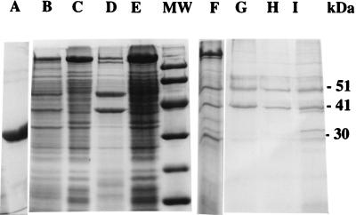
Cell lysis was complete after 72 to 90 h of culture for all recombinant B. sphaericus strains, whereas the parental recipient strains were totally lysed within 72 h. A large additional crystal was observed in the recombinant B. sphaericus strain 2297 (pcyt1Ab1) by light microscopy. It was shown to contain protein by Coomassie blue staining. A large amount of Cyt1Ab1 protein (30 kDa) was produced in B. sphaericus 2297 (Fig. 1). This additional 30-kDa protein could only be detected by specific antibodies in recombinant B. sphaericus strains 2362(pcyt1Ab1) and IAb872(pcyt1Ab1) and the nonsporulating strain 2297(pcyt1Ab1), indicating a low level of expression of the cyt1Ab1 gene (Fig. 2).
FIG. 1.
Protein analysis in wild-type and recombinant B. sphaericus strains. Fifteen micrograms of protein of the washed FWC was subjected to electrophoresis on an SDS-PAGE gel (12% polyacrylamide) followed by staining with Coomassie brilliant blue. Lanes: A, purified inclusion bodies of Cyt1Ab1 from B. thuringiensis SPL407(pcyt1Ab1); B, wild-type strain 2297; C, recombinant nonsporulating mutant strain 2297(pcyt1Ab1); D, recombinant strain 2297(pMK3); E, mutant asporulated strain 2297; F, recombinant strain 2297(pcyt1Ab1); G, recombinant strain Iab872(pcyt1Ab1); H, recombinant strain Iab872(pMK3); I, recombinant strain 2362(pcyt1Ab1); MW, low-molecular-mass kit standard protein markers from Pharmacia.
FIG. 2.
Western blot of 15 μg of protein from FWC or inclusion bodies of recombinant and wild-type B. sphaericus strains and of 5 μg of purified Cyt1Ab1 inclusion bodies from B. thuringiensis SPL407(pcyt1Ab1). The filter was incubated with antiserum (dilution 1/2,000) raised against Cyt1Ab1 protein purified from B. thuringiensis SPL407(pcyt1Ab1). Lanes: 1, 2297(pcyt1Ab1); 2, wild-type strain 2297; 3, nonsporulating mutant 2297(pcyt1Ab1); 4, nonsporulating mutant 2297(pMK3); 5, purified inclusion bodies of 2297(pcyt1Ab1); 6 and 7, purified inclusion bodies of nonsporulating mutant 2297(pcyt1Ab1) with 15 and 30 μg of protein, respectively; 8, Iab872(pcyt1Ab1); 9, 2362(pMK3); 10, 2362(pcyt1Ab1); 11, purified Cyt1Ab1 inclusion bodies; 12, wild-type strain Iab872; 13, Iab872(pMK3); 14, Iab872(pcyt1Ab1).
Inclusion body purification.
Ultracentrifugation of the washed pellet of the recombinant and wild-type versions of strain 2297 through a biphasic sucrose gradient gave two main phases: a pellet and interlayer material. The pellet consisted mostly of free spores and spore-crystal complexes (linked by intact exosporium), whereas the interlayer material contained mostly inclusion bodies and was less than 10% spores. For the wild-type strain, 2297, there were 2 × 10⁷ spores/mg of protein in the purified inclusion body layer, whereas there were 5 × 10⁹ spores/mg of protein in the pellet. A total of 2.6 × 10⁶ spores/mg of protein were found in the purified inclusion body suspension, and 1.7 × 10⁸ spores/mg of protein were found in the pellet from the recombinant B. sphaericus strain 2297(pcyt1Ab1). Thus for both strains, the interlayer material was 100 times enriched in inclusion bodies compared to the pellet.
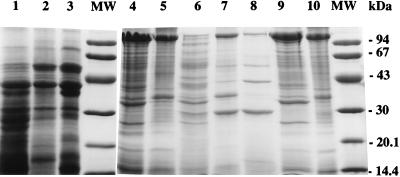
The protein content of each phase was analyzed by SDS-PAGE (Fig. 3). The Cyt1Ab1 protein produced a band on SDS-PAGE gels and was detected in both spore-pellet and inclusion body phases obtained from the recombinant strain. Similar results were obtained for the P51-P41 binary toxin in recombinant and wild-type strains.
FIG. 3.
Protein analysis after sonication of FWC, inclusion body, and spore-pellet (after ultracentrifugation) suspensions from wild-type and recombinant B. sphaericus 2297 strains. Fifteen micrograms of protein per well was subjected to SDS-PAGE (12% polyacrylamide). Lanes: 1 to 3, wild-type strain 2297 (lane 1, FWC; lane 2, inclusion body suspension; lane 3, spore pellet); 4 and 5, mutant nonsporulating strain 2297(pMK3) (lane 4, FWC; lane 5, inclusion body suspension); 6 to 8, recombinant 2297(pcyt1Ab1) (lane 6, FWC; lane 7, inclusion body suspension; lane 8, spore pellet); 9 and 10, mutant nonsporulating strain 2297(pcyt1Ab1) (lane 9, FWC; lane 10, inclusion body suspension); MW, standard protein markers from Pharmacia low-molecular-mass kit.
Electron microscopy.
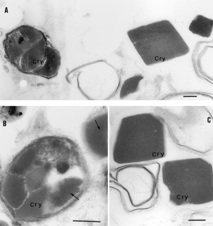
The Cyt1Ab1 protein, purified from the B. thuringiensis crystal-negative strain SPL407 (23), was produced in two types of crystalline lattice inclusion bodies (Fig. 4A): a more or less round body, with several parts differing in density, surrounded by an envelope (Fig. 4B), and a cuboid body of uniform density with no membrane (Fig. 4C).
FIG. 4.
Electron micrographs of ultrathin sections of two crystalline inclusions of purified Cyt1Ab1 crystals from B. thuringiensis SPL407(pcyt1Ab1) (A) and inclusion bodies with various dense areas (B). (C) Cuboid crystalline inclusion body. Bar, 200 nm; Cry, crystal. The arrows indicate the crystalline lattice.
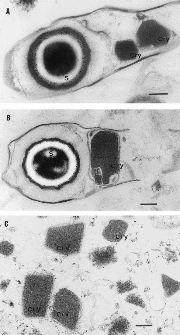
Two crystalline lattice inclusion bodies were present in the exosporium (Fig. 5A) when the cytolytic protein was produced with binary toxin in the recombinant B. sphaericus strain 2297(pA7), whereas only one inclusion body was present in the wild-type 2297 strain (Fig. 5B). We were unable to determine which crystal contained the cytolytic protein in the recombinant strain, but the B. sphaericus binary toxin crystal was clearly surrounded by an envelope, as seen in the parental strain and as observed by Charles (5a). We purified the inclusion bodies from the recombinant strain 2297(pMK3) and found that the strong sonication used to break the exosporium and release the inclusion bodies (Fig. 5C) destroyed or destabilized the inclusion body lattice.
FIG. 5.
Electron micrographs of ultrathin sections of B. sphaericus 2297. (A) Spore crystal of recombinant B. sphaericus 2297(pcyt1Ab1) after cell lysis. (B) Spore crystal of wild-type strain 2297. (C) Purified inclusion bodies from recombinant strain 2297(pMK3). Bar, 200 nm. S, spore; Cry, crystal.
Effect of the synthesis of the Cyt1Ab1 protein on the toxicity of B. sphaericus.
Final whole cultures (FWCs) of the wild-type strain and each recombinant B. sphaericus strain were assayed for toxicity to fourth-instar larvae of C. pipiens subsp. pipiens. They gave similar LC50s, a dilution of the FWC of about 10−6. The nonsporulating cultures were, as expected, only very weakly toxic (data not shown). Moreover the LC50 of the recombinant B. thuringiensis strain SPL407(pcyt1Ab1) FWC for C. pipiens and Aedes aegypti larvae was a ca. 2 × 10⁻² to 7 × 10⁻² dilution. The toxicity of these recombinant cultures to A. aegypti larvae was no higher than that of the wild type (data not shown). The larvicidal activity per unit of protein of the recombinant strains 2297, 2362, and Iab872 was determined with C. pipiens larvae (Table 1). The toxicity of the FWC was compared to those of the spore-crystal pellets and the purified inclusion bodies obtained by ultracentrifugation, when sufficient quantities of protein were obtained for bioassays. The LC50s of the parental or recombinant strain FWCs were 15 to 73 ng of protein/ml, whereas those of the ultracentrifugation pellets were about 10 times lower, as were those of purified inclusion bodies from the 2297 parental strain (2 to 8 ng of protein per ml). Inclusion bodies purified from the recombinant strain 2297(pcyt1Ab1), which produced large amounts of the Cyt1Ab1 protein, were 10 times less toxic than those from the parental strain.
TABLE 1.
Larvicidal activity of B. sphaericus recombinant strains against C. pipiens subsp. pipiens larvae
| B. sphaericus strain | Suspension (all sonicated)a | Larvicidal activityb
|
|
|---|---|---|---|
| LC50c | LC90 | ||
| 2297 (parental) | Spore pelletd | 2.1 ± 0.4 | 12.6 ± 8.8 |
| Purified inclusion bodies | 8 ± 0.05 | 15.5 ± 1.3 | |
| 2297(pMK3) | Washed FWC | 31 ± 2.5 | 59.7 ± 7.3 |
| 2297(pcyt1Ab1) | Washed FWC | 73.3 ± 17.3 | 181.3 ± 75.3 |
| Spore pellet | 7.1 ± 0.9 | 18.6 ± 2.8 | |
| Purified inclusion bodies | 72.4 ± 4.8 | 306 ± 222 | |
| 2362 | Washed FWC | 15.9 ± 1.9 | 28.6 ± 4.4 |
| 2362(pcyt1Ab1) | Washed FWC | 28.5 ± 4.5 | 64.5 ± 0.4 |
| Iab872(pcyt1Ab1) | Washed FWC | 27.9e | 194 |
| Iab872(pcyt1Ab1) | Spore pellet | 5.8 ± 3.5 | 27.4 ± 19 |
| Iab872(pMK3) | Spore pellet | 2.6e | 7.1 |
| 2297 asporulated(pMK3) | Washed FWC | 95.8 ± 45 μg/ml | 526.9 ± 299 μg/ml |
| 2297 asporulated(pcyt1Ab1) | Washed FWC | 109 ± 29.4 μg/ml | 360.0 ± 180 μg/ml |
| B. thuringiensis SPL 407(pcyt1Ab1) | Purified crystals | 5.7 ± 3.1 μg/ml | 29.7 ± 17.3 μg/ml |
All B. sphaericus suspensions were subjected to sonication two times for 10 min each before the experiments.
Unless otherwise noted, values are in nanograms per milliliter.
Lethal concentrations after 48 h of larval exposure.
Spores pelleted after ultracentrifugation.
The result of one experiment.
The toxicities of the two nonsporulating 2297 strains were, as expected, very low—1,000 times less than that of the sporulated strains and about 20 times less than that of the Cyt1Ab1 protein alone. Purified cytolytic crystals were about 1,000 times less toxic than the binary toxin.
Populations of C. pipiens larvae resistant to B. sphaericus were exposed to FWC of wild-type strains 2297 and 2362 (Table 2). They were 5,000 to 90,000 times less susceptible than susceptible C. pipiens populations (Table 1). The strain producing large amounts of Cyt1Ab1 protein [recombinant strain 2297(pcyt1Ab1)] was 10 times more toxic than the parental strain to this resistant Culex (SPHAE) population, whereas the strain producing small amounts of the protein [recombinant strain 2362(pcyt1Ab1)] was not significantly more toxic than the parental strain. Moreover, a resistant C. quinquefasciatus (GeoR) larval population was 20 times more susceptible to strain 2297(pcyt1Ab1) than to the parental strain. The nonsporulating 2297(pMK3) strain was five times less toxic to the resistant population (SPHAE [Table 2]) than to the susceptible population (Table 1). Similarly the toxicity of the nonsporulating strain 2297(pcyt1Ab1) to the resistant population (SPHAE) was half that to the susceptible population. The purified Cyt1Ab1 crystals alone were ca. 8 to 10 times less toxic to the resistant populations than to the susceptible populations.
TABLE 2.
Larvicidal activity of B. sphaericus recombinant strains against C. pipiens subsp. pipiens (SPHAE) and C. quinquefasciatus (GeoR) larval populations resistant to B. sphaericus
| Mosquito strain | Strain (FWC)a | Larvicidal activity (μg/ml)
|
|
|---|---|---|---|
| LC50b | LC90 | ||
| C. quinquefasciatus | 2297 (parental) | 134.0 ± 6.7c | 383.6 ± 28.4 |
| 2297(pcyt1Ab1) | 6.0 ± 2.8 | 16.4 ± 8.8 | |
| C. pipiens subsp. pipiens | 2297 (parental) | 193.2 ± 46 | 611.4 ± 42 |
| 2297(pcyt1Ab1) | 14.1 ± 7.4 | 38.8 ± 18 | |
| 2362 (parental) | 66.8 ± 24.9 | 115.2 ± 65 | |
| 2362(pcyt1Ab1) | 45.8 ± 7.8 | 122.3 ± 67 | |
| 2297 asporulated(pMK3) | 595d | 7,806 | |
| 2297 asporulated(pcyt1Ab1) | 215 ± 78 | 1,346 ± 944 | |
| B. thuringiensis SPL 407 (pcyt1Ab1)e | 43.2 ± 21 | 416 ± 219 | |
All strains are B. sphaericus, except as noted.
LC after 48 h of larval exposure ± standard error; mean of three experiments.
Result of two experiments.
Result of one experiment.
Purified crystals of Cyt1Ab1 protein.
DISCUSSION
We introduced a plasmid containing the cyt1Ab1 gene encoding the cytolytic protein from B. thuringiensis subsp. medellin into toxic B. sphaericus strains by electroporation. The level of expression of the gene differed according to the strain into which the gene was transferred. Only strain 2297(pcyt1Ab1) produced Cyt1Ab1 protein in amounts large enough to be detected as a band by SDS-PAGE. The recombinant strain 2297(pcyt1Ab1) produced two cuboid crystalline inclusion bodies inside the exosporium: a large crystal surrounded by an envelope which probably contains the binary toxin (5a) and an additional inclusion body thought to contain the Cyt1Ab1 cytolytic protein (Fig. 5A). Immunocytochemical studies are required to confirm the contents of the inclusion bodies.
The B. sphaericus recombinant strains Iab872 and 2362 did not produce any additional crystals. This is consistent with the work of Bar et al. (1), who observed no extra crystals after introducing the genes encoding the Cyt1Aa1 (and/or Cry11A) from B. thuringiensis subsp. israelensis into B. sphaericus 2362. Other Cry4B and Cry11A toxins from B. thuringiensis subsp. israelensis have been synthesized in B. sphaericus 2297 (17), and Cry4B has been synthesized in strains 2362 and 1593 (24). However, an additional crystal was reported only for the recombinant B. sphaericus 2297 strain producing the Cry11A protein (16), although whether the protein accumulated inside or outside the exosporium was not determined. The production of an additional crystal may be recipient strain dependent, because it is only observed in B. sphaericus 2297, whether the gene is located on a plasmid or the chromosome. The introduction of the p19 or p21 gene at the same time may affect the crystallization of the Cry11A and Cyt1Ab1 proteins in B. sphaericus 2297. This effect could be studied by the deletion or interruption of these genes.
The production of the Cyt1Ab1 protein in B. thuringiensis SPL407 led to the synthesis of two crystals, one cuboid, with a very homogeneous lattice, and another, with areas of various densities surrounded by an envelope. B. sphaericus 2297(pcyt1Ab1) expressing the cyt1Ab1 gene produced only the cuboid crystal. Furthermore, production of the Cyt1Ab1 protein was much more efficient than that of the binary toxin: twice as much of the 30-kDa polypeptide (13% of total protein) as of the 41- or 56-kDa polypeptides was produced, as assessed by densitometry of Coomassie blue-stained gels (data not shown). This suggests that cyt1Ab1 expression is under the control of a strong promoter in strain 2297 or that the stability of Cyt1Ab1 is increased by P21 (a chaperone-like protein). In contrast, Poncet et al. (16) found that twice as much binary toxin was synthesized in the recombinant strain 2297 producing Cry11A protein as in the parental strain.
We separated the inclusion bodies from the spores. The preparation contained high concentrations of inclusion bodies and 100 times fewer spores than the spore pellet of the parental and recombinant 2297 strains. Most exosporia were disrupted by the passage through a French press before ultracentrifugation (15), but the toxic fraction obtained was not pure. Strong sonication broke up not only the exosporium but also many of the crystalline lattices of inclusion bodies (as observed under the electron microscope). It probably also caused degradation of the binary protein but did not affect the larvicidal activity, despite the heating during sonication. The spore pellet and inclusion bodies were more toxic than the washed FWC culture of the parental 2297 strain. Purified inclusion bodies from the recombinant strain 2297, producing the Cyt1Ab1 crystal, were less toxic than the spore pellet. This may reflect different ratios of toxic proteins in the inclusion bodies and spore pellet or different particle sizes in the two extracts. Note that particle size is important in larval feeding behavior.
The Cyt1Ab1 protein did not increase the toxicity of the binary toxin to A. aegypti or C. pipiens larvae. This may be due to the low level of activity of Cyt1Ab1 alone: it was 1,000 times less toxic than the binary toxin. Further experiments, involving mixtures of various quantities of these two proteins, are required to determine whether there is any positive effect on toxicity to the mosquito population. These results contrast with those of Bar et al. (1), who observed a higher level of toxicity to Aedes larvae in their recombinant 2362 strain producing the Cyt1Aa1 protein. In our study, the level of expression of the cyt1Ab1 gene in recombinant strain 2362 was probably too low to have any significant effect on toxicity. The toxicity of strain 2297 to A. aegypti larvae was very low and dose independent, and, thus, an LC50 could not be calculated and a negative slope was generally obtained (not shown).
The production of the Cyt1Ab1 protein in strain 2297 partly overcame the resistance to B. sphaericus binary toxin in resistant mosquito populations. The two resistant mosquito populations tested differ in their capacity to bind the toxin to the gut brush border membranes. The GeoR strain of C. quinquefasciatus has no functional receptor for B. sphaericus toxin, and no specific binding is observed (13). The midgut receptor for the binary toxin in the resistant strain of C. pipiens SPHAE is normal, suggesting that the resistant mechanism does not involve the binding step (14). The reduction in resistance of these two populations, caused by Cyt1Ab1, may be due to this cytolytic protein inducing additional pores in the gut membrane (11), enabling the binary toxin to pass through the membrane. It has been suggested previously that the Cyt1A protein is responsible for suppressing resistance to Cry toxins (7, 25), but the mechanism involved is unknown.
We compared the activities of the nonsporulating strains [(2297(pMK3) and 2297(pcyt1Ab1)] and Cyt1Ab1 crystals against susceptible and resistant C. pipiens strains. We found that the resistant SPHAE mosquito strain was more resistant (two to five times) to these nonsporulating strains and to the cytolytic protein itself. It is unclear whether this is due to factors produced during the vegetative phase. If this were the case, it would suggest that the mosquito population (SPHAE), which is resistant to binary toxin, may also have mechanisms limiting its susceptibility to these other factors. No such mechanisms have been identified. Alternatively, this resistance may be due to the SPHAE mosquito population being less susceptible generally than the C. pipiens subsp. pipiens population reared for 15 years in our laboratory. Given the very low level of toxicity, 50,000 to 100,000 less than that of the binary toxin, we conclude that this resistance results from the differences in the intrinsic susceptibilities of the populations.
Our results support the conclusions of other investigations demonstrating that the cytolytic protein is central to expression of resistance by the mosquito population.
ACKNOWLEDGMENTS
We are indebted to P. Gounon, Central Station of Electron Microscopy, for free access to the ultramicrotome and electron microscope and C. Rolin for micrograph printing. We thank J.-F. Charles and B. Chavinier for help with electron microscopy. We thank N. Pasteur, Montpellier, France, for providing eggs of the B. sphaericus-resistant C. pipiens mosquito population (SPHAE strain) and C. Nielsen-LeRoux for larvae of the resistant C. quinquefasciatus population (GeoR strain).
This investigation received financial support from Colchiencas, Medellin, Colombia.
REFERENCES
- 1.Bar E, Lieman-Hurwitz J, Rahamim E, Keynan A, Sandler N. Cloning and expression of Bacillus thuringiensis israelensis δ-endotoxin DNA in B. sphaericus. J Invertebr Pathol. 1991;57:149–158. doi: 10.1016/0022-2011(91)90110-c. [DOI] [PubMed] [Google Scholar]
- 2.Baumann P, Clark M A, Baumann L, Broadwell A H. Bacillus sphaericus as a mosquito pathogen: properties of the organism and its toxins. Microbiol Rev. 1991;55:425–436. doi: 10.1128/mr.55.3.425-436.1991. [DOI] [PMC free article] [PubMed] [Google Scholar]
- 3.Bradford M M. A rapid and sensitive method for the quantitation of microgram quantities of protein utilizing the principle of protein-dye binding. Anal Biochem. 1976;72:248–254. doi: 10.1016/0003-2697(76)90527-3. [DOI] [PubMed] [Google Scholar]
- 4.Charles J-F, Hamon S, Baumann P. Inclusion bodies and crystals of Bacillus sphaericus mosquitocidal proteins expressed in various bacterial hosts. Res Microbiol. 1993;144:411–416. doi: 10.1016/0923-2508(93)90199-c. [DOI] [PubMed] [Google Scholar]
- 5.Charles J-F, Nielsen-LeRoux C, Delécluse A. Bacillus sphaericus toxins: molecular biology and mode of action. Annu Rev Entomol. 1996;41:451–472. doi: 10.1146/annurev.en.41.010196.002315. [DOI] [PubMed] [Google Scholar]
- 5a.Charles, J.-F. Personal communication.
- 6.Delécluse A, Bourgouin C, Klier A, Rapoport G. Specificity of action on mosquito larvae of Bacillus thuringiensis israelensis toxins encoded by two different genes. Mol Gen Genet. 1988;214:42–47. doi: 10.1007/BF00340177. [DOI] [PubMed] [Google Scholar]
- 7.Federici B, Bauer L S. Abstracts of the Society for Invertebrate Pathology 30th Annual Meeting, Banff, Alberta, Canada, 24 to 29 August 1997. 1997. Mosquitocidal Cyt1A of Bacillus thuringiensis is toxic to the leaf beetle, Chrysomela scripta, and overcomes high levels of resistance to Cry3A; p. 18. [Google Scholar]
- 7a.Frachon, E. Personal communication.
- 8.Georghiou G P, Malik J I, Wirth M, Sainato K. Characterization of resistance of Culex quinquefasciatus to the insecticidal toxins of Bacillus sphaericus (strain 2362) University of California, Mosquito Control Research; 1992. pp. 34–35. , Annual Report. [Google Scholar]
- 9.Georghiou G P, Wirth M C. Influence of exposure to single versus multiple toxins of Bacillus thuringiensis subsp. israelensis on development of resistance in the mosquito Culex quinquefasciatus (Diptera: Culicidae) Appl Environ Microbiol. 1997;63:1095–1101. doi: 10.1128/aem.63.3.1095-1101.1997. [DOI] [PMC free article] [PubMed] [Google Scholar]
- 10.Kalfon A, Larget-Thiéry I, Charles J-F, de Barjac H. Growth, sporulation and larvicidal activity of Bacillus sphaericus. Eur J Appl Microbiol Biotechnol. 1983;18:168–173. [Google Scholar]
- 11.Li J, Koni P A, Ellar D J. Structure of the mosquitocidal delta-endotoxin CytB from Bacillus thuringiensis subspecies kyushuensis and implications for membrane pore formation. J Mol Biol. 1996;257:129–152. doi: 10.1006/jmbi.1996.0152. [DOI] [PubMed] [Google Scholar]
- 12.Nielsen-Leroux C, Charles J-F. Binding of Bacillus sphaericus binary toxin to a specific receptor on midgut brush-border membranes from mosquito larvae. Eur J Biochem. 1992;210:585–590. doi: 10.1111/j.1432-1033.1992.tb17458.x. [DOI] [PubMed] [Google Scholar]
- 13.Nielsen-LeRoux C, Charles J-F, Thiéry I, Georghiou G P. Resistance in a laboratory population of Culex quinquefasciatus (Diptera: Culicidae) to Bacillus sphaericus binary toxin is due to a change in the receptor on midgut brush-border membranes. Eur J Biochem. 1995;228:206–210. doi: 10.1111/j.1432-1033.1995.tb20251.x. [DOI] [PubMed] [Google Scholar]
- 14.Nielsen-LeRoux C, Pasquier F, Charles J-F, Sinègre G, Gaven B, Pasteur N. Resistance to Bacillus sphaericus involves different mechanisms in Culex pipiens (Diptera: Culicidae) J Med Entomol. 1997;34:312–327. doi: 10.1093/jmedent/34.3.321. [DOI] [PubMed] [Google Scholar]
- 15.Payne J M, Davidson E W. Insecticidal activity of crystalline parasporal inclusions and other components of the Bacillus sphaericus 1593 spore complex. J Invertebr Pathol. 1984;43:383–388. doi: 10.1016/0022-2011(84)90084-3. [DOI] [PubMed] [Google Scholar]
- 16.Poncet S, Bernard C, Dervyn E, Cayley J, Klier A, Rapoport G. Improvement of Bacillus sphaericus toxicity against dipteran larvae by integration, via homologous recombination, of the Cry11A toxin gene from Bacillus thuringiensis subsp. israelensis. Appl Environ Microbiol. 1997;63:4413–4420. doi: 10.1128/aem.63.11.4413-4420.1997. [DOI] [PMC free article] [PubMed] [Google Scholar]
- 17.Poncet S, Delécluse A, Anello G, Klier A, Rapoport G. Transfer and expression of the cryIVB and cryIVD genes of Bacillus thuringiensis subspecies israelensis in Bacillus sphaericus 2297. FEMS Microbiol Lett. 1994;117:91–96. [Google Scholar]
- 18.Sambrook J, Frisch E F, Maniatis T. Molecular cloning: a laboratory manual. 2nd ed. Cold Spring Harbor, N.Y: Cold Spring Harbor Laboratory; 1989. [Google Scholar]
- 19.Sharif F A, Alaeddinoglu N G. A rapid and simple method for staining of the crystal protein of Bacillus thuringiensis. J Ind Microbiol. 1988;3:227–229. [Google Scholar]
- 20.Sinègre G, Babinot M, Quermel J M, Gaven B. Proceedings of the VIII European Meeting of the Society for Vector Ecology, Barcelona, Spain, 5 to 8 September 1994. 1994. First field occurrence of Culex pipiens resistance to Bacillus sphaericus in Southern France; p. 17. [Google Scholar]
- 21.Sullivan M A, Yasbin R E, Young F E. New shuttle vectors for Bacillus subtilis and Escherichia coli which allow rapid detection of inserted fragments. Gene. 1984;29:21–26. doi: 10.1016/0378-1119(84)90161-6. [DOI] [PubMed] [Google Scholar]
- 22.Taylor L D, Burke W F., Jr Transformation of an entomopathogenic strain of Bacillus sphaericus by high voltage electroporation. FEMS Microbiol Lett. 1990;66:125–128. [Google Scholar]
- 23.Thiéry I, Delécluse A, Tamayo M C, Orduz S. Identification of a gene for Cyt1A-like hemolysin from Bacillus thuringiensis subsp. medellin and expression in a crystal-negative B. thuringiensis strain. Appl Environ Microbiol. 1997;63:468–473. doi: 10.1128/aem.63.2.468-473.1997. [DOI] [PMC free article] [PubMed] [Google Scholar]
- 24.Trisrisook M, Pantuwatana S, Bhumiratana A, Panbangred W. Molecular cloning of the 130-kilodalton mosquitocidal δ-endotoxin gene of Bacillus thuringiensis subsp. israelensis in Bacillus sphaericus. Appl Environ Microbiol. 1990;56:1710–1716. doi: 10.1128/aem.56.6.1710-1716.1990. [DOI] [PMC free article] [PubMed] [Google Scholar]
- 25.Wirth M C, Georghiou G P. Cross-resistance among CryIV toxins of Bacillus thuringiensis subspecies israelensis in Culex quinquefasciatus (Diptera: Culicidae) J Econ Entomol. 1997;90:1471–1477. [Google Scholar]
- 26.Wirth M C, Georghiou G P, Federici B A. CytA enables CryIV endotoxins of Bacillus thuringiensis to overcome high levels of CryIV resistance in the mosquito Culex quinquefasciatus. Proc Natl Acad Sci USA. 1997;94:10536–10540. doi: 10.1073/pnas.94.20.10536. [DOI] [PMC free article] [PubMed] [Google Scholar]


