An official website of the United States government
Here's how you know
Official websites use .gov
A
.gov website belongs to an official
government organization in the United States.
Secure .gov websites use HTTPS
A lock (
) or https:// means you've safely
connected to the .gov website. Share sensitive
information only on official, secure websites.
As a library, NLM provides access to scientific literature. Inclusion in an NLM database does not imply endorsement of, or agreement with,
the contents by NLM or the National Institutes of Health.
Learn more:
PMC Disclaimer
|
PMC Copyright Notice
1Servicio de Medicina General. Burdwan Medical College and Hospital. Burdwan, Burdwan Medical College and Hospital, Burdwan Medical College and Hospital,
KolkataIndia
3Sección de Neurofisiología Clínica. Servicio de Neurología. Hospital Universitario La Paz, Hospital Universitario La Paz, Hospital Universitario La Paz,
MadridEspaña
2Servicio de Neuromedicina. Bangur Institute of Neurosciences. Institute of Post Graduate Medical Education and Research & SSKM Hospital. Kolkata, India, Institute of Post Graduate Medical Education and Research & SSKM Hospital, Institute of Post Graduate Medical Education and Research & SSKM Hospital,
KolkataIndia
4Servicio de Neurología. Hospital Universitario 12 de Octubre, Hospital Universitario 12 de Octubre, Hospital Universitario 12 de Octubre,
MadridEspaña
5Centro de Investigación Biomédica en Red Sobre Enfermedades Neurodegenerativas (CIBERNED), Centro de Investigación Biomédica en Red Sobre Enfermedades Neurodegenerativas (CIBERNED), Centro de Investigación Biomédica en Red Sobre Enfermedades Neurodegenerativas (CIBERNED),
MadridEspaña
6Departamento de Medicina. Facultad de Medicina. Universidad Complutense de Madrid. Madrid, España, Universidad Complutense de Madrid, Universidad Complutense de Madrid,
MadridEspaña
1Servicio de Medicina General. Burdwan Medical College and Hospital. Burdwan, Burdwan Medical College and Hospital, Burdwan Medical College and Hospital,
KolkataIndia
2Servicio de Neuromedicina. Bangur Institute of Neurosciences. Institute of Post Graduate Medical Education and Research & SSKM Hospital. Kolkata, India, Institute of Post Graduate Medical Education and Research & SSKM Hospital, Institute of Post Graduate Medical Education and Research & SSKM Hospital,
KolkataIndia
3Sección de Neurofisiología Clínica. Servicio de Neurología. Hospital Universitario La Paz, Hospital Universitario La Paz, Hospital Universitario La Paz,
MadridEspaña
4Servicio de Neurología. Hospital Universitario 12 de Octubre, Hospital Universitario 12 de Octubre, Hospital Universitario 12 de Octubre,
MadridEspaña
5Centro de Investigación Biomédica en Red Sobre Enfermedades Neurodegenerativas (CIBERNED), Centro de Investigación Biomédica en Red Sobre Enfermedades Neurodegenerativas (CIBERNED), Centro de Investigación Biomédica en Red Sobre Enfermedades Neurodegenerativas (CIBERNED),
MadridEspaña
6Departamento de Medicina. Facultad de Medicina. Universidad Complutense de Madrid. Madrid, España, Universidad Complutense de Madrid, Universidad Complutense de Madrid,
MadridEspaña
✉
Correspondencia: Dr. Julián Benito León. Servicio de Neurología. Hospital Universitario 12 de Octubre. Avda. Córdoba, s/n. E-28041 Madrid. E-mail: jbenitol67@gmail.com
El trastorno de la marcha frontal/apraxia de la marcha es un déficit motor del nivel superior con diversas causas, caracterizado por dificultades en el inicio de la marcha (congelación). Nuestro objetivo es presentar una paciente con un trastorno de la marcha del nivel superior con episodios de caídas como manifestaciones iniciales de una parálisis supranuclear progresiva (PSP). Sus datos se obtuvieron de los registros médicos del Servicio de Medicina General del Burdwan Medical College & Hospital (Burdwan, Bengala Occidental, India).
Caso clínico.
Mujer de 58 años sana que consultó por un trastorno de la marcha con caídas. La exploración neurológica mostró una apariencia facial característica (mirada fija, ojos muy abiertos, ceño fruncido y expresión fija hemifacial inferior), e hipocinesia-rigidez simétrica de predominio axial (postura retrocólica del tronco y el cuello). La exploración de la marcha reveló un trastorno de la marcha del nivel superior, caracterizado por una significativa vacilación inicial, que precisaba ayuda de objetos/personas cercanos. Al iniciar la marcha, los pasos mejoraban relativamente, pero reaparecía una deambulación inefectiva al girar. Presentaba zancadas cortas, congelación, base amplia de sustentación, desequilibrio, movimiento lento de las piernas, arrastre de los pies, y pérdida de la cadencia normal del tronco y las extremidades. Los reflejos posturales estaban alterados. La resonancia magnética cerebral desveló atrofia mesencefálica, dilatación de acueducto de Silvio y III ventrículo, atrofia frontal bilateral y el signo típico del colibrí. Finalmente, la paciente fue diagnosticada de una PSP probable.
Conclusiones.
Varias etiologías, incluida la PSP, deben considerarse, en el contexto clínico apropiado, si la exploración de la deambulación demuestra un trastorno de la marcha del nivel superior.
Palabras clave: Forma de presentación, Marcha, Parálisis supranuclear progresiva, Trastorno de la marcha del nivel superior, Trastorno del movimiento, Vídeo caso
Introducción
La parálisis supranuclear progresiva (PSP), un subtipo de taupatía, es un trastorno neurodegenerativo rápidamente progresivo que cursa con un parkinsonismo rigidoacinético caracterizado por un inicio insidioso y afectación predominantemente axial simétrica. Las caídas hacia atrás no provocadas y las dificultades para caminar son las manifestaciones iniciales más frecuentes de la PSP. Otras características típicas son el enlentecimiento motor generalizado, el deterioro cognitivo, las alteraciones motoras oculares, los trastornos del sueño y la mala respuesta a la levodopa [1]. Aunque la oftalmoplejía supranuclear de la mirada vertical es un sello distintivo de la PSP, puede aparecer varios años después de la enfermedad, retrasando el diagnóstico [1,2]. Asimismo, los movimientos sacádicos pueden ser la única manifestación temprana del movimiento ocular [3].
El trastorno de la marcha frontal/apraxia de la marcha es un déficit motor del nivel superior con diversas causas (Tabla I), caracterizado por dificultades en el inicio de la marcha, como congelación o fallo en el inicio [1,4].
Tabla I.
Principales diagnósticos diferenciales de los trastornos de la marcha del nivel superior.
1. Hidrocefalia crónica del adulto
2. Otros tipos de hidrocefalia comunicante en adultos
3. Parkinsonismo vascular
4. Parálisis supranuclear progresiva
5. Degeneración corticobasal
6. Atrofia multisistémica
7. Enfermedad de Parkinson avanzada
8. Tumores del lóbulo frontal
9. Demencia frontotemporal
10. Enfermedad de Alzheimer
11. Enfermedad de Creutzfeldt-Jakob
12. Enfermedad de Huntington juvenil (variante de Westphal)
Nuestro objetivo es informar sobre una mujer de 58 años, previamente sana, que presentó un trastorno progresivo de la marcha del nivel superior y episodios de caídas como manifestación inicial de una PSP.
Los datos de la paciente fueron obtenidos de los registros médicos del Servicio de Medicina General del Burdwan Medical College & Hospital (Burdwan, Bengala Occidental, India).
Caso clínico
La exploración neurológica exhaustiva puso de manifiesto una apariencia facial característica: mirada fija con los ojos muy abiertos, fruncimiento de la frente con una expresión de ceño fruncido (signo del procerus) y expresión fija de la parte inferior de la cara. La paciente presentaba hipocinesia-rigidez simétrica de predominio axial con postura retrocólica del tronco y el cuello. El examen de la deambulación reveló un patrón de la marcha del nivel superior caracterizado por una llamativa vacilación inicial que requería ayuda de objetos/personas cercanas. Una vez que comenzaba a caminar, los pasos mejoraban relativamente, pero volvía a aparecer una marcha inefectiva cuando intentaba girar. Exhibía zancadas cortas, congelación, base amplia de sustentación, desequilibrio, movimiento lento de los miembros inferiores, arrastre de los pies y pérdida de la fluidez normal del tronco y las extremidades. Los reflejos posturales estaban alterados. Asimismo, existía una dificultad grave para la bipedestación tras la sedestación y dificultad para darse la vuelta en la cama (Vídeo). Sin embargo, los signos frontales, como la rigidez paratónica, los reflejos de prensión, la incontinencia urinaria y las características de los déficits cognitivos frontales (por ejemplo, cambios disejecutivos y de personalidad e impulsividad) estaban ausentes, excepto por un estado apático progresivo. Los movimientos oculares sacádicos verticales estaban alterados, con amplitudes relativamente normales y movimientos sacádicos horizontales. Se evidenciaron sacadas de onda cuadrada y una oftalmoplejía supranuclear. La resonancia magnética del cerebro reveló atrofia del mesencéfalo, dilatación del acueducto de Silvio y del III ventrículo y atrofia de los lóbulos frontales, y los signos típicos del colibrí y de la gloria de la mañana (Figura). Finalmente, la paciente fue diagnosticada de una PSP probable [2,5].
La evaluación de la marcha reveló un patrón de marcha del nivel superior (complejo e integrador) caracterizado por una signicativa vacilación inicial (como si los pies estuvieran pegados al suelo) que requería asistencia y apoyo de objetos/personas cercanas con movimientos reducidos de la parte superior del cuerpo. La paciente presenta zancadas cortas, congelación, base de sustentación amplia, desequilibrio, movimiento lento de las piernas, arrastre de los pies y pérdida de la cadencia normal del tronco y las extremidades. También tiene una gran dificultad para sentarse en una silla. Una vez que comienza a caminar, los pasos mejoran relativamente, pero la marcha inefectiva vuelve a surgir cuando intenta girar. (Clicar en la imagen para reproducir el vídeo).
Resonancia magnética cerebral que revela el signo del colibrí en las imágenes ponderadas en T1 sagital media (a) y las imágenes ponderadas en T2 (b), y el signo de la gloria de la mañana en imágenes ponderadas en T2 axiales (c). La posición de la cabeza con respecto a la columna cervical nos da una idea de que la paciente sufre un retrocollis activo.
Discusión y conclusiones
La marcha siempre debe examinarse meticulosamente en todo paciente con antecedentes de caídas no provocadas. La exploración clínica de los patrones de la marcha puede ser una ayuda clínica confiable en entornos clínicos y de investigación, y puede ayudar a formular diferentes enfoques de rehabilitación para estos pacientes. Todas las etiologías descritas en la tabla I deben considerarse en contextos clínicos apropiados si la exploración de la deambulación demuestra un trastorno de la marcha del nivel superior. En nuestro caso, una exploración clínica completa, incluyendo los patrones de marcha y los hallazgos de la resonancia magnética cerebral, nos aportaron pistas esenciales para llegar al diagnóstico de PSP. Cabe destacar que la evaluación cuantitativa de la marcha puede distinguir a los pacientes con PSP de los pacientes con enfermedad de Parkinson incluso en las primeras etapas de la enfermedad (Tabla II) [6].
Tabla II.
Diagnóstico diferencial de parkinsonismo y trastorno de la marcha frontal con inspección visual de la postura y la marcha.
Enfermedad de Parkinson
Parkinsonismo atípico
Trastorno de la marcha frontal
Apoyo y postura
Inclinación hacia delante, temblor de reposo
Cuello y tronco extendidos Distónico (axial) en algunos pacientes
Parte superior del cuerpo respetada Base de sustentación amplia
Fallo del inicio
Tardío
Inicial
Muy inicial
Braceo
Reducido (asimétrico)
Reducido-normal (simétrico)
Movimientos superfluos compensatorios
Longitud de la zancada
Corta
Más corta
Muy corta
Fase de apoyo
Variable
La más larga
Larga
Velocidad de la marcha
Progresivamente rápida (festinación)
Más lenta que la enfermedad de Parkinson
Cadencia
Muy alta
Relativamente menor
Inexistente
Caídas
Tardías
Precoces
Precoces
Congelación de la marcha
Tardía
Frecuente
Muy frecuente
Apraxia de la marcha
Rara
Frecuencia variable
Frecuente
Sustrato anatómico primario
Vía nigroestriada
Ganglios de la base, tálamo, mesencéfalo
Área motora suplementaria, área premotora, circunvolución frontal medial y conexiones subcorticales frontales
Con respecto al curso de los diferentes subtipos de PSP, los estudios de la historia natural se han centrado principalmente en el subtipo PSP-síndrome de Richardson (disfunción motora ocular vertical, inestabilidad postural de aparición temprana y caídas) [1]. En una serie de 100 casos de PSP que incluyó a pacientes con PSP-síndrome de Richardson, PSP-parkinsonismo, PSP-inestabilidad postural, PSP-motora ocular, PSP-síndrome corticobasal, PSP-demencia frontotemporal y fenotipos no clasificados, la duración media de la enfermedad (± error estándar de la media) para todos los subtipos fue de 8,7 (± 0,4) años, con un rango de 2 a 28 años [7]. Los pacientes con el fenotipo PSP-síndrome de Richardson tuvieron la duración media más corta de la enfermedad (7,3 ± 0,6; rango de 4 a 17 años) [7]. Con respecto a los pacientes con el fenotipo PSP-síndrome de Richardson, el subtipo de PSP-parkinsonismo (fenotipo clínico que se asemeja a la enfermedad de Parkinson, con desarrollo posterior de clínica de PSP-síndrome de Richardson) tuvo la duración más prolongada de la enfermedad (12,8 ± 1,5; rango de 4 a 28 años). Es probable que el subtipo PSP-congelación progresiva de la marcha (presentación con un trastorno de la marcha aislado con vacilación al comienzo y bloqueo progresivo de la marcha) también tenga una duración más prolongada de la enfermedad, según los datos de una serie de casos anatomopatológicos (duración media de la enfermedad de 13 años; rango de 5 a 21 años) [8] y de informes de casos que revelan duraciones de la enfermedad hasta de 15 años [9].
Los predictores de una supervivencia más corta en la PSP, derivados de cohortes de pacientes con PSP anatomopatológicamente probada o diagnósticos de PSP-síndrome de Richardson en vida utilizando los criterios de diagnóstico previos de PSP, incluyen el subtipo PSP-síndrome de Richardson (frente a PSP-parkinsonismo), disfagia temprana, síntomas cognitivos y caídas [10].
Finalmente, se requieren más estudios que aborden los subtipos de criterios de la Movement Disorder Society para la PSP para comprender la historia natural de estos diferentes subtipos de PSP, y ayudar a asesorar a los pacientes y a sus familiares sobre la progresión y el pronóstico esperados.
Rev Neurol. 2023 Aug 16;77(4):101–104. [Article in English]
Higher-level gait disorder as a presenting manifestation of progressive supranuclear palsy: a video case report
1Department of General Medicine. Burdwan Medical College and Hospital. Burdwan, Burdwan Medical College and Hospital, Burdwan Medical College and Hospital,
KolkataIndia
3Section of Clinical Neurophysiology. Department of Neurology. Hospital Universitario La Paz, Hospital Universitario La Paz, Hospital Universitario La Paz,
MadridSpain
2Department of Neuromedicine. Bangur Institute of Neurosciences. Institute of Post Graduate Medical Education and Research & SSKM Hospital. Kolkata, India, Institute of Post Graduate Medical Education and Research & SSKM Hospital, Institute of Post Graduate Medical Education and Research & SSKM Hospital,
KolkataIndia
4Department of Neurology. Hospital Universitario 12 de Octubre, Hospital Universitario 12 de Octubre, Hospital Universitario 12 de Octubre,
MadridSpain
5Centro de Investigación Biomédica en Red Sobre Enfermedades Neurodegenerativas (CIBERNED), Centro de Investigación Biomédica en Red Sobre Enfermedades Neurodegenerativas (CIBERNED), Centro de Investigación Biomédica en Red Sobre Enfermedades Neurodegenerativas (CIBERNED),
MadridSpain
6Department of Medicine. Faculty of Medicine. Universidad Complutense de Madrid. Madrid, Spain, Universidad Complutense de Madrid, Universidad Complutense de Madrid,
MadridSpain
1Department of General Medicine. Burdwan Medical College and Hospital. Burdwan, Burdwan Medical College and Hospital, Burdwan Medical College and Hospital,
KolkataIndia
2Department of Neuromedicine. Bangur Institute of Neurosciences. Institute of Post Graduate Medical Education and Research & SSKM Hospital. Kolkata, India, Institute of Post Graduate Medical Education and Research & SSKM Hospital, Institute of Post Graduate Medical Education and Research & SSKM Hospital,
KolkataIndia
3Section of Clinical Neurophysiology. Department of Neurology. Hospital Universitario La Paz, Hospital Universitario La Paz, Hospital Universitario La Paz,
MadridSpain
4Department of Neurology. Hospital Universitario 12 de Octubre, Hospital Universitario 12 de Octubre, Hospital Universitario 12 de Octubre,
MadridSpain
5Centro de Investigación Biomédica en Red Sobre Enfermedades Neurodegenerativas (CIBERNED), Centro de Investigación Biomédica en Red Sobre Enfermedades Neurodegenerativas (CIBERNED), Centro de Investigación Biomédica en Red Sobre Enfermedades Neurodegenerativas (CIBERNED),
MadridSpain
6Department of Medicine. Faculty of Medicine. Universidad Complutense de Madrid. Madrid, Spain, Universidad Complutense de Madrid, Universidad Complutense de Madrid,
MadridSpain
✉
Corresponding author: Dr. Julián Benito León. Servicio de Neurología. Hospital Universitario 12 de Octubre. Avda. Córdoba, s/n. E-28041 Madrid. E-mail: jbenitol67@gmail.com
Frontal gait disorder/gait apraxia is a higher-order motor deficit with various causes, characterized by difficulties with gait initiation, such as freezing or ignition failure. We aimed to report a patient who presented with progressive higher-level gait disorder and fall episodes as the initial manifestations of progressive supranuclear palsy (PSP). Patient data were obtained from medical records from the Department of General Medicine, Burdwan Medical College & Hospital (Burdwan, West Bengal, India).
Case report.
A 58-year-old previously healthy woman presented with a gait disorder and fall episodes. Detailed neurological examination highlighted characteristic facial appearance (wide-eyed staring, furrowing of the forehead with a frowning expression, and fixed expression of the lower face). She was hypokinetic-rigid with symmetrical signs and predominant axial rigidity with retrocolic trunk and neck posture. Gait examination revealed a higher-level gait pattern characterized by an exhibition of profound start hesitation requiring assistance from nearby objects/persons. Once walking was underway, steps became relatively better, but ineffective gait re-emerged when she attempted turning. She had short strides, freezing, broad stance base, disequilibrium, slow leg movement, shuffling, and loss of normal fluidity of trunk and limbs. Postural reflexes were impaired. Brain magnetic resonance imaging revealed atrophy of the midbrain, dilated aqueduct of Sylvius and third ventricle, atrophy of frontal lobes and typical hummingbird sign. Diagnosis of probable PSP was finally made.
Conclusions.
Several etiologies, including PSP, should be considered in appropriate clinical contexts if gait examination demonstrates a higher-order gait disorder.
Key words: Gait, Higher-level gait disorder, Movement disorder, Presenting manifestation, Progressive supranuclear palsy, Video case report
Introduction
Progressive supranuclear palsy (PSP), a tauopathy subtype, is a rapidly progressive neurodegenerative disorder with akinetic-rigid parkinsonism characterized by insidious onset and predominantly symmetrical axial involvement. Backward unprovoked falls and gait difficulties are PSP’s most common initial manifestation. Other standard features are generalized motor slowing, cognitive impairment, ocular motor disturbances, sleep disorders, and poor response to levodopa [1]. Although vertical supranuclear gaze palsy is a hallmark of PSP, it can appear several years into the disease delaying the diagnosis [1,2]; slowed vertical saccades may be the only eye movement manifestation early on [3].
Frontal gait disorder/gait apraxia is a higher-order motor deficit with various causes (Table I), characterized by difficulties with gait initiation, such as freezing or ignition failure [1,4].
Table I.
Main differential diagnoses of higher-level gait disorders.
1. Normal-pressure hydrocephalus
2. Other subtypes of communicating hydrocephalus in adults
We aimed to report a 58-year-old previously healthy woman who presented with progressive higher-level gait disorder and fall episodes as the presenting manifestation of PSP.
Patient data were obtained from medical records from the Department of General Medicine, Burdwan Medical College & Hospital (Burdwan, West Bengal, India).
Case report
Detailed neurological examination highlighted characteristic facial appearance: wide-eyed staring, furrowing of the forehead with a frowning expression (procerus sign), and fixed expression of the lower face. The patient was hypokinetic-rigid with symmetrical signs and predominant axial rigidity with retrocolic trunk and neck posture. Gait examination revealed a higher-level gait pattern characterized by an exhibition of profound start hesitation requiring assistance from nearby objects/persons. Once walking was underway, steps became relatively better, but ineffective gait re-emerged when she attempted turning. She had short strides, freezing, broad stance base, disequilibrium, slow leg movement, shuffling, and loss of normal fluidity of trunk and limbs. Postural reflexes were impaired. There was severe difficulty standing from sitting and difficulty turning over in bed (Video). However, frontal signs such as paratonic rigidity, grasp reflexes, urinary incontinence, and features of frontal cognitive impairments (e.g., dysexecutive and personality changes and impulsivity) were absent except for a progressive apathetic state. Vertical saccades were impaired, with relatively normal amplitudes and horizontal saccadic movements. Square wave jerks and vertical supranuclear gaze palsy were evident. Brain magnetic resonance imaging revealed atrophy of the midbrain, dilated aqueduct of Sylvius and third ventricle, and atrophy of frontal lobes, and typical hummingbird and morning glory signs (Figure). Diagnosis of probable PSP was finally made [2,5].
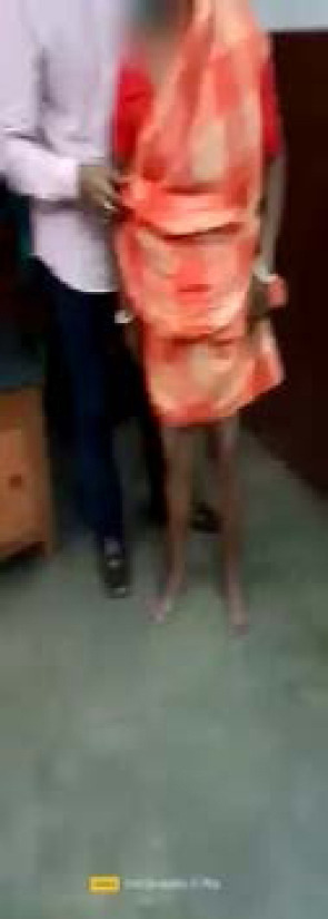
Gait examination revealed a higher (complex, integrative) order gait pattern characterized by an exhibition of profound start hesitation (as if the feet were glued to the floor) requiring assistance and support from nearby objects/persons with reduced upper body movements. The patient has short strides, freezing, broad stance base, disequilibrium, slow leg movement, shuffling, and loss of normal fluidity of the trunk and limbs. There is also severe difficulty in sitting over a chair. Once walking is underway, steps become relatively better, but ineffective gait re-emerges when she attempts turning. (Click on the image to play video).
Magnetic resonance imaging of the brain revealing hummingbird sign-on mid-sagittal T1 weighted imaging (a) and T2 weighted imaging (b) and morning glory sign on axial T2 weighted imaging (c). The head position concerning the cervical spine gives us an idea that the patient has active retrocollis.
Discussion and conclusions
Gait should always be examined meticulously in every patient with a history of unprovoked falls. Clinical examination of gait patterns can be a reliable clinical aid in clinical and research settings and may help formulate different rehabilitation approaches for these patients. All the etiologies described in table I should be considered in appropriate clinical contexts if gait examination demonstrates a higher-order gait disorder. In our case, a comprehensive clinical examination, including the gait patterns and MRI findings, gave us essential clues to reach a diagnosis of PSP. Of note is that quantitative gait evaluation can distinguish PSP patients from Parkinson’s disease patients even at the earliest stages of the disease (Table II) [6].
Table II.
Differential diagnosis of parkinsonism and frontal gait disorder with a visual inspection of posture and gait.
Parkinson’s disease
Atypical parkinsonism
Frontal gait disorder
Stance and posture
Forward stoop, rest tremor
Extended neck and back Dystonic (axial) in some
Upper body spared. Wide based
Ignition failure
Late
Early
Very early
Arm swing
Reduced (asymmetric)
Reduced to normal (symmetric)
Compensatory superfluous movements
Stride length
Short
Shorter
Very short
Stance phase
Variable
Longest
Long
Gait speed
Progressively rapid (festination)
Slower than Parkinson’s disease
Cadence
Higher
Relatively less
None
Falls
Late
Early
Early
Freezing of gait
Late
Common
Very common
Apraxia of gait
Rare
Variable frequency
Common
Primary anatomical substrate
Nigrostriatal pathmway
Basal ganglia, thalamus, midbrain
The supplementary motor area, premotor area, medial frontal gyrus, and frontal subcortical connections
Regarding the course of the different PSP subtypes, natural history studies have mainly focused on the PSP-Richardson’s syndrome subtype (vertical ocular motor dysfunction, early onset postural instability, and falls) [1]. In one series of 100 pathologically confirmed PSP cases that included patients with PSP-Richardson’s syndrome, PSP- parkinsonism, PSP-postural instability, PSP-ocular motor, PSP-corticobasal syndrome, PSP-frontotemporal dementia, and unclassified phenotypes, the mean disease duration (± standard error of the mean) for all subtypes was 8.7 (±0.4) years, ranging from 2 to 28 years [7]. Individuals with the PSP-Richardson’s syndrome phenotype had the shortest mean disease duration (7.3 ± 0.6; range 4 to 17 years). Meanwhile, individuals with the PSP- parkinsonism subtype (clinical phenotype resembling Parkinson’s disease, later development of symptoms of PSP-Richardson’s syndrome) had the longest disease duration (12.8 ± 1.5; range 4 to 28 years). It is likely that those with the PSP-progressive gait freezing subtype (presentation with an isolated gait disorder with start hesitation and progressive freezing of gait) also have a longer disease duration, according to data from a pathologic case series (mean disease duration of 13 years; range 5 to 21 years) [8], and from case reports revealing disease durations up to 15 years [9].
Predictors of shorter survival in PSP, derived from cohorts of individuals with pathologically proven PSP or in-life PSP-Richardson’s syndrome diagnoses using prior PSP diagnostic criteria, include the PSP-Richardson’s syndrome subtype (versus PSP-parkinsonism) and early dysphagia, cognitive symptoms, and falls [10].
Further studies addressing the Movement Disorder Society PSP criteria subtypes are warranted to understand the natural history of these different PSP subtypes and assist in counselling patients and families about expected progression and prognosis.
2.Höglinger GU, Respondek G, Stamelou M, Kurz C, Josephs KA, Lang AE, et al. Clinical diagnosis of progressive supranuclear palsy:the movement disorder society criteria. Mov Disord. 2017;32:853–64. doi: 10.1002/mds.26987. [DOI] [PMC free article] [PubMed] [Google Scholar]
3.Choi JH, Kim H, Shin JH, Lee JY, Kim HJ, Kim JM, et al. Eye movements and association with regional brain atrophy in clinical subtypes of progressive supranuclear palsy. J Neurol. 2021;268:967–77. doi: 10.1007/s00415-020-10230-w. [DOI] [PubMed] [Google Scholar]
4.León-Ruiz M, García-Soldevilla MÁ, García-Albea Ristol E. Primary progressive freezing gait with impressive response to laser light visual cueing:a video case report. J Neurol. 2018;265:2146–8. doi: 10.1007/s00415-018-8966-9. [DOI] [PubMed] [Google Scholar]
5.Respondek G, Kurz C, Arzberger T, Compta Y, Englund E, Ferguson LW, et al. Which ante mortem clinical features predict progressive supranuclear palsy pathology? Mov Disord. 2017;32:995–1005. doi: 10.1002/mds.27034. [DOI] [PMC free article] [PubMed] [Google Scholar]
6.Amboni M, Ricciardi C, Picillo M, De Santis C, Ricciardelli G, Abate F, et al. Gait analysis may distinguish progressive supranuclear palsy and Parkinson's disease since the earliest stages. Sci Rep. 2021;11:9297. doi: 10.1038/s41598-021-88877-2. [DOI] [PMC free article] [PubMed] [Google Scholar]
7.Respondek G, Stamelou M, Kurz C, Ferguson LW, Rajput A, Chiu WZ, et al. Movement Disorder Society-endorsed PSP Study Group. The phenotypic spectrum of progressive supranuclear palsy:a retrospective multicenter study of 100 definite cases. Mov Disord. 2014;29:1758–66. doi: 10.1002/mds.26054. [DOI] [PubMed] [Google Scholar]
8.Williams DR, Holton JL, Strand K, Revesz T, Lees AJ. Pure akinesia with gait freezing:a third clinical phenotype of progressive supranuclear palsy. Mov Disord. 2007;22:2235–41. doi: 10.1002/mds.21698. [DOI] [PubMed] [Google Scholar]
9.Compta Y, Valldeoriola F, Tolosa E, Rey MJ, Martí MJ, Valls-Solé J. Long lasting pure freezing of gait preceding progressive supranuclear palsy:a clinicopathological study. Mov Disord. 2007;22:1954–8. doi: 10.1002/mds.21612. [DOI] [PubMed] [Google Scholar]
10.Glasmacher SA, Leigh PN, Saha RA. Predictors of survival in progressive supranuclear palsy and multiple system atrophy:a systematic review and meta-analysis. J Neurol Neurosurg Psychiatry. 2017;88:402–11. doi: 10.1136/jnnp-2016-314956. [DOI] [PubMed] [Google Scholar]