

Abstract
Introduction
Gallbladder volvulus is a rare disease whose presentation usually overlaps with that of typical calculous cholecystitis. It's diagnosis is critical as it is associated with high morbidity and mortality and therefore should be managed urgently with cholecystectomy.
Case presentation
85-year-old female patient presented with right upper quadrant pain of one day duration that is associated with nausea and vomiting, but no fever or jaundice. She was tachycardiac and had severe abdominal right upper quadrant tenderness with positive Murphy's sign. Laboratory results showed only increase in inflammatory markers. Both ultrasound and computed tomography scan of the abdomen were done and acalculous cholecystitis was diagnosed. Open cholecystectomy was planned and performed, but gallbladder volvulus as the cause of cholecystitis was noted intraoperatively.
Discussion
There is still no consensus on the exact cause of gallbladder volvulus. Even though it presents mostly in elderly patients, different ages have been already reported. It's diagnosis can be suspected based on the appearance, symptoms, and examination of the patient. Laboratory tests and imaging might provide some clues for it's diagnoses. It's ischemic process prompts urgent surgical intervention and does not improve conservatively. Our patient did not present with all of it's typical symptoms and the diagnosis was made intraoperatively.
Conclusion
Gallbladder volvulus could have been missed had we not opt for an urgent surgery. More studies should be done to further define its presentation, and accurately know when to consider it up in our differential diagnosis.
Keywords: Gallbladder volvulus, Acalculous cholecystitis, Case report
Highlights
-
•
Gallbladder volvulus is a rare disease whose presentation overlaps with typical calculous cholecystitis.
-
•
Gallbladder volvulus diagnosis can be suspected based on the appearance, symptoms, and examination of the patient.
-
•
Gallbladder volvulus ischemic process prompts urgent surgical intervention and does not improve conservatively.
-
•
Gallbladder volvulus should be further studied to define it and know when to consider it in our different diagnosis.
1. Introduction
Gallbladder volvulus (GBV) is a rare occurrence described in about 500 cases in the English literature [1]. Its symptoms usually overlap with that of typical acute calculus cholecystitis; therefore, the majority of cases are diagnosed intraoperatively and only about 25 % of cases are diagnosed preoperatively [2]. The diagnosis of GBV is very critical, as complications such as ischemia, necrosis, gangrene, perforation, or sepsis are reported in about 16 % of cases, and mortality in 6 % of cases; therefore, once diagnosed, cholecystectomy should be done as soon as possible [3]. Herein, we present a case of GBV presenting to our university hospital with severe right upper quadrant pain and diagnosed intraoperatively. To note, our case is reported in line with the SCARE criteria [4].
2. Case presentation
85-year-old female Caucasian patient with history of hypertension, Sjogren's syndrome, and pudental neuralgia presented to our emergency department for severe right upper quadrant pain of one day duration. The pain started as mild, intermittent, non-radiating right upper quadrant pain, which exacerbated 9 h prior to presentation to become constant and severe. Her pain was associated with nausea and two episodes of food content vomiting, but no fever or chills, no diarrhea or constipation, and no jaundice or change in urine or stool color. To note, the patient has previously undergone open left hemicolectomy for recurrent diverticulitis and hysterectomy for uterine fibroids. Upon presentation, the patient's systolic blood pressure was 110 mmHg. She was tachycardiac with a regular heart rate of 110 beats per minute and was afebrile. On physical exam she was awake, alert, and a midline incision scar was noted. She had no abdominal guarding and positive bowel sounds; however, she had severe tenderness, rebound tenderness, rigidity, and a palpable lump in the right upper quadrant with positive Murphy's sign, but otherwise soft abdomen in the other abdominal quadrants. Laboratory results (Table 1) showed increase in inflammatory markers, but otherwise normal including normal liver function tests. Ultrasound of the abdomen (Fig. 1) followed by CT (Computer Tomography) scan of the abdomen with intravenous contrast (Fig. 2) were done and showed a very distended gallbladder with a very thickened wall, no visible calculi, and presence of pericholecystic fluids compatible with acalculous cholecystitis. No intra- or extra- hepatic bile duct dilatation was noted with common bile duct measuring 6.7 mm. Taking into consideration the severity of the patient's pain; her physical, laboratory, and radiologic findings; in-addition to her past surgical history, the patient was started on intravenous antibiotics (ceftriaxone and metronidazole) and urgent open cholecystectomy was planned. Intraoperatively, mild bloody ascites was noted and aspirated. A twisted severely distended, necrotic, and engorged gallbladder (Fig. 3) was noted that was partially held to the liver bed and the diagnosis of GBV was made. Bloody content of the gallbladder was aspirated. Detorsion of the gallbladder was done followed by dissection of its pedicle. Cystic duct and artery were identified, liberated, ligated, and dissected followed by the dissection of the gallbladder from its minimal liver attachment. The specimen was removed and sent for histopathology. Adequate lavage and drainage was done, and a laminated drain draining the liver bed was placed. Histopathology showed a gallbladder with hemorrhagic necrosis and no signs of malignancy. Post operation the patient was transferred to a regular floor, and her hospital stay was not complicated. She was discharged on day two post operation on antibiotics and pain medication, but her drain was giving 40 ml serosanguenous non-bilious content per 24 h; therefore, she was discharged with her drain that was removed three days later, when it was giving <10 ml clear serous secretions per 24 h.
Table 1.
Patient's laboratory results on admission.
| WBC | Neutrophils | Hemoglobin | Hematocrit | Platelets | Creatinine | Sodium | Potassium | Chloride | CO2 |
|---|---|---|---|---|---|---|---|---|---|
| 14 × 10^3/ul | 90.3 % | 14.0 g/dl | 40.9 % | 301 × 10^3/ul | 0.55 mg/dl | 127 mmol/l | 3.9 mmol/l | 91 mmol/l | 22 mmol/l |
| ALP | GGT | AST | ALT | Lipase | Bilirubin Direct | Bilirubin Total | PT INR | Troponin-I | CRP |
|---|---|---|---|---|---|---|---|---|---|
| 66 IU/l | 15 IU/l | 22 IU/l | 15 IU/l | 12 IU/l | 0.33 mg/dl | 1.02 mg/dl | 1.00 | <0.010 ng/ml | 44 mg/l |
WBC: White Blood Cells, ALP: Alkaline Phosphatase, GGT: Gamma-Glutamyl Transferase, AST: Aspartate Aminotransferase, ALT: Alanine Transaminase, PT INR: Prothrombin Time International Normalized Ratio, CRP: C-Reactive Protein.
Fig. 1.

Ultrasound of the abdomen showing gallbladder with signs of acalculous cholecystitis.
Fig. 2.

CT scan of the abdomen and pelvis with intravenous contrast.
Blue arrows showing the distended gallbladder with thickened wall with findings compatible with acalculous cholecystitis. (For interpretation of the references to color in this figure legend, the reader is referred to the web version of this article.)
Fig. 3.
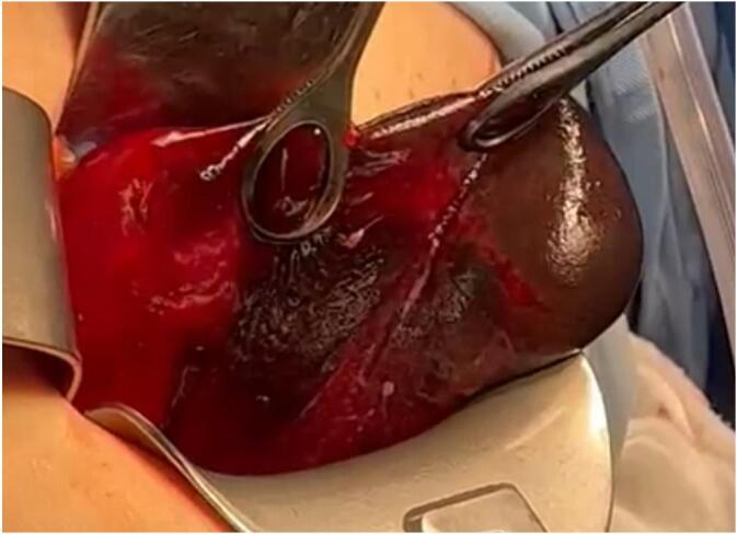
Volvulus of gallbladder with gallbladder ischemia identified intraoperatively.
3. Discussion
The exact cause of a GBV is still ambiguous, yet the most acceptable theory goes back to embryological origins. The liver diverticulum, one of the two liver primordia, has a solid cranial and hollow caudal hepatic component. During migration, if the cranial bud moves faster than the caudal bud, the peritoneum covers the gallbladder causing a long mesentery gallbladder, consequently leading to a “floating gallbladder” [5,6]. Other proposed causes include liver atrophy, kyphosis, loss of fatty visceral surrounding tissue, and increased gallbladder elasticity [7]. The cause of GBV in our case might be multifactorial. For instance, minimal attachment of the gallbladder to the liver bed was noted; therefore, embryological factor might be present. Moreover, our patient was elderly and thin, hence loss of fatty visceral surrounding tissue might have also played a role. Nevertheless, interestingly, our patient has a history of Sjogren's syndrome; however, no relation between it and GBV exists in the literature.
The median age of occurrence of GBV is about 77 years, yet cases have been described between ages of 5 days to 100 years [2]. Patients most commonly present with abdominal pain, with or without nausea and vomiting, right upper quadrant palpable lump, fever, or jaundice [8]. If a volvulus of <180 degrees of rotation occurs (incomplete torsion), patients typically present with biliary colic-like symptoms, while if complete torsion (>180 degree of rotation) occurs, patients present with a picture of acute cholecystitis or an acute abdomen [9]. The difficulty of diagnosing a volvulus gallbladder has led Lau et al. to the creation of the “triad of triads” which might help in identifying a GBV. Patient features described include 1) appearance: elderly, thin, spinal deformities; 2) symptoms: sudden onset, right upper quadrant pain, early emesis; and 3) examination: non-toxic presentation, palpable abdominal mass, and pulse-temperature discrepancy [10]. Our reported case presented with several features of the “triad of triads” where she was elderly and thin but had no spinal deformities, had sudden right upper quadrant pain with two episodes of vomiting, a non-toxic presentation with palpable abdominal mass, and she was tachycardiac yet afebrile. Her presentation was a picture of acute acalculous cholecystitis, and a complete torsion of the gallbladder was noted intra-operatively, which is consistent with what's described in the literature.
Laboratory tests and imaging might provide some clues to diagnose GBV preoperatively. Inflammatory markers such as white blood cell count and C-reactive protein (CRP) are not always elevated and are only reported in about 55 % of cases, and liver function tests (LFTs) are within normal range in 85 % of cases [3]. Ultrasound findings include a floating gallbladder lying horizontally with surrounding fluid accumulation, a gallbladder located outside the fossa and inferior to the liver, edematous and thickened gallbladder wall, cystic duct enhancement to the right of the gallbladder, and a hypoechoic zone between the two echogenic walls of the gallbladder. A Doppler ultrasound can be used and it shows an impaired blood flow in the cystic pedicle [11]. Suspicion of GBV is warranted when a clinical presentation of acute cholecystitis is found with an acalculous gallbladder as about 68 % to 78 % of cases present with such ultrasonic findings [2]. CT findings suggestive of GBV also include a gallbladder outside its anatomical location, a horizontally rotated gallbladder, pericholecystic fluid, and a swirl sign (also called beak appearance or whirlpool sign) which represents a twisted cystic artery [12]. Other imaging modalities such as Magnetic Resonance Cholangiopancreatography (MRCP) might also be of use as it shows duct traction leading to what is seen as a V-shaped distortion of the extrahepatic bile ducts; gallbladder distention and deviation towards the midline; tapering a twisting interruption of the cystic duct; and uneven intensities of the gallbladder, extrahepatic bile ducts, and cystic duct [13]. In our case, the patient had elevated inflammatory markers (Table 1) but normal LFTs, consistent with the literature. Radiologically, in our case both ultrasound and CT scan of the abdomen were done and from the radiologic features of GBV described in the literature, only gallbladder distention, wall thickening, and pericholecystic fluids existed; as such, our working diagnosis was acalculous cholecystitis and the final diagnosis of GBV was only done intraoperatively.
Unlike typical cases of acute cholecystitis where conservative and antibiotic treatment may be successful when the patient is inoperable, the ischemic process in GBV prompts urgent surgical intervention and does not improve conservatively [8]. Moreover, while typical acute calculus or acalculous cholecystitis may be treated in specific conditions by percutaneous drainage, the last is not recommended in GBV since the pathophysiology here is not due to an infectious process and therefore will not be treated. In addition, since in GBV cases the gallbladder has minimal attachment to the fossa and has a necrotic wall, transhepatic cholecystostomy would increase the risk of bile leak and peritonitis [8,14]. Taking into consideration the high morbidity and mortality in GBV, Tokyo Guidelines 2018 indicated that surgical intervention is warranted within 72 h after the onset of symptoms [15]. It was found that cholecystectomy done within this window for GBV is associated with lower complications and mortality rates, lower incidence of bile duct injury, and less conversion to open surgery [16]. Cholecystectomy for GBV can be performed by laparotomy or laparoscopically, but there are no studies that compare the outcomes among the two [17]. Either way, decompression, detorsion, and gallbladder resection are the three most important steps in cholecystectomy for GBV to avoid injury of the tented common bile duct [18,19]. Nevertheless, as there are no reports published, a concern remains whether detorsion results in reperfusion damage with systemic effects due to toxins released from the necrotic tissue in case of gallbladder necrosis in GBV [11]. In our case, due to the severity of the patient's acute pain, her increase in inflammatory markers, and radiologic findings, a decision was made to go for urgent cholecystectomy. The surgery was decided to be open due to the patient's past surgical history of midline laparotomy and the diagnosis of GBV was not made until intraoperatively. Moreover, detorsion of the gallbladder was done in our case after the diagnosis of GBV and no reperfusion damage was noted both intra- or post-operatively.
4. Conclusion
Even though GBV is a rare disease, when dealing with patients presenting with severe right upper quadrant pain and turn to have acalculous cholecystitis - with or without the full typical presentation mentioned above - GBV should be considered. The importance of the last stems from the fact that GBV is associated with high morbidity and mortality; therefore, it should not be missed and should be treated urgently with cholecystectomy. Our patient did not present with all the typical features of GBV described in the literature, and the diagnosis of GBV could have been missed or been late had we not opt for an urgent surgery. Therefore, more cases of GBV should be reported and more studies should be done to further define its presentation, and accurately know when to consider it up in our differential diagnosis.
Informed consent
An informed consent was signed by the patient in order to authorise access on her medical records and for the completion of this work.
Ethical approval
Ethical approval for this case report was provided by the Mount Lebanon Hospital Balamand University Medical Center ethical committee, and Head of General Surgery division at Mount Lebanon Hospital Balamand University Medical Center on August 16, 2023.
Funding
Faculty of Medicine and Medical Sciences, University of Balamand.
Author contribution
Omar Tabbikha (first author and corresponding author), Daniel El Israwi, Hussein Yehya, Jad Al Bitar, and Henri Bitar.
Guarantor
Henri Bitar.
Research registration number
-
1)
Name of the registry: Clinicaltrials.gov
-
2)
Unique identifying number or registration ID: NCT06050876
-
3)
Hyperlink to your specific registration (must be publicly accessible and will be checked): www.https://classic.clinicaltrials.gov/ct2/show/NCT06050876.
Conflict of interest statement
The authors report no conflict of interest.
Contributor Information
Omar Tabbikha, Email: omartabbikha@gmail.com.
Daniel El Israwi, Email: daniel.israwi@std.balamand.edu.lb.
References
- 1.Yokoi T., Miyata K., Yuasa N., et al. Twisted cystic artery disclosed by 3-dimensional computed tomography angiography for torsion of the gallbladder. Am. J. Surg. 2011;201:e33–e34. doi: 10.1016/j.amjsurg.2010.04.026. [DOI] [PubMed] [Google Scholar]
- 2.Reilly D.J., Kalogeropoulos G., Thiruchelvam D. Torsion of the gallbladder: a systematic review. HPB (Oxford) 2012;14:669–672. doi: 10.1111/j.1477-2574.2012.00513.x. [DOI] [PMC free article] [PubMed] [Google Scholar]
- 3.Moser L., Joliat G.R., Tabrizian P., et al. Gallbladder volvulus. Hepatobiliary Surg. Nutr. 2021;10:249–253. doi: 10.21037/hbsn-20-771. [DOI] [PMC free article] [PubMed] [Google Scholar]
- 4.Agha R.A., Franchi T., Sohrab C., Mathew G., Kirwan A., Thomas A., et al. The SCARE 2020 guideline: updating consensus Surgical Case Report (SCARE) guidelines. Int. J. Surg. 2020;84(1):226–230. doi: 10.1016/j.ijsu.2020.10.034. [DOI] [PubMed] [Google Scholar]
- 5.Adkins R.B., Jr., Chapman W.C., Reddy V.S. Embryology, anatomy, and surgical applications of the extrahepatic biliary system. Surg. Clin. North Am. 2000;80:363–379. doi: 10.1016/s0039-6109(05)70410-2. [DOI] [PubMed] [Google Scholar]
- 6.McEvoy C.F., Suchy F.J. Biliary tract disease in children. Pediatr. Clin. N. Am. 1996;43:75–98. doi: 10.1016/s0031-3955(05)70398-9. [DOI] [PubMed] [Google Scholar]
- 7.Nakao A., Matsuda T., Funabiki S., et al. Gallbladder torsion: case report and review of 245 cases reported in the Japanese literature. J. Hepato-Biliary-Pancreat. Surg. 1999;6:418–421. doi: 10.1007/s005340050143. [DOI] [PubMed] [Google Scholar]
- 8.Armistead C.W., Favors L.E., Mejia V.A. Gangrenous cholecystitis from gallbladder volvulus with a hiatal hernia. Am. Surg. 2022;88:804–806. doi: 10.1177/00031348211054704. [DOI] [PubMed] [Google Scholar]
- 9.Lau W.Y., Fan S.T., Wong S.H. Acute torsion of the gall bladder in the aged: a re- emphasis on clinical diagnosis. Aust. N. Z. J. Surg. 1982;52:492–494. doi: 10.1111/j.1445-2197.1982.tb06036.x. [DOI] [PubMed] [Google Scholar]
- 10.Kachi A., Nicolas G., Nasser J., Hashem M., Abou Sleiman C. A rare presentation of gall bladder volvulus: a case report. Am. J. Case Rep. 2019;20:1466–1470. doi: 10.12659/AJCR.916234. [DOI] [PMC free article] [PubMed] [Google Scholar]
- 11.Younan G., Schumm M., Ali F., et al. Case Reports in Surgery 2016. 2016. Gallbladder volvulus in a patient with type I choledochal cyst: a case report and review of the literature. [DOI] [PMC free article] [PubMed] [Google Scholar]
- 12.Usui M., Matsuda S., Suzuki H., Ogura Y. Preoperative diagnosis of gallbladder torsion by magnetic resonance cholangiopancreatography. Scand. J. Gastroenterol. 2000;35:218–222. doi: 10.1080/003655200750024425. [DOI] [PubMed] [Google Scholar]
- 13.Cecire J., Sutherland A., Das K.K. Gallbladder torsion masking as acalculus cholecystitis: a review of two cases including unsuccessful management with percutaneous cholecystostomy. J. Med. Cases. 2021;12(223–5) doi: 10.14740/jmc3683. [DOI] [PMC free article] [PubMed] [Google Scholar]
- 14.Wakabayashi G., Iwashita Y., Hibi T., et al. Tokyo Guidelines 2018: surgical management of acute cholecystitis: safe steps in laparoscopic cholecystectomy for acute cholecystitis (with videos) J. Hepatobiliary Pancreat. Sci. 2018;25:73–86. doi: 10.1002/jhbp.517. [DOI] [PubMed] [Google Scholar]
- 15.Cao A.M., Eslick G.D., Cox M.R. Early laparoscopic cholecystectomy is superior to delayed acute cholecystitis: a meta-analysis of case-control studies. Surg. Endosc. 2016;30:1172–1182. doi: 10.1007/s00464-015-4325-4. [DOI] [PubMed] [Google Scholar]
- 16.Keus F., Gooszen H.G., Van Laarhoven C.J. Systematic review: open, small-incision or laparoscopic cholecystectomy for symptomatic cholecystolithiasis. Aliment. Pharmacol. Ther. 2009;29:359–378. doi: 10.1111/j.1365-2036.2008.03894.x. [DOI] [PubMed] [Google Scholar]
- 17.Garciavilla P.C., Alvarez J.F., Uzqueda G.V. Diagnosis and laparoscopic approach to gallbladder torsion and cholelithiasis. JSLS. 2010;14:147–151. doi: 10.4293/108680810X12674612765588. [DOI] [PMC free article] [PubMed] [Google Scholar]
- 18.Barrett M., Asbun H.J., Chien H.L., Brunt L.M., Telem D.A. Bile duct injury and morbidity following cholecystectomy: a need for improvement. Surg. Endosc. 2018;32:1683–1688. doi: 10.1007/s00464-017-5847-8. [DOI] [PubMed] [Google Scholar]
- 19.Price E.E., DiMarco L. An unusual presentation of acute cholecystitis: gallbladder volvulus. J. Surg. Case Rep. 2019;2019 doi: 10.1093/jscr/rjz221. [DOI] [PMC free article] [PubMed] [Google Scholar]