Abstract
Simple Summary
Unicellular blood parasites are common in amphibians all over the world. It is poorly understood whether the hemoparasite load can cause disease or affect life-history traits. Our study on the neotropical treefrog Boana cordobae from Argentina quantified the hemoparasite load in 37 individuals and focused on their cellular immune response (leukocyte profile) and the potential effects on the life-history traits size, body condition, and age. Thirty frogs were infected by either hemogregarines or trypanosomes or both in high intensities, a prevalence unprecedented in other anuran hosts. Yet, none of them showed externally visible signs of disease or aberrant behavior. The leukocyte profile did not differ significantly between parasite-free controls and infected frogs. Age-adjusted size, body condition, and age were not affected significantly by hemoparasite load, but most parasite-free frogs were young first-breeders, suggesting that infections take place after sexual maturation of the hosts. Vectors transmitting trypanosomes and/or hemogregarines to B. cordobae remain to be identified and studied in detail.
Abstract
We provide the first evidence for hemoparasites in the endemic Cordoba treefrog Boana cordobae. We collected 37 adult frogs at 1200 m a.s.l. in the Comechingones Mountains in the Córdoba province (Argentina). Each individual was sexed, then snout–vent length and body mass were recorded, a toe was collected for skeletochronological age determination, and a slide with a blood smear was prepared for hemoparasite screening, before releasing the frogs in situ. A total of 81% (n = 30) of the frogs were infected by hemogregarines and trypanosomes with a high intensity of infections. Dactylosoma was found for the first time in Argentina. Hemoparasites had no significant effect on the leukocyte profile, which we assessed from the May–Grünwald–Giemsa-stained blood smears. The neutrophils/lymphocytes ratio, indicative of stress, was insignificantly higher (0.06) in parasitized frogs than in parasite-free individuals (0.04). Infected frogs were larger than the controls, but this effect vanished when correcting size data for age. Young frogs (first-breeders) dominated the age distribution of parasite-free individuals, suggesting that infection of frogs takes usually place after sexual maturation. Vectors transmitting hemoparasites to B. cordobae remain to be identified. We demonstrate that moderate to high intensities of hemoparasites do not significantly affect the cellular immune response of B. cordobae, or any of the life-history traits studied, nor did they show any external sign of disease.
Keywords: extracellular and intraerythrocytic hemoparasites, Dactylosoma, Trypanosoma, life-history traits, leukocyte profile
1. Introduction
In the current biodiversity crisis, amphibians are one of the most threatened taxa, and populations of many species are declining rapidly [1,2]. The major threats among them are habitat loss and infectious diseases, including parasitism [3]. Fungal infections such as chytridiomycosis have gained global attention, but viruses, protozoan, and metazoan parasites may negatively affect life-history traits as well [4,5,6,7,8]. Yet, hemoparasites have received little attention [9], though anurans host a wide variety of hemoparasites, such as protozoans, microfilariae, nematodes, rickettsians, and viral inclusions [10,11,12,13,14,15,16]. The impact of blood parasites on the life-history traits of amphibian hosts is poorly known but there is an agreement that pathological effects are rare [12,17,18,19,20,21]. Trypanosomes are common hemoparasites of amphibians, but only a few species are considered to harm their host significantly [12,22,23]. In trypanosomiasis, clinical signs include anorexia, lethargy, pallor, splenomegaly, and splenic necrosis, occasionally leading to death [23,24]. The life cycles of trypanosomes is heteroxenous and includes often insects as vector species. The prevalence of trypanosomes is often considerably greater in males than in females because frog-biting midges, the vectors of some trypanosomes, are attracted by the males’ advertisement calls [25,26,27]. Intracellular parasites, like hemogregarines, have also been detected within the erythrocytes of many anuran species [17,18,28,29,30]. In natural amphibian hosts, clinical diseases caused by hemogregarines, such as anemia, have not been demonstrated so far [20,31]. On a subclinical level, acute or permanent stress reactions in the presence of hemoparasites resulting in characteristic ratio alterations of the leukocyte profile have not been reported yet for amphibians [32,33,34].
In vertebrates, one line of defense against pathogens such as hemoparasites are leukocytes [35,36]. Consequently, leukocyte profiles assessed in wild populations cannot only provide information on the physiological health and the immune status of their members but are also reliable indicators of permanent stress, unlike glucocorticoid levels [37,38]. This is because a stressed individual has increased numbers of circulating neutrophils, while their lymphocytes decrease. Thus, the relative proportions of neutrophils and lymphocytes (N/L ratio) serve as reliable biomarkers of the physiological response, specifically to permanent stress [32,38].
The Cordoba treefrog Boana cordobae (Barrio, 1965), formerly described as Hyla pulchella cordobae, is an endemic anuran of the provinces of Córdoba and San Luis, Argentina [39,40]. The species status of this treefrog has been recognized by Faivovich et al. in 2004 [41], followed by a reconstruction of its phylogenetic relationships and a generic reassignment [42,43]. These frogs inhabit rocky mountain streams and rivers and are only known to exist at elevations of 808–2310 m a.s.l., in mountainous areas [44]. They exhibit a variable dorsal color pattern. There is a sexual size dimorphism in favor of the females, but males and females attain sexual maturity at the same age of two years [45,46]. Their longevity is five years at lower altitudes and seven years at altitudes higher than 2000 m [46]. From autumn to spring southern hemisphere, i.e., September to March, adults bask on rocks near streams, rivers, and springs. Reproduction is associated with vocal advertisement from the males and takes place in the rock pools of springs, streams, and rivers, where the water flow is slow [47,48,49]. Egg masses are attached to aquatic, mostly submerged vegetation. In cold water, tadpoles need up to 10 months to complete their larval development. The nektonic tadpoles feed on a wide diversity of phytoplankton species, predominantly on diatoms [50,51]. At their metamorphic climax, their total length is 60–90 mm. As the blood markers of tadpoles vary with the fluorite concentration of the streams, they may serve as bioindicators for this pollutant [51,52].
This research was designed to address the still unexplored aspects of life history, the impact of blood parasites on life-history traits, and immune responses to pathogen stress, in B. cordobae. We hypothesize that hemoparasites cause metabolic costs for the host, i.e., modify its resource allocation to increase the immune response. Severe consequences for hosts do not seem likely, but detectable variations of morphological, immunological, and demographic host traits are expected to occur [19,21,53]. To evaluate the potential impact of hemoparasites on life-history traits and the immune response, we focused our study on the prevalence and intensity of hemoparasite infection and quantified the corresponding host traits such as size, age, body condition, and leukocyte profile as an indicator of permanent stress.
2. Materials and Methods
2.1. Frog Collection and Processing
We collected 37 adult frogs (32 males, 5 females) from September 2015 to March 2016 in the central region of the Comechingones Mountains, Córdoba, Argentina (32°50′34″ S, 64°79′30″ W, 1200 m a.s.l.). The sample size was small and sex-biased, likely due to a local population decline as indicated by the fact that a similar survey in 2013/14 during the same season yielded 71 frogs (39 males, 21 females, 11 juveniles) [45]. The study area was a lotic system including several high-altitude streams flowing undisturbed within metamorphic rock. Adults were detected during visual encounter surveys and captured by hand. In situ, we recorded three life-history traits: (1) sex, according to external secondary sexual characters; (2) snout–vent length (SVL), using a digital caliper Mahr (0.01 mm); and (3) body mass, using a Mettler balance (P11N0-1000 g). To determine hematological parameters and hemoparasite prevalence, we collected blood samples from the vena angularis [54] and prepared blood smears. For age determination, we clipped a phalanx of each frog. Before releasing the frogs in situ, antifungal/antibacterial healing agents were added at the puncture site and at the clipped toe to prevent infections.
2.2. Microscopical Screening of Blood Smears
In the laboratory, the blood smears were fixed in methanol for 3 min and stained for 10 min with May–Grünwald solution, followed by 10 min in Giemsa solution [55,56]. Slides were examined using a Carl Zeiss trinocular Primo Star (Pack 5) microscope and photographed using an Axiocam ERc 5s digital camera, with ZEN 2.3 lite software. One slide per individual frog was examined. The overall leukocyte titer of each frog was estimated by classifying at least 100 cells as lymphocytes, monocytes, neutrophils, eosinophils, or basophils [35,36]. To obtain an index of immune status, we calculated the proportion of each cell type and the ratio of neutrophils to lymphocytes [34]. Microscopic screening was conducted under equal conditions, i.e., the time spans used for each individual were about the same.
Hemoparasites were classified as morphotypes. We distinguished between extracellular and intraerythrocytic parasites. Extracellular hemoparasites were studied at a magnification of 400×, the intraerythrocytic ones under immersion oil (1000×). Morphological identification of the parasites was based on previous studies [13,20,57,58,59,60,61]. Please note that molecular tools for species identification were not available for this study.
The quantitative descriptors of parasitism were chosen according to Bush et al. [62]: (1) Prevalence, as the proportion of infected individual hosts in relation to the total sample analyzed. (2) Intensity, as the number of extracellular hemoparasites per infected host in 100 microscope fields (magnification 400×). For intraerythrocytic hemoparasites, the intensity was calculated as the percentage of infected erythrocytes per host out of 1000 cells examined. (3) Mean intensity, as the arithmetic mean of all individual densities.
2.3. Skeletochronological Age Determination
The demographic life-history trait of age was assessed using the standard protocol of skeletochronology [63,64], as follows: (1) fixation in formaldehyde 4% (at least 12 h); (2) decalcification of bones (10% formic acid, 24 h); (3) paraffin embedding; (4) cross-sectioning of the diaphysis 10–12 μm using a rotary microtome (Leica® RM2125 RTS); (5) staining with Ehrlich’s hematoxylin (2 min); (6) light microscopic count of the number of lines of arrested growth (LAGs) using a light microscope (Zeiss Axiophot-Axiolab, 100×); (7) documenting the most informative cross sections with a digital camera (Axiocam ERc 5s, software ZEN 2.3 lite. 4.3). Age was estimated by examining 10 cross sections per individual and counting the number of LAGs in the periosteal part of the bone, performed independently by two observers (M.B. and M.O.). We tested for potential endosteal resorption following the protocol outlined by Lai et al. [65]. Age was defined as the number of LAGs, plus one to account for the year following the last LAG formation during winter. We refrained from estimating growth parameters based on the von Bertalanffy equation [66] because the low number of parasite-free individuals, and the male bias, did not allow for reliable estimates.
2.4. Data Analysis
Descriptive statistics are given as the mean ± standard error. The Kolmogorov–Smirnov test was used to compare the shape of distributions. Analysis of Variance (ANOVA) was used to compare statistically sex-specific parasite loads and levels of infections with parasite-free controls based on log10-normalized data. As the age, SVL, and condition data of the males were normally distributed, we used untransformed data to compare the life-history traits of parasite-free and infected individuals in Analyses of Variance (ANOVA). In additional analyses of covariance (ANCOVA), we considered the usually correlated SVL and age parameters as covariables. We fitted regression models (model selection based on maximum R2) to the relationships between SVL and age, and SVL and body mass, respectively. The body condition of an individual was calculated as the studentized residual of the SVL–mass relationship using a multiplicative model, as follows: ln(mass) = a + b × ln(SVL), where a = intercept and b = slope (residual index, as detailed in [67]). The significance level used was p < 0.05. Tests were performed using the statistical package STAGRAPHICS Centurion XVIII (Statpoint, Inc., Warrenton, VA, USA, 2018).
3. Results
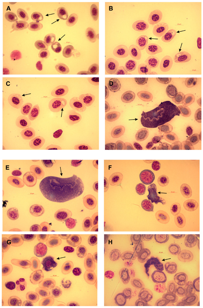
Most of the B. cordobae (n = 30, 81.1%) that we collected were infected (25 males, 5 females) by hemoparasites (Figure 1). Both extracellular and intraerythrocytic hemoparasites were detected in twenty-three individuals (twenty males, three females), exclusively extracellular parasites were detected in two males and two females, and exclusively intraerythrocytic ones were detected in three males. None of the frogs infected with hemoparasites showed external morphological signs of disease, or deviated in behavior from parasite-free individuals.
Figure 1.
Hemoparasites detected in blood smears of Cordoba treefrog (Boana cordobae). (A–C) Dactylosoma spec.; (D–H) Trypanosoma spp. (D,E) Morphotype “A” cells with rounded or elliptical trypomastigotes; (F,G) Morphotype “B” elongated trypomastigotes with pointed ends; (H) Morphotype “C” elongated trypomastigotes with pointed ends. Arrows indicate hemoparasites. Scale bar is 10 μm.
3.1. Taxonomic Assessment and Features of Hemoparasites
The intraerythrocytic parasites were morphologically identified as hemogregarines belonging to the genus Dactylosoma (Apicomplexa, Dactylosomatidae) (Figure 1A–C). The light microscopic examination did not allow for species identification, and molecular assessment was not available. All extracellular blood parasites detected were trypanosomes (Euglenozoa, Trypanosomatidae), standing out among the blood cells because of their large size and characteristic morphology. We found classical fusiform trypomastigotes as well as rounded, oval, fan-shaped, leaf-shaped, or irregular cells, with or without a free flagellum. The trypanosomes included three distinct morphotypes: rounded or elliptical cells represent morphotype “A” (Figure 1D,E), rounded trypomastigotes with pointed ends represent morphotype “B” (Figure 1F,G), and elongated trypomastigotes with pointed ends represent morphotype “C” (Figure 1H). The prevalence, intensity, and average intensity of Dactylosoma and Trypanosoma hemoparasites did not differ significantly between males and females (Table 1).
Table 1.
Quantitative descriptors of hemoparasites infecting B. cordobae. Statistical comparison of the mean intensity between males and females refers to an ANOVA with log10-normalized data.
| Hemoparasites | Prevalence (%) |
Intensity (Range) |
Mean Intensity (n ± SE) |
ANOVA |
|---|---|---|---|---|
| Intraerythrocytic (Dactylosoma) | ||||
| Males | 72 | 1–40 | 10.5 ± 2.3 | p = 0.9841 |
| Female | 60 | 1–22 | 8.7 ± 6.7 | |
| All adults | 70 | 1–40 | 10.3 ± 2.2 | |
| Extracellular (Trypanosoma) | ||||
| Males | 69 | 1–50 | 10.0 ± 2.5 | p = 0.0907 |
| Female | 100 | 2–28 | 8.6 ± 5.0 | |
| All adults | 73 | 1–50 | 9.8 ± 2.2 | |
3.2. Leukocyte Profiles
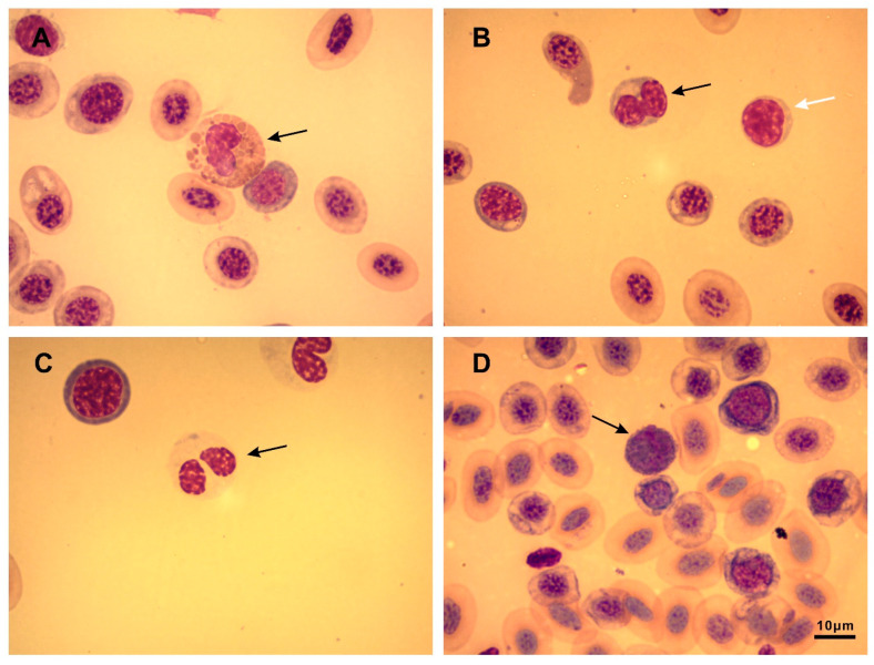
For this analysis, we pooled data of the males and females. Lymphocytes were the dominant group of leukocytes, followed by neutrophils (Figure 2, Table 2). Parasite-free frogs tended to have more lymphocytes and less neutrophils and basophils, but this difference was not statistically significant. The average N/L ratio of the parasite-free frogs was only about half of that of the infected ones, but again, this difference was not statistically significant.
Figure 2.
Different types of leukocytes in blood smears of Cordoba treefrogs (Boana cordobae). (A) Eosinophil (arrow); (B) lymphocyte (black arrow), monocyte (white arrow); (C) neutrophil (arrow); (D) basophil (arrow). Scale bar is10 μm.
Table 2.
Leukocyte profiles measured in parasite-free B. cordobae, and in those infected with exclusively Dactylosoma (D) or Trypanosoma spp. (T), and in those with both simultaneously (D + T). n: sample size; N/L ratio: neutrophils/lymphocytes ratio. Statistical comparison of groups (ANOVA) refers to log10-normalized data.
| Leukocytes [%] | Parasite-Free Individuals (n = 7) |
Infected Individuals (D) (n = 3) |
Infected Individuals (T) (n = 4) |
Infected Individuals (D + T) (n = 23) |
ANOVA |
|---|---|---|---|---|---|
| Lymphocytes | 94.1 | 89.4 | 89.3 | 89.9 | p = 0.5994 |
| Neutrophils | 3.3 | 6.6 | 5.6 | 5.2 | p = 0.5269 |
| Eosinophils | 0.9 | 2.3 | 2.0 | 2.6 | p = 0.4838 |
| Basophils | 0.6 | 0.3 | 2.7 | 1.2 | p = 0.0288 |
| Monocytes | 1.1 | 1.3 | 0.5 | 0.3 | p = 0.6328 |
| N/L ratio | 0.036 | 0.063 | 0.076 | 0.066 | p = 0.8610 |
3.3. Influence of Hemoparasites on Life-History Traits
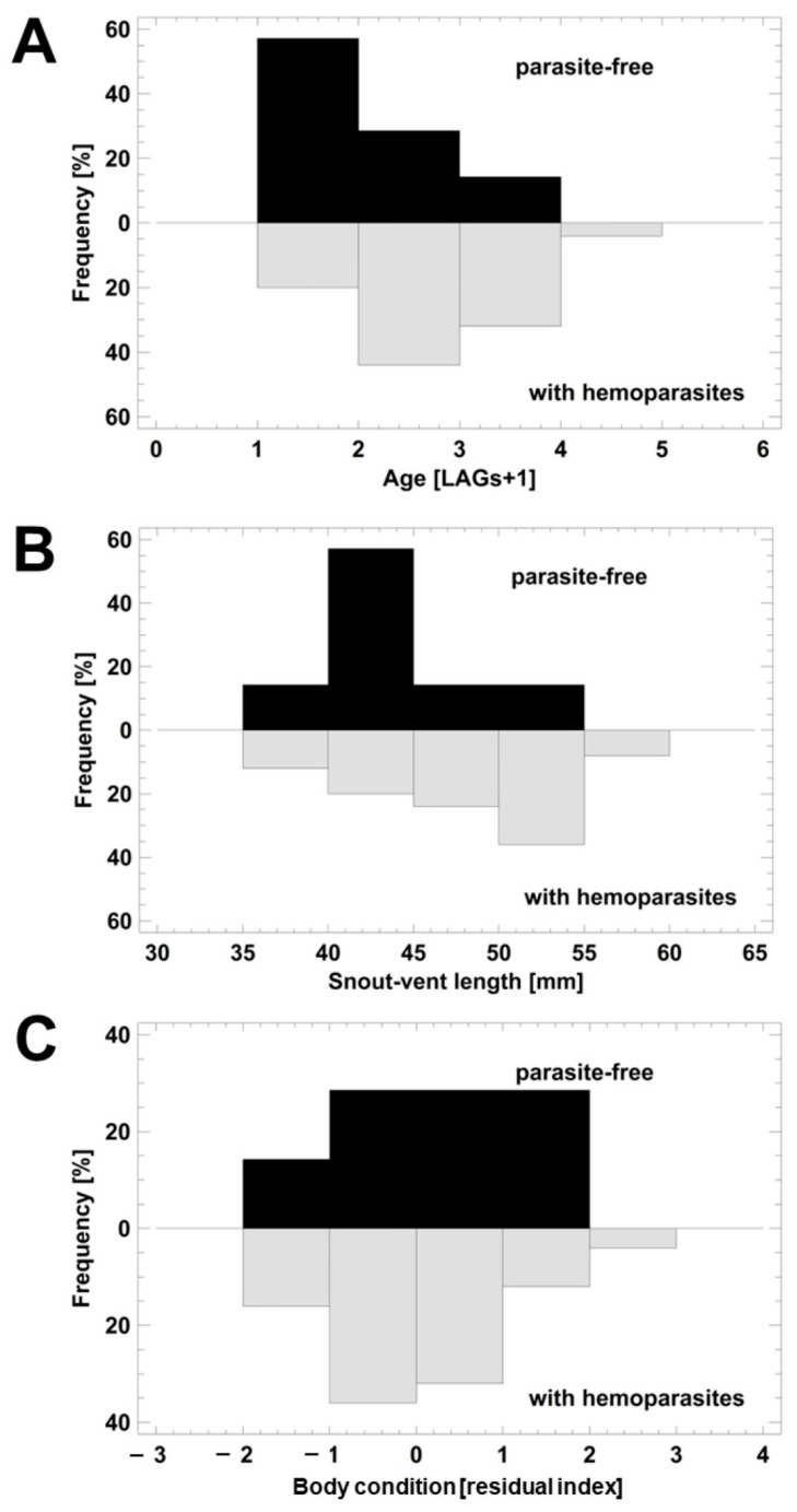
Because of the low number of females available and the fact that all were infected, we limited the analysis of the impact of parasitism on life-history traits to exclusively males. Their age ranged between 2 and 5 years (Figure 3A). Age did not differ significantly between the infected and parasite-free males (ANOVA, F1,30 = 3.49; p = 0.0798). Still, the shape of the age distribution was biased significantly toward younger individuals in parasite-free frogs (Kolmogorov–Smirnov test, p = 0.01777; Figure 3A). Snout–vent length varied between 38 and 58 mm. SVLs of the parasite-free frogs were significantly smaller than those of the infected ones, if uncorrected for age (ANOVA, F1,3 = 4.77; p = 0.0369; Figure 3B). This statistical significance disappeared when considering age as a covariable (ANCOVA, F1,29 = 1.81; p = 0.1886). As indicated by the ANCOVA, SVL was significantly correlated to age (double-reciprocal regression model, R2 = 36.6%; F1,30 = 17.30; p = 0.0002). Body condition (as a residual index) did not differ between the two groups (ANOVA, F1,30 = 0.34; p = 0.5660; Figure 3C). Adjusting the body condition results using age and SVL as covariables did not change the absence of this statistical significance (ANCOVA, F1,28 = 0.35; p = 0.5584).
Figure 3.
Life-history traits of parasite-free and infected Cordoba treefrogs (Boana cordobae). (A) Age; (B) SVL; (C) body condition. Details on statistical comparison are in the text.
4. Discussion
Our study provides the first evidence that the endemic treefrog Boana cordobae is a new host for hemogregarines of the genus Dactylosoma and flagellates of the genus Trypanosoma. An extraordinary high proportion of 81.1% of wild free-living B. cordobae were found to carry at least one type of hemoparasite. In Argentina, limited research has been conducted on anuran amphibians affected by hemoparasites. To our knowledge, we report the first Dactylosoma ever detected in Argentina. Cabagna-Zenkluzen et al. [68] presented in 2009 the first records of hemoparasites (Trypanosoma, microfilariae) in leptodactylid and hylid frogs from Argentina. Trypanosomes were found in Leptodactylus chaquensis (5–22% prevalence), L. ocellatus (9%), and Trachycephalus venulosus (17%), i.e., in prevalences far below the levels reported here. The prevalence in anurans infected with trypanosomes was also lower in Brazil (10% in Boana multifasciata, 43% in B. geographica), in Panama (40% in male and 7% in female Engystomops pustulosus), in South Africa (11%), and in Thailand (34% in Hoplobatrachus rugulosus) [15,27,69,70]. This geographical review on trypanosome-infected anurans suggests that their prevalence in B. cordobae is exceptionally high. With respect to Dactylosoma infections, its 70% prevalence in B. cordobae also contrasts with the reported prevalences of 6% in Rhinella major, 16% in Leptodactylus ocellatus/labyrinthicus in Brazil, and 28% of Ptychadena anchietae in South Africa [15,29,71,72]. Further investigations will be needed to elucidate the reasons for the remarkably high prevalence of hemoparasites in B. cordobae.
Multiple infections with high pathogen intensities seem to be the trend in B. cordobae, as has been observed in many Palearctic and Neotropical anuran hosts as well [13,61,68,69,70]. Still, external manifestations of pathogenic impacts were absent, as they are in other anurans carrying hemoparasites, but inflammatory lesions, predominantly in the liver, have been reported in other frog hosts infected with hemogregarines [12,70].
An activation of the cellular immune system in response to Rickettsia hemoparasites is indicated by changes in the leukocyte profile of parasitized salamander individuals [34]. The main response seemed to be an increase in the total leukocyte number, whereas the single fractions did not show significant intergroup differences, except for eosinophils [34]. Similarly, we observed an increase in eosinophils to levels more than twice as high in parasitized frogs compared to those of the parasite-free controls; however, this was not statistically significant. This lack of significance may be an effect of the small sample size or, alternatively, may indicate that there is no adaptive response.
In vertebrates, an immunological stress indicator is the ratio between their neutrophil and lymphocyte densities, as during infections, their neutrophils increase and their lymphocytes decrease [34,73]. Neutrophils are known to phagocytize and target foreign particles and microbes and they increase numerically in response to injuries or infections [73]. Yet, recent studies on parasitized amphibians have found only minor changes in their leukocyte profile [34,74]. Our study is in line with the observation that trypanosomes and hemogregarines do not cause significant changes in the N/L ratio, as we noticed only a slight and insignificant increase from 0.03 to 0.06 in B. cordobae.
A high prevalence and intensity of hemoparasites may negatively affect the life history and fitness of a reptile host by reducing its capability to transport oxygen and changing its allocation of resources [75,76,77], whereas similar effects in amphibian hosts have not been reported so far. In B. cordobae, we could not confirm any difference in the life-history traits we studied between infected and parasite-free individuals, suggesting that parasitism could potentially exert a limited or negligible impact on the host’s fitness. At first glance, the surprising fact that the infected frogs were significantly larger than the parasite-free controls seems to be due to the dominance of young and subsequently small individuals in the controls (this size–age relationship has been established in a previous study [32]). Thus, infection seems to occur at the post-metamorphic stage of life, in agreement with similar observations of Lithobates clamitans and L. catesbeianus [17]. The stage of life in which individuals are most often infected might be the first breeding period of an individual and the infection may remain for life, like in Notophthalmus viridescens [78]. Potential vectors of hemoparasites are leeches and hematophagous dipterans [79,80,81]. Mosquitoes (Uranotaenia spp.) and frog-biting midges (Corethrella spp.) are known to be attracted by anuran advertisement calls and to transmit a wide range of hemoparasites [26,27,82]. In this case, the prevalence of infection is sex-biased toward males, as shown in Engystomops pustulosus and in Dryophytes cinereus [25,26,27]. We do not know the local vectors responsible for transmitting parasites to B. cordobae hosts, but the fact that all females were infected does not suggest a role of vocalization in vector attraction.
5. Conclusions
In conclusion, we have demonstrated for the first time that moderate to high densities of hemoparasites do not affect significantly the cellular immune response of B. cordobae hosts or any of the life-history traits studied. Infection of the frogs seems to occur usually following sexual maturation and seems to persist until death, but its vector and mode of transmission remain to be identified. Further studies are needed in which other measures of the host’s fitness are considered, such as growth rate, feeding rate, locomotor performance, reproductive status, and reproductive output, to understand this complex host–parasite relationship.
Acknowledgments
Our study was authorized by the Cordoba Environmental Agency (A.C.A.S.E.), Environmental Secretary of the Cordoba Government. The manuscript benefited from the comments of anonymous reviewers.
Author Contributions
F.P., Z.S., M.B. and M.A.O. carried out the field and laboratory work and drafting of the manuscript; N.S. supervised the microscopic analyses and drafting of the manuscript; P.R.G. and A.L.M. designed the study and provided field and laboratory facilities; U.S. designed the study, analyzed the data, and wrote the final version of the manuscript. All authors have read and agreed to the published version of the manuscript.
Institutional Review Board Statement
The Ethical Committee of Investigation of the Universidad Nacional de Río Cuarto (file number 241/21) authorized our study.
Data Availability Statement
All data used for this manuscript are given in the text.
Conflicts of Interest
The authors declare no conflict of interest. The funders had no role in the design of the study; in the collection, analyses, or interpretation of data; in the writing of the manuscript; or in the decision to publish the results.
Funding Statement
Financial support was provided by the Secretary of Research and Technology of the National University of Río Cuarto (PPI 18/C475) and National Agency for Scientific and Technological Promotion FONCYT (BID-PICT 0932-2012; PICT 2533-2014). FP, ZS, MB, MO, and PG thank CONICET Argentina (Argentinean National Research Council for Science and Technology) for the fellowships granted.
Footnotes
Disclaimer/Publisher’s Note: The statements, opinions and data contained in all publications are solely those of the individual author(s) and contributor(s) and not of MDPI and/or the editor(s). MDPI and/or the editor(s) disclaim responsibility for any injury to people or property resulting from any ideas, methods, instructions or products referred to in the content.
References
- 1.Stuart S.N., Chanson J.S., Cox N.A., Young B.E., Rodrigues A.S.L., Fischman D.L., Waller R.W. Status and trends of amphibian declines and extinctions worldwide. Science. 2004;306:1783–1786. doi: 10.1126/science.1103538. [DOI] [PubMed] [Google Scholar]
- 2.Grant E.H.C., Miller D.A.W., Schmidt B.R., Adams M.J., Amburgey S.M., Chambert T., Cruickshank S.S., Fisher R.N., Green D.M., Hossack B.R., et al. Quantitative evidence for the effects of multiple drivers on continental-scale amphibian declines. Sci. Rep. 2016;6:25625. doi: 10.1038/srep25625. [DOI] [PMC free article] [PubMed] [Google Scholar]
- 3.Hof C., Araujo M.B., Jetz W., Rahbek C. Additive threats from pathogens, climate and land-use change for global amphibian diversity. Nature. 2011;480:516–522. doi: 10.1038/nature10650. [DOI] [PubMed] [Google Scholar]
- 4.Koprivnikar J., Marcogliese D.J., Rohr J.R., Orlofske S.A., Raffel T.R., Johnson P.T.J. Macroparasite Infections of Amphibians: What Can They Tell Us? EcoHealth. 2012;9:342–360. doi: 10.1007/s10393-012-0785-3. [DOI] [PubMed] [Google Scholar]
- 5.Murray K.A., Skerratt L.F. Predicting Wild Hosts for Amphibian Chytridiomycosis: Integrating Host Life-History Traits with Pathogen Environmental Requirements. Hum. Ecol. Risk Assess. Int. J. 2012;18:200–224. doi: 10.1080/10807039.2012.632310. [DOI] [Google Scholar]
- 6.Spitzen-van der Sluijs A., van den Broek J., Kik M., Martel A., Janse J., van Asten F., Pasmans F., Grone A., Rijks J.M. Monitoring ranavirus-associated mortality in a Dutch heathland in the aftermath of a ranavirus disease outbreak. J. Wildl. Dis. 2016;52:817–827. doi: 10.7589/2015-04-104. [DOI] [PubMed] [Google Scholar]
- 7.Azat C., Alvarado-Rybak M., Solano-Iguaran J.J., Velasco A., Valenzuela-Sánchez A., Flechas S.V., Peñafiel-Ricaurte A., Cunningham A.A., Bacigalupe L.D. Synthesis of Batrachochytrium dendrobatidis infection in South America: Amphibian species under risk and areas to focus research and disease mitigation. Ecography. 2022;2022:e05977. doi: 10.1111/ecog.05977. [DOI] [Google Scholar]
- 8.Towe A.E., Gray M.J., Carter E.D., Wilber M.Q., Ossiboff R.J., Ash K., Bohanon M., Bajo B.A., Miller D.L. Batrachochytrium salamandrivorans Can Devour More than Salamanders. J. Wildl. Dis. 2021;57:942–948. doi: 10.7589/JWD-D-20-00214. [DOI] [PubMed] [Google Scholar]
- 9.Bower D.S., Brannelly L.A., McDonald C.A., Webb R.J., Greenspan S.E., Vickers M., Gardner M.G., Greenlees M.J. A review of the role of parasites in the ecology of reptiles and amphibians. Aust. Ecol. 2019;44:433–448. doi: 10.1111/aec.12695. [DOI] [Google Scholar]
- 10.Davies A.J., Johnston M.R.L. The biology of some intraerythrocytic parasites of fishes, amphibia and reptiles. Adv. Parasitol. 2000;45:1–107. doi: 10.1016/s0065-308x(00)45003-7. [DOI] [PubMed] [Google Scholar]
- 11.Duszynski D.W., Bolek M.G., Upton S.J. Coccidia (Apicomplexa: Eimeriidae) of amphibians of the world. Zootaxa. 2007;1667:1–77. [Google Scholar]
- 12.Densmore C.L., Green D.E. Diseases of amphibians. ILAR J. 2007;48:235–254. doi: 10.1093/ilar.48.3.235. [DOI] [PubMed] [Google Scholar]
- 13.Ferreira R., Campaner M., Viola L.B., Takata C.S.d.A., Takeda G., Teixeira M.M.G. Morphological and molecular diversity and phylogenetic relationships among anuran trypanosomes from the Amazonia, Atlantic Forest and Pantanal biomes in Brazil. Parasitology. 2007;134:1623–1638. doi: 10.1017/S0031182007003058. [DOI] [PubMed] [Google Scholar]
- 14.Leal D.D.M., Dreyer C.S., da Silva R.J., Ribolla P.E.M., Paduan K.D., Bianchi I., O’Dwyer L.H. Characterization of Hepatozoon spp. in Leptodactylus chaquensis and Leptodactylus podicipinus from two regions of the Pantanal, state of Mato Grosso do Sul, Brazil. Parasitol. Res. 2015;114:1541–1549. doi: 10.1007/s00436-015-4338-x. [DOI] [PubMed] [Google Scholar]
- 15.Netherlands E.C., Cook C.A., Kruger D.J.D., du Preez L.H., Smit N.J. Biodiversity of frog haemoparasites from sub-tropical northern KwaZulu-Natal, South Africa. Int. J. Parasitol. Parasites Wildl. 2015;4:135–141. doi: 10.1016/j.ijppaw.2015.01.003. [DOI] [PMC free article] [PubMed] [Google Scholar]
- 16.Bilhalva L.C., de Almeida B.A., Colombo P., de Faria Valle S., Soares J.F. Hematologic variables of free-living Leptodactylus luctator with and without hemoparasites and thrombidiform mites in southern Brazil. Vet. Parasitol. Reg. Stud. 2023;38:100834. doi: 10.1016/j.vprsr.2023.100834. [DOI] [PubMed] [Google Scholar]
- 17.Barta J.R., Desser S.S. Blood parasites of amphibians from Algonquin Park, Ontario. J. Wildl. Dis. 1984;20:180–189. doi: 10.7589/0090-3558-20.3.180. [DOI] [PubMed] [Google Scholar]
- 18.Barta J.R. The Dactylosomatidae. J. Adv. Parasitol. 1991;30:1–37. doi: 10.1016/S0065-308X(08)60305-X. [DOI] [PubMed] [Google Scholar]
- 19.Barnard S.M., Upton S.J. A Veterinary Guide to the Parasites of Reptiles. Volume 1 Krieger Publishing Company; Malabar, FL, USA: 1994. Protozoa. [Google Scholar]
- 20.Spodareva V.V., Grybchuk-Ieremenko A., Losev A., Votýpka J., Lukeš J., Yurchenko V., Kostygov A.Y. Diversity and evolution of anuran trypanosomes: Insights from the study of European species. Parasites Vectors. 2018;11:447. doi: 10.1186/s13071-018-3023-1. [DOI] [PMC free article] [PubMed] [Google Scholar]
- 21.Mutschmann F. Erkrankungen der Amphibien. Georg Thieme Verlag; New York, NY, USA: 2010. p. 344. [Google Scholar]
- 22.Franca C. Le Trypanosoma inopinatum. Arch. Protistenk. 1915;36:1–12. [Google Scholar]
- 23.Willette-Frahm M., Wright K.M., Thode B.C. Select Protozoal Diseases in Amphibians and Reptiles: A Report for the Infectious Diseases Committee, American Association of Zoo Veterinarians. Bull. Assoc. Reptil. Amphib. Vet. 1995;5:19–29. doi: 10.5818/1076-3139.5.1.19. [DOI] [Google Scholar]
- 24.Wright K.M. Reptile Medicine and Surgery. Volume 2. Saunders; St. Louis, MO, USA: 2006. Overview of amphibian medicine; pp. 941–971. [Google Scholar]
- 25.Johnson R.N., Young D.G., Butler J.F. Trypanosome Transmission by Corethrella wirthi (Diptera, Chaoboridae) to the Green Treefrog, Hyla cinerea (Anura, Hylidae) J. Med. Entomol. 1993;30:918–921. doi: 10.1093/jmedent/30.5.918. [DOI] [PubMed] [Google Scholar]
- 26.Bernal X.E., de Silva P. Cues used in host-seeking behavior by frog-biting midges (Corethrella spp. Coquillet) J. Vector Ecol. 2015;40:122–128. doi: 10.1111/jvec.12140. [DOI] [PubMed] [Google Scholar]
- 27.Bernal X.E., Pinto C.M. Sexual differences in prevalence of a new species of trypanosome infecting tungara frogs. Int. J. Parasitol. Parasites Wildl. 2016;5:40–47. doi: 10.1016/j.ijppaw.2016.01.005. [DOI] [PMC free article] [PubMed] [Google Scholar]
- 28.Calil P.R., Gonzalez I.H.L., Salgado P., Cruz J.d., Ramos P.L., Chagas C.R.F. Hemogregarine parasites in wild captive animals, a broad study in São Paulo Zoo. J. Entomol. Zool. Stud. 2017;5:1378–1387. [Google Scholar]
- 29.Coêlho T.A., De Souza D.C., da Costa Oliveira E., Correa L.L., Viana L.A., Kawashita-Ribeiro R.A. Haemogregarine of Genus Dactylosoma (Adeleorina: Dactylosomatidae) in Species of Rhinella (Anura: Bufonidae) from the Brazilian Amazon. Acta Parasitol. 2021;66:1574–1580. doi: 10.1007/s11686-021-00399-z. [DOI] [PubMed] [Google Scholar]
- 30.Cotes-Perdomo A., Santodomingo A., Castro L.R. Hemogregarine and Rickettsial infection in ticks of toads from northeastern Colombia. Int. J. Parasitol. Parasites Wildl. 2018;7:237–242. doi: 10.1016/j.ijppaw.2018.06.003. [DOI] [PMC free article] [PubMed] [Google Scholar]
- 31.González L.P., Vargas-Leon C.M., Fuentes-Rodriguez G.A., Calderon-Espinosa M.L., Matta N.E. Do blood parasites increase immature erythrocytes and mitosis in amphibians? Rev. Biol. Trop. 2021;69:615–625. doi: 10.15517/rbt.v69i2.45459. [DOI] [Google Scholar]
- 32.Davis A.K., Maney D.L., Maerz J.C. The use of leukocyte profiles to measure stress in vertebrates: A review for ecologists. Funct. Ecol. 2008;22:760–772. doi: 10.1111/j.1365-2435.2008.01467.x. [DOI] [Google Scholar]
- 33.Davis A.K., Hopkins W.A. Widespread trypanosome infections in a population of eastern hellbenders (Cryptobranchus alleganiensis alleganiensis) in Virginia, USA. Parasitol. Res. 2013;112:453–456. doi: 10.1007/s00436-012-3076-6. [DOI] [PubMed] [Google Scholar]
- 34.Davis A.K., Golladay C. A survey of leukocyte profiles of red-backed salamanders from Mountain Lake, Virginia, and associations with host parasite types. Comp. Clin. Path. 2019;28:1743–1750. doi: 10.1007/s00580-019-03015-9. [DOI] [Google Scholar]
- 35.Allender M.C., Fry M.M. Amphibian Hematology. Vet. Clin. N. Am. Exot. Anim. Pract. 2008;11:463–480. doi: 10.1016/j.cvex.2008.03.006. [DOI] [PubMed] [Google Scholar]
- 36.Bain P., Harr K.E. Schalm’s Veterinary Hematology. Wiley; Hoboken, NJ, USA: 2022. Hematology of Amphibians; pp. 1228–1232. [DOI] [Google Scholar]
- 37.Davis A.K., Maney D.L. The use of glucocorticoid hormones or leucocyte profiles to measure stress in vertebrates: What’s the difference? Methods Ecol. Evol. 2018;9:1556–1568. doi: 10.1111/2041-210X.13020. [DOI] [Google Scholar]
- 38.Goessling J.M., Kennedy H., Mendonça M.T., Wilson A.E. A meta-analysis of plasma corticosterone and heterophil:lymphocyte ratios—Is there conservation of physiological stress responses over time? Funct. Ecol. 2015;29:1189–1196. doi: 10.1111/1365-2435.12442. [DOI] [Google Scholar]
- 39.Barrio A. Las subespecies de Hyla pulchella Dumeril y Bibron (Anura, Hylidae) Physis. 1965;25:115–128. [Google Scholar]
- 40.Cei J.M. Amphibians of Argentina. Monit. Zool. Ital. N.S. Monogr. 1980;2:1–609. [Google Scholar]
- 41.Faivovich J., García P.C.A., Ananias F., Lanari L., Basso N.G., Wheeleri W.C. A molecular perspective on the phylogeny of the Hyla pulchella species group (Anura, Hylidae) Mol. Phyl. Evol. 2004;32:938–950. doi: 10.1016/j.ympev.2004.03.008. [DOI] [PubMed] [Google Scholar]
- 42.Dubois A. The nomenclatural status of Hysaplesia, Hylaplesia, Dendrobates and related nomina (Amphibia, Anura), with general comments on zoological nomenclature and its governance, as well as on taxonomic databases and websites. Bionomina. 2017;11:1–48. doi: 10.11646/bionomina.11.1.1. [DOI] [Google Scholar]
- 43.Ferro J.M., Cardozo D.E., Suárez P., Boeris J.M., Blasco-Zúñiga A., Barbero G., Gomes A., Gazoni T., Costa W., Nagamachi C.Y., et al. Chromosome evolution in Cophomantini (Amphibia, Anura, Hylinae) PLoS ONE. 2018;13:e0192861. doi: 10.1371/journal.pone.0192861. [DOI] [PMC free article] [PubMed] [Google Scholar]
- 44.Baraquet M., Otero M.A., Grenat P.R., Babini M.S., Martino A.L. Effect to age on the geographic variation in morphometric traits among populations of Boana cordobae (Anura: Hylidae) Rev. Biol. Trop. 2018;66:1401–1411. doi: 10.15517/rbt.v66i4.32365. [DOI] [Google Scholar]
- 45.Otero M.A., Baraquet M., Pollo F., Grenat P., Salas N.E., Martino A.L. Sexual Size Dimorphism in Relation to Age and Growth in Hypsiboas cordobae (Anura: Hylidae) from Córdoba, Argentina. Herpetol. Conserv. Biol. 2017;12:141–148. [Google Scholar]
- 46.Baraquet M., Otero M.A., Valetti J.A., Grenat P.R., Martino A.L. Age, Body Size, And growth of Boana cordobae (Anura: Hylidae) along an elevational gradient in Argentina. Herpetol. Conserv. Biol. 2018;13:391–398. [Google Scholar]
- 47.di Tada I.E., Zavattieri M.V., Martino A.L. Análisis estructural del canto nupcial de Hyla pulchella cordobae (Amphibia: Hylidae) en la provincia de Córdoba (Argentina) Rev. Esp. Herp. 1996;10:7–11. [Google Scholar]
- 48.Baraquet M., Salas N.E., Martino A.L. Advertisement Calls and Interspecific Variation in Hypsiboas cordobae and Hypsiboas pulchellus (Anura, Hylidae) from Central Argentina. Acta Zool. Bulg. 2013;65:479–486. [Google Scholar]
- 49.Baraquet M., Grenat P., Salas N., Martino A. Geographic variation in the advertisement call of Hypsiboas cordobae (Anura, Hylidae) Acta Ethol. 2015;18:79–86. doi: 10.1007/s10211-014-0188-2. [DOI] [Google Scholar]
- 50.Pollo F.E., Cibils M.L., Bionda C.L., Salas N.E., Martino A.L. Trophic ecology of syntopic anuran larvae, Rhinella arenarum (Anura: Bufonidae) and Hypsiboas cordobae (Anura: Hylidae): Its relation to the structure of periphyton. Ann. Limnol. Int. J. Lim. 2015;51:211–217. doi: 10.1051/limn/2015015. [DOI] [Google Scholar]
- 51.Pollo F.E., Cibils-Martina L., Otero M.A., Baraquet M., Grenat P.R., Salas N.E., Martino A.L. Anuran tadpoles inhabiting a fluoride-rich stream: Diets and morphological indicators. Heliyon. 2019;5:e02003. doi: 10.1016/j.heliyon.2019.e02003. [DOI] [PMC free article] [PubMed] [Google Scholar]
- 52.Pollo F.E., Grenat P.R., Otero M.A., Salas N.E., Martino A.L. Assessment in situ of genotoxicity in tadpoles and adults of frog Hypsiboas cordobae (Barrio 1965) inhabiting aquatic ecosystems associated to fluorite mine. Ecotoxicol. Environ. Saf. 2016;133:466–474. doi: 10.1016/j.ecoenv.2016.08.003. [DOI] [PubMed] [Google Scholar]
- 53.de la Navarre B. Common Parasitic Diseases of Reptiles and Amphibians. In Proceedings of CVC in San Diego, 2008. [(accessed on 1 September 2023)]. Available online: https://www.dvm360.com/view/common-parasitic-diseases-reptiles-amphibians-proceedings.
- 54.Nöller H.G. Eine einfache Technik der Blutentnahme beim Frosch. Pflügers Arch. Physiol. 1959;269:98–100. doi: 10.1007/BF00362975. [DOI] [PubMed] [Google Scholar]
- 55.Dacie S.J., Lewis S.M. Practical Haematology. 8th ed. Churchill & Livingstone; Edinburgh, Scotland: 1995. [Google Scholar]
- 56.Pessier A.P. Cytologic diagnosis of disease in amphibians. Vet. Clin. Exot. Anim. Pract. 2007;10:187–206. doi: 10.1016/j.cvex.2006.10.006. [DOI] [PubMed] [Google Scholar]
- 57.Bardsley J., Harmsen R. The trypanosomes of anura. J. Adv. Parasitol. 1973;11:1–73. doi: 10.1016/s0065-308x(08)60184-0. [DOI] [PubMed] [Google Scholar]
- 58.Manwell R.D. The Genus Dactylosoma. J. Protozool. 1964;11:526–530. doi: 10.1111/j.1550-7408.1964.tb01792.x. [DOI] [PubMed] [Google Scholar]
- 59.Smith T.G. The genus Hepatozoon (Apicomplexa: Adeleina) J. Parasitol. 1996;82:565–585. doi: 10.2307/3283781. [DOI] [PubMed] [Google Scholar]
- 60.Desser S.S. The blood parasites of anurans from Costa Rica with reflections on the taxonomy of their trypanosomes. J. Parasitol. 2001;87:152–160. doi: 10.1645/0022-3395(2001)087[0152:TBPOAF]2.0.CO;2. [DOI] [PubMed] [Google Scholar]
- 61.Žičkus T. The First Data on the Fauna and Distribution of Blood Parasites of Amphibians in Lithuania. Acta Zool. Litu. 2002;12:197–202. doi: 10.1080/13921657.2002.10512506. [DOI] [Google Scholar]
- 62.Bush A., Lafferty K., Lotz J., Shostak A. Parasitology meets ecology on its own terms: Margolis et al. revisited. J. Parasitol. 1997;83:575–583. doi: 10.2307/3284227. [DOI] [PubMed] [Google Scholar]
- 63.Castanet J., Francillon-Vieillot H., Meunier F.J., de Ricqles A. Bone and individual aging. In: Hall B.K., editor. Bone Growth. Volume 7. CRC Press; Boca Raton, FL, USA: 1993. pp. 245–283. [Google Scholar]
- 64.Sinsch U. Review: Skeletochronological assessment of demographic life-history traits in amphibians. Herpetol. J. 2015;25:5–13. [Google Scholar]
- 65.Lai Y.C., Lee T.H., Kam Y.C. A skeletochronological study on a subtropical, riparian ranid (Rana swinhoana) from different elevations in Taiwan. Zool. Sci. 2005;22:653–658. doi: 10.2108/zsj.22.653. [DOI] [PubMed] [Google Scholar]
- 66.von Bertalanffy L. A quantitative theory of organic growth (Inquires on growth laws. II) Hum. Biol. 1938;10:181–213. [Google Scholar]
- 67.Băncilă R.I., Hartel T., Plaiasu R., Smets J., Cogălniceanu D. Comparing three body condition indices in amphibians: A case study of yellow-bellied toad Bombina variegata. Amphib. Reptil. 2010;31:558–562. doi: 10.1163/017353710x518405. [DOI] [Google Scholar]
- 68.Cabagna-Zenklusen M.C., Lajmanovich R.C., Peltzer P.M., Attademo A.M., Fiorenza Biancucci G.S., Bassó A. Primeros registros de endoparásitos en cinco especies de anfibios anuros del litoral argentino. Cuad. herpetol. 2009;23:33–40. [Google Scholar]
- 69.Pinho S.R.C., Rodriguez-Malaga S., Lozano-Osorio R., Correa F.S., Silva I.B., Santos-Costa M.C. Effects of the habitat on anuran blood parasites in the Eastern Brazilian Amazon. An. Acad. Bras. Cienc. 2021;93:e20201703. doi: 10.1590/0001-3765202120201703. [DOI] [PubMed] [Google Scholar]
- 70.Sailasuta A., Satetasit J., Chutmongkonkul M. Pathological study of blood parasites in rice field frogs, Hoplobatrachus rugulosus (Wiegmann, 1834) Vet. Med. Int. 2011;2011:850568. doi: 10.4061/2011/850568. [DOI] [PMC free article] [PubMed] [Google Scholar]
- 71.da Costa S.C.G., Pereira N.d.M. Lankesterella alencari n. sp., a toxoplasma-like organism in the central nervous system of Amphibia (Protozoa, Sporozoa) Mem. Inst. Oswaldo Cruz. 1971;69:397–411. doi: 10.1590/S0074-02761971000300002. [DOI] [PubMed] [Google Scholar]
- 72.Úngari L.P., Netherlands E.C., Santos A.L.Q., de Alcantara E.P., Emmerich E., da Silva R.J., O’Dwyer L.H. Diversity of Haemogregarine Parasites Infecting Brazilian Anurans, with a Description of New Species of Dactylosoma (Apicomplexa: Adeleorina: Dactylosomatidae) Acta Parasitol. 2022;67:1740–1755. doi: 10.1007/s11686-022-00624-3. [DOI] [PubMed] [Google Scholar]
- 73.Davis A.K., Keel M.K., Ferreira A., Maerz J.C. Effects of chytridiomycosis on circulating white blood cell distributions of bullfrog larvae (Rana catesbeiana) Comp. Clin. Path. 2010;19:49–55. doi: 10.1007/s00580-009-0914-8. [DOI] [Google Scholar]
- 74.Salinas Z.A., Biole F.G., Grenat P.R., Pollo F.E., Salas N.E., Martino A.L. First report of Lernaea cyprinacea (Copepoda: Lernaeidae) in tadpoles and newly-metamorphosed frogs in wild populations of Lithobates catesbeianus (Anura: Ranidae) in Argentina. Phyllomedusa. 2016;15:43–50. doi: 10.11606/issn.2316-9079.v15i1p43-50. [DOI] [Google Scholar]
- 75.Oppliger A., Celerier M., Clobert J. Physiological and behaviour changes in common lizards parasitized by haemogregarines. Parasitology. 1996;113:433–438. doi: 10.1017/S003118200008149X. [DOI] [Google Scholar]
- 76.Martín J., Amo L., López P. Parasites and health affect multiple sexual signals in male common wall lizards, Podarcis muralis. Naturwissenschaften. 2008;95:293–300. doi: 10.1007/s00114-007-0328-x. [DOI] [PubMed] [Google Scholar]
- 77.Brown G., Shilton C., Shine R. Do parasites matter? Assessing the fitness consequences of haemogregarine infection in snakes. Can. J. Zool. 2006;84:668–676. doi: 10.1139/z06-044. [DOI] [Google Scholar]
- 78.Mock B.A., Gill P. The infrapopulation dynamics of trypanosomes in red-spotted newts. Parasitology. 1984;88:267–282. doi: 10.1017/S0031182000054524. [DOI] [Google Scholar]
- 79.Ramos B., Urdaneta-Morales S. Hematophagous insects as vectors for frog trypanosomes. Rev. Biol. Trop. 1977;25:209–217. [PubMed] [Google Scholar]
- 80.Ferguson L.V., Smith T.G. Reciprocal trophic interactions and transmission of blood parasites between mosquitoes and frogs. Insects. 2012;3:410–423. doi: 10.3390/insects3020410. [DOI] [PMC free article] [PubMed] [Google Scholar]
- 81.Toledo L.F., Ruggeri J., de Campos L.L.F., Martins M., Neckel-Oliveira S., Breviglieri C.P.B. Midges not only sucks, but may carry lethal pathogens to wild amphibians. Biotropica. 2021;53:722–725. doi: 10.1111/btp.12928. [DOI] [Google Scholar]
- 82.Legett H.D., Aihara I., Bernal X.E. Within host acoustic signal preference of frog-biting mosquitoes (Diptera: Culicidae) and midges (Diptera: Corethrellidae) on Iriomote Island, Japan. Entomol. Sci. 2021;24:116–122. doi: 10.1111/ens.12455. [DOI] [Google Scholar]
Associated Data
This section collects any data citations, data availability statements, or supplementary materials included in this article.
Data Availability Statement
All data used for this manuscript are given in the text.


