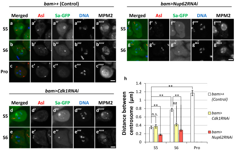
Figure 4.
Cdk1-mediated centrosome separation before the initiation of meiotic division. (a–g) Observation of mature spermatocytes (growth phase at S5 and S6 in (a,b,d–g); at prophase I (Pro) in (c)) immunostained with anti-Asl and anti-MPM2 antibodies. Normal control (bam>+) (a–c), Cdk1-silenced (d,e), and Nup62-silenced (f,g) cells. Images show anti-Asl immunofluorescence to visualize centrosomes (red in (a–g), white in (a’–g’)), Sa-GFP fluorescence for visualizing the nucleolus (green in (a–g), white in (a’’–g’’)), and DNA staining with DAPI (blue in a-g, white in (a’’’–g’’’)) and anti-MPM2 epitopes (white in (a’’’’–g’’’’)). Scale bar: 10 μm. (h) Quantification of the distance between paired centrosomes in S5 to ProI spermatocytes. The lengths between paired centrosomes were measured in each spermatocyte and the mean length was displayed as a white bar (control cells, bam>+), a yellow bar (Cdk1RNAi; bam>Cdk1RNAi), or a red bar (Nup62RNAi; bam>Nup62RNAi). Data are presented as means ± SEMs (n > 29 cells). No ProI cells were observed in bam>Cdk1RNAi or bam>Nup62RNAi testes. Significance was tested by one-way ANOVA followed by Bonferroni’s post hoc comparison tests. ** p < 0.01. n.s.; not significant.

