

Abstract
Palisade cells from fully expanded leaves from irrigated and nonirrigated, field grown cotton (Gossypium hirsutum L. cv. Paymaster 266) were subjected to a microscopic examination to evaluate the effect of water stress on subcellular structures. The water potential difference between the two treatments was 13 bars at the time of sampling. The dimensions of the palisade cells and their density per unit leaf area were determined by light microscopy. Palisade cells from stressed plants had the same diameter, but were taller than their counterparts in irrigated plants. The density of the palisade cells was the same in both treatments as was the fractional volume of the intercellular space. It was concluded that the reduced leaf area observed in the stressed plants resulted primarily from a mitotic sensitivity to water stress. Further, expansion of palisade cells was not inhibited by the stress imposed in this study.
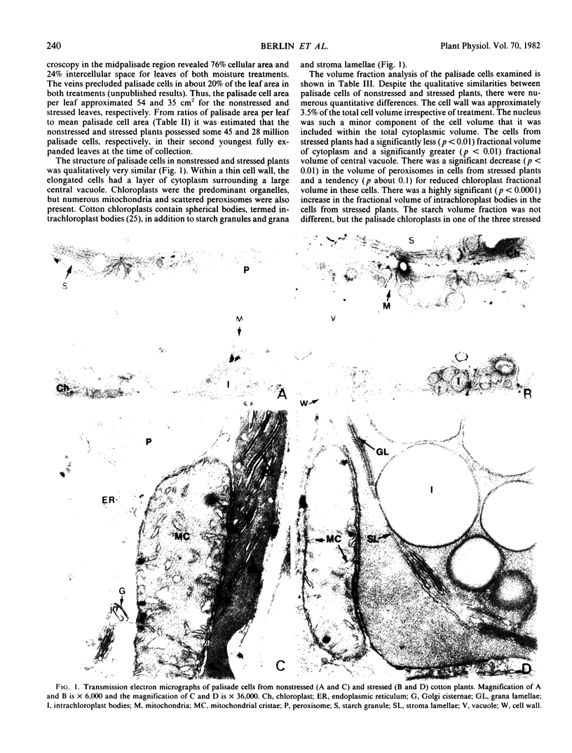
Morphometric analysis of electron micrographs was used to evaluate the subcellular structure of palisade cells from nonstressed and stressed plants. The fractional volumes of cell walls, total cytoplasm, chloroplasts, starch granules, intrachloroplast bodies, mitochondria, peroxisomes, and central vacuoles were determined. The surface densities of grana and stroma lamellae, outer chloroplast membranes, mitochondrial cristae, endoplasmic reticulum and Golgi cisternae were also measured. The number of chloroplasts, mitochondria, and peroxisomes were determined. These data were expressed as actual volumes, areas, and numbers per palisade cell for each treatment. Palisade cells from stressed plants had thinner cell walls, larger central vacuoles and approximately the same amount of cytoplasm compared to cells from nonstressed plants. Within the cytoplasm, stressed plants had more but smaller chloroplasts with increased grana and stroma lamellae surfaces, larger mithchondria with reduced cristae surfaces, smaller peroxisomes and reduced membrane surfaces of endoplasmic reticulum and Golgi cisternae.
Full text
PDF
Images in this article
Selected References
These references are in PubMed. This may not be the complete list of references from this article.
- Ackerson R. C., Hebert R. R. Osmoregulation in Cotton in Response to Water Stress : I. ALTERATIONS IN PHOTOSYNTHESIS, LEAF CONDUCTANCE, TRANSLOCATION, AND ULTRASTRUCTURE. Plant Physiol. 1981 Mar;67(3):484–488. doi: 10.1104/pp.67.3.484. [DOI] [PMC free article] [PubMed] [Google Scholar]
- Boyer J. S. Relationship of water potential to growth of leaves. Plant Physiol. 1968 Jul;43(7):1056–1062. doi: 10.1104/pp.43.7.1056. [DOI] [PMC free article] [PubMed] [Google Scholar]
- Da Silva J. V., Naylor A. W., Kramer P. J. Some ultrastructural and enzymatic effects of water stress in cotton (gossypium hirsutum L.) leaves. Proc Natl Acad Sci U S A. 1974 Aug;71(8):3243–3247. doi: 10.1073/pnas.71.8.3243. [DOI] [PMC free article] [PubMed] [Google Scholar]
- Giles K. L., Beardsell M. F., Cohen D. Cellular and ultrastructural changes in mesophyll and bundle sheath cells of maize in response to water stress. Plant Physiol. 1974 Aug;54(2):208–212. doi: 10.1104/pp.54.2.208. [DOI] [PMC free article] [PubMed] [Google Scholar]
- Jordan W. R., Ritchie J. T. Influence of soil water stress on evaporation, root absorption, and internal water status of cotton. Plant Physiol. 1971 Dec;48(6):783–788. doi: 10.1104/pp.48.6.783. [DOI] [PMC free article] [PubMed] [Google Scholar]
- Loud A. V. A quantitative stereological description of the ultrastructure of normal rat liver parenchymal cells. J Cell Biol. 1968 Apr;37(1):27–46. doi: 10.1083/jcb.37.1.27. [DOI] [PMC free article] [PubMed] [Google Scholar]
- Weibel E. R., Stäubli W., Gnägi H. R., Hess F. A. Correlated morphometric and biochemical studies on the liver cell. I. Morphometric model, stereologic methods, and normal morphometric data for rat liver. J Cell Biol. 1969 Jul;42(1):68–91. doi: 10.1083/jcb.42.1.68. [DOI] [PMC free article] [PubMed] [Google Scholar]