Abstract
There are known pathophysiologic and clinical differences according to sex in patients with aortic stenosis (AS). To evaluate if these differences persist after valve replacement, we conducted an observational study including 451 patients with symptomatic AS who survived aortic valve intervention (AVI) in two centers. Clinical data and mortality were evaluated at a mean follow-up of 5 years. 56% of patients were women. At baseline, women were older (80.6 vs. 78 years, p = 0.013), presented higher mean gradient (48 vs. 45 mmHg, p = 0.023), lower aortic valve area (0.70 vs. 0.74 cm2, p = 0.002) and higher systolic pulmonary artery pressure (36 vs. 33 mmHg, p = 0.016). They underwent percutaneous aortic valve replacement more frequently than men (47 vs. 35.9%, p = 0.017). At 5 years follow-up, women required more admissions due to heart failure (23 vs. 9%, p = 0.046) but they did not present higher cardiovascular nor overall mortality (27.7% vs. 29.8%, p = 0.741; 11.1 vs. 10.1%, p = 0.619, respectively). Female sex was an independent predictor of heart failure hospitalization at follow-up (HR 95% 1.16–4.22, p = 0.016). Women undergo AVI at a more advanced stage than men, resulting in a higher frequency of readmissions due to heart failure during the follow-up period, but not in higher mortality.
Keywords: aortic stenosis, sex, long-term prognosis
1. Introduction
Aortic stenosis (AS) is one of the most common valvular heart diseases in Western countries due to the increasing aging of their population [1]. Given the longer life expectancy in women, they represent a significant proportion of severe AS patients, which has led to great interest in the scientific community to identify whether there are specific gender differences regarding AS.
While epidemiological studies do not demonstrate differences in the incidence of AS between sexes, distinctions have been identified in nearly all other aspects of the disease. From a morphological perspective, women have a smaller aortic annulus in accordance with their smaller body surface area (BSA), while men exhibit a three to four times higher prevalence of bicuspid aortic valve [2]. Physiopathologically, it has been demonstrated that men exhibit greater valve calcification, while women have a higher degree of valve fibrosis [3]. Additionally, there are differences in ventricular remodeling secondary to pressure overload. Most cardiac resonance studies show that, for the same degree of aortic stenosis, men develop greater myocardial mass, greater wall thickness, and larger ventricular volume [4,5]. However, it is not clear whether these morphological changes in the ventricle translate into a higher degree of fibrosis since studies looking for differences in myocardial fibrosis present conflicting results [4,5,6]. Regarding clinical presentation, women tend to be older and more fragile when they first experience symptoms. They also report dyspnea more frequently and are often in a more advanced functional class. In a consecutive cohort of 408 patients with severe AS referred to AVI, Fusch et al. report that 41% of women were in New York Heart Association (NYHA) functional class III or IV, compared to 18.9% of men [7]. The associated comorbidities are also different, with women having more hypertension, while men are more likely to be smokers and have a higher atherosclerotic burden [8].
There are many studies analyzing gender differences in prognosis following aortic valve replacement (AVI), either surgical (SAVR) or percutaneous (TAVR). In the short term, the majority of studies and meta-analyses agree that women have a higher rate of complications following the procedure, both with TAVR and surgery. Meta-analyses indicate that there are no sex differences in short-term mortality after TAVR but show a non-significant trend towards higher short-term mortality in women after SAVR [9,10]. Regarding long-term mortality, the evidence indicates that women have a worse prognosis than men after SAVR, while they exhibit greater survival after TAVR. Researchers attribute these differences to the lower degree of patient-prosthesis mismatch achieved with TAVR, a complication that is more relevant in women due to their smaller size. Additionally, they are also attributed to the more favorable outcome with TAVR in frail and older patients [9,10,11].
Most studies and meta-analyses show that women experience a higher degree of immediate complications after both types of procedures, SAVR and TAVR. Women also present a higher mortality after surgical replacement but not with TAVR, where long-term survival tends to be better due to their longer life expectancy [9,10,11]. Despite the extensive literature analyzing sex differences in AS, there is scarce evidence regarding the long-term symptomatic status of patients who undergo AVI and whether sex has an impact on the evolution of symptoms once the stenosis is relieved. With this in mind, we have conducted a study to investigate whether there are differences between both sexes in clinical manifestations and long-term prognosis following valve replacement.
2. Materials and Methods
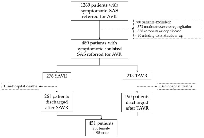
We conducted a two-center retrospective observational study on 451 consecutive patients referred for AVI due to symptomatic severe AS (aortic valve area ≤ 1 cm2 and/or mean gradient > 40 mm Hg) who were discharged from the hospital alive after the intervention between 2013 and 2017. We excluded patients with concurrent moderate or severe valvular regurgitation and significant coronary stenosis on coronary angiography. Both centers are tertiary referral hospitals for cardiology and cardiovascular surgery. Figure 1 shows the flow chart of the patients included.
Figure 1.
Patient inclusion flow chart. SAS: severe aortic stenosis; AVI: aortic valve intervention; SAVR: surgical aortic valve replacement; TAVR: transcatheter aortic valve replacement.
The median follow-up duration was 56 months (IQR: 40–73). The follow-up was conducted through in-person visits at the intervention center. In cases where in-person visits were not feasible, electronic medical records were reviewed, direct contact was established with the patient’s responsible physician or the patient was contacted by phone to collect necessary follow-up data. The study endpoints were the overall all-cause mortality and hospitalization for heart failure, defined as any event requiring hospital admission and intravenous diuretic therapy administration.
Statistical analysis: categorical variables are presented as frequencies, and comparisons between groups were performed using the χ2 or Fisher’s exact test when necessary. Continuous variables are expressed as median (25th–75th IQR). The normal distribution of continuous variables was tested using the Kolmogorov–Smirnov test. Comparisons between groups were performed using Mann–Whitney U. We identified that women had higher rates of admission due to heart failure at follow-up. To assess the influence of sex on heart failure admission, we performed an explanatory multivariable Cox proportional hazard regression with Fine and Gray competing risks using mortality as a competing event and reports estimations as subdistribution hazard ratios (sHR). Kaplan–Meier tests in the absence of competing risk and adjusted cumulative incidence function (CIF) were performed to show the effect on this variable. Possible collinearity among the different introduced variables was evaluated and controlled for overfitting in the model. Verification of proportional hazard assumption was performed by Schoenfeld residuals. All tests were two-sided at the 0.05 significance level. The errors and confidence intervals for the models were calculated using 500 bootstrap samples. All analyses were performed using R software, V.3.6.1 (R Project for Statistical Computing).
3. Results
Table 1 shows the comparison between males and females. At baseline, we identified some epidemiological differences between the two sexes. Women were older and more frequently hypertensive, while men were significantly more likely to be current or former smokers. Additionally, men had higher rates of chronic obstructive pulmonary disease and peripheral arterial disease. We did not identify differences in the proportion of diabetic patients, those with renal insufficiency, or those with atrial fibrillation between the two groups. The clinical manifestations also showed marked differences based on gender. Angina and syncope were significantly more frequent among men. Women exhibited a more advanced dyspnea functional class, although the differences did not reach statistical significance (functional class III–IV 45% vs. 36.5%, p = 0.067). In the baseline echocardiogram, women showed signs of more advanced valvular disease, with a higher mean gradient, a smaller aortic valve area, and higher pulmonary artery systolic pressure values. The type of intervention was TAVR in 47% of women, while only in 35.9% of men, p = 0.017. There were no significant differences in the prescribed treatment between the two sexes. At discharge, 43% of the patients were prescribed ACE inhibitors, 58.3% beta-blockers, and up to 71% received diuretic treatment. At that time, neither neprilysin inhibitors nor sodium-glucose co-transporter 2 inhibitors were part of the therapeutic options. Therefore, it can be considered that the cohort had a fairly optimized treatment for heart failure, as patients with an ejection fraction below 45% were less than 10%. Table 2 shows these data.
Table 1.
Clinical, echocardiographic, and follow-up data of the cohort.
| Total Population (n = 451) |
Female (n = 253) |
Male (n = 198) |
p-Value | |
|---|---|---|---|---|
| Clinical characteristics | ||||
| Age—years | 79.7 [72.0–84.6] | 80.6 [73.4–85.0] | 78.0 [70.4–84.0] | 0.013 |
| TAVR—n (%) | 190 (42.1) | 119 (47.0) | 71 (35.9) | 0.017 |
| BMI | 27.8 [25.3–30.9] | 27.5 [25.0–30.8] | 28.2 [25.8–30.8] | 0.174 |
| BSA | 1.74 [1.62–1.87] | 1.66 [1.54–1.76] | 1.85 [1.75–1.95] | <0.001 |
| Smoker—n (%) | ||||
| No | 337 (74.7) | 233 (92.1) | 104 (52.5) | <0.001 |
| Yes | 25 (5.5) | 3 (1.2) | 22 (11.1) | |
| Former | 89 (19.7) | 17 (6.7) | 72 (36.4) | |
| Hypertension—n (%) | 334 (74.1) | 198 (78.3) | 136 (68.7) | 0.021 |
| Dyslipemia—n (%) | 269 (59.6) | 154 (60.9) | 115 (58.4) | 0.592 |
| Diabetes mellitus—n (%) | 138 (30.6) | 71 (28.1) | 67 (33.8) | 0.187 |
| Peripheral vascular disease—n (%) | 22 (4.9) | 7 (2.8) | 15 (7.6) | 0.018 |
| Atrial fibrillation—n (%) | 124 (27.5) | 68 (27.3) | 56 (29.0) | 0.692 |
| COPD—n (%) | 50 (11.1) | 13 (5.1) | 37 (18.7) | <0.001 |
| Chronic kidney disease—n (%) | 89 (19.7) | 54 (21.3) | 35 (17.7) | 0.332 |
| Previous Stroke/TIA—n (%) | 34 (7.5) | 20 (7.9) | 14 (7.1) | 0.730 |
| Dyspnea on effort—n (%) | 419 (92.9) | 238 (94.1) | 181 (91.4) | 0.275 |
| Heart Failure—n (%) | 131 (29.0) | 78 (30.8) | 53 (26.8) | 0.346 |
| NYHA functional class—n (%) | ||||
| I | 6 (1.4) | 4 (1.7) | 2 (1.1) | 0.134 |
| II | 239 (57.0) | 126 (52.9) | 113 (62.4) | |
| III | 162 (38.7) | 101 (42.4) | 61 (33.7) | |
| IV | 12 (2.9) | 7 (2.9) | 5 (2.8) | |
| NYHA functional class III–IV—n (%) | 174 (41.6) | 108 (45.4) | 66 (36.5) | 0.067 |
| Angina—n (%) | 129 (28.6) | 60 (23.7) | 69 (34.8) | 0.009 |
| CCS class—n (%) | ||||
| 1 | 34 (26.4) | 13 (21.7) | 21 (30.4) | 0.321 |
| 2 | 83 (64.3) | 41 (68.3) | 42 (60.9) | |
| 3 | 12 (9.3) | 6 (10.0) | 6 (8.7) | |
| Syncope—n (%) | 59 (13.1) | 26 (10.3) | 33 (16.7) | 0.046 |
| Type of syncope—n (%) | ||||
| At rest | 24 (40.7) | 13 (50.0) | 11 (33.3) | 0.999 |
| On Exercise | 32 (54.2) | 12 (46.2) | 20 (60.6) | |
| Unknown | 3 (5.1) | 1 (3.8) | 2 (6.1) | |
| Echocardiographic parameters | ||||
| Mean aortic gradient—mmHg | 47.0 [40.0–57.0] | 48.0 [40.0–58.5] | 45.0 [40.0–55.0] | 0.023 |
| Max Velocity—m/seg | 4.40 [4.00–4.80] | 4.46 [4.09–4.85] | 4.36 [4.03–4.80] | 0.123 |
| Valve area—cm2 | 0.70 [0.60–0.90] | 0.70 [0.54–0.85] | 0.74 [0.60–0.94] | 0.002 |
| AVA index cm2/m2 | 0.41 [0.32–0.51] | 0.41 [0.32–0.52] | 0.40 [0.32–0.50] | 0.380 |
| LVEF—% | 60.0 [58.0–65.0] | 62.0 [59.0–66.0] | 60.0 [56.0–65.0] | 0.106 |
| LVEF < 45% | 42 (9.3) | 21 (8.3) | 21 (10.6) | 0.403 |
| EDLVD—mm | 46 [42.0–50.0] | 45.0 [41.0–50.0] | 47.0 [43.0–51.0] | <0.001 |
| ESLVD—mm | 29.0 [25.0–33.0] | 28.0 [24.0–32.0] | 30.0 [27.0–34.0] | <0.001 |
| IVS—mm | 14.0 [13.0–16.0] | 14.0 [13.0–16.0] | 14.0 [13.0–17.0] | 0.126 |
| PW—mm | 12.0 [11.0–14.0] | 12.0 [10.0–13.0] | 12.0 [11.0–14.0] | 0.009 |
| PASP—mmHg | 33.0 [25.0–45.0] | 36.0 [25.0–47.0] | 33.0 [25.0–42.5] | 0.016 |
| TAPSE—mm | 20.0 [18.0–22.0] | 20.0 [18.0–22.0] | 20.0 [19.0–23.0] | 0.066 |
| Blood analysis | ||||
| Hemoglobin—g/dL | 13.1 [11.8–14.1] | 12.8 [11.6–13.6] | 13.6 [12.0–14.7] | <0.001 |
| Creatinine—μmol/L | 0.97 [0.80–1.20] | 0.89 [0.70–1.11] | 1.04 [0.90–1.29] | <0.001 |
| Symptoms at Follow-up | ||||
| Any symptoms at Follow-up | 259 (57.4) | 151 (59.7) | 108 (54.5) | 0.273 |
| Dyspnea—n (%) | 253 (56.1) | 149 (58.9) | 104 (52.5) | 0.176 |
| Angina—n (%) | 9 (2.0) | 4 (1.6) | 5 (2.5) | 0.515 |
| Syncope—n (%) | 19 (4.2) | 12 (4.7) | 7 (3.5) | 0.526 |
| Death—n (%) | 129 (28.6) | 70 (27.7) | 59 (29.8) | 0.619 |
| Cardiovascular death—n (%) | 48 (10.6) | 28 (11.1) | 20 (10.1) | 0.741 |
| Heart Failure Hospitalization—n (%) | 67 (14.8) | 47 (18.6) | 20 (10.1) | 0.012 |
BMI: body mass index. BSA: body surface area. EDLVD: end-diastolic left ventricular diameter; ESLVD: end-systolic left ventricular diameter; IVS: interventricular septum; PW: posterior wall; LVEF: left ventricular ejection fraction; PASP: pulmonary artery systolic pressure; TAPSE: tricuspid annular plane systolic excursion; TIA: transient ischemic attack.
Table 2.
Medication prescribed at discharge.
| Medication Discharge | N | Total Population (n = 451) |
Female (n = 253) |
Male (n = 198) |
p-Value |
|---|---|---|---|---|---|
| ASA—n (%) | 337 | 206 (57.4) | 109 (58.0) | 97 (56.7) | 0.810 |
| Clopidogrel—n (%) | 359 | 56 (16.2) | 33 (18.2) | 23 (13.9) | 0.279 |
| Warfarin—n (%) | 370 | 193 (52.2) | 104 (52.3) | 89 (52.0) | 0.967 |
| DOACs—n (%) | 282 | 13 (4.6) | 7 (4.8) | 6 (4.4) | 0.878 |
| ACE inhibitors—n (%) | 336 | 145 (43.2) | 73 (42.0) | 72 (44.4) | 0.645 |
| Diuretics—n (%) | 336 | 240 (71.4) | 131 (75.3) | 109 (67.3) | 0.105 |
| Betablockers—n (%) | 336 | 196 (58.3) | 106 (60.9) | 90 (55.6) | 0.319 |
| Nitrates—n (%) | 331 | 1 (0.3) | 0 (0.0) | 1 (0.6) | 0.477 |
| Statins—n (%) | 315 | 190 (60.3) | 102 (62.2) | 88 (58.3) | 0.478 |
| Calcium antagonist—n (%) | 307 | 31 (10.1) | 18 (11.2) | 13 (8.9) | 0.509 |
ASA: acetylsalicylic acid. DOACs: direct oral anticoagulants. ACE: angiotensin-converting enzyme.
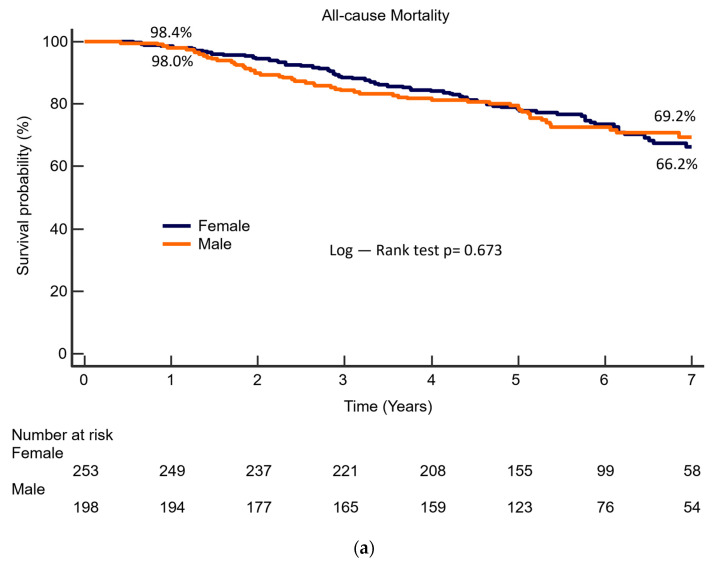
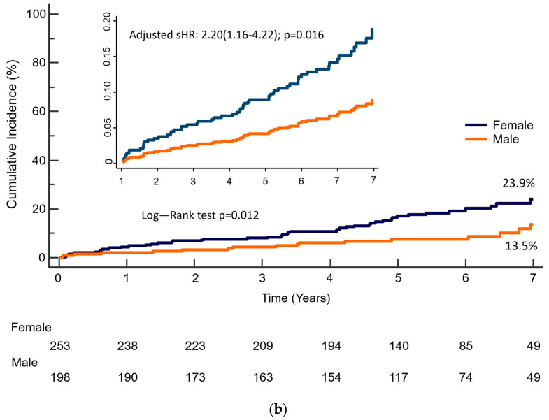
During a 5-year follow-up period, 57% of the patients presented cardiac symptoms after AVI. There were no differences between men and women in the frequency of symptom occurrence. In contrast to what was observed before the intervention, there were no differences in the types of symptoms reported between the two groups. The overall mortality was 28.6%, with cardiovascular mortality at 10.6%, and there were no differences between genders. However, women required hospitalization due to heart failure significantly more often than men (18.6% vs. 10.1%, p = 0.012). No relevant differences were identified between the two centers.
Predictors of Heart Failure Hospitalization during Follow-Up
To better characterize the role of gender in the development of heart failure during follow-up, we conducted a multivariate analysis to identify independent predictors of hospitalization due to heart failure. Table 3 presents the results of this analysis. Female sex is independently associated with hospitalization for heart failure during follow-up (HR 95% CI: 1.16–4.22, p = 0.016), as are age, prior heart failure, body mass index, left ventricular end-diastolic diameter, and a history of atrial fibrillation. The type of intervention (TAVR vs. SAVR) is not independently related to the development of heart failure during follow-up. Figure 2 shows the Kaplan–Meier curve for long-term survival (Figure 2a) and adjusted cumulative incidence estimates for heart failure rehospitalization after AVI (Figure 2b), according to sex.
Table 3.
Univariate and multivariate competing risk analysis of predictors of heart failure admission at follow-up.
| Univariate Analysis | Multivariate Analysis | |||||||
|---|---|---|---|---|---|---|---|---|
| Sub-Hazard Ratio | Bootstrap 95.0% CI | p-Value | Sub-Hazard Ratio | Bootstrap 95.0% CI | p-Value | |||
| Lower | Upper | Lower | Upper | |||||
| Tavi Intervention | 1.87 | 1.20 | 2.92 | 0.005 | ||||
| Female | 2.00 | 1.17 | 3.44 | 0.012 | 2.20 | 1.16 | 4.22 | 0.016 |
| Age | 1.05 | 1.02 | 1.08 | 0.002 | 1.04 | 1.01 | 1.07 | 0.018 |
| Previous Heart Failure | 2.51 | 1.72 | 3.65 | <0.001 | 1.77 | 1.05 | 2.96 | 0.031 |
| BMI | 1.04 | 1.00 | 1.09 | 0.035 | ||||
| Previous AF | 2.92 | 1.79 | 4.77 | <0.001 | 2.99 | 1.93 | 4.65 | <0.001 |
| Baseline EDLVD | 1.03 | 1.01 | 1.06 | 0.013 | 1.04 | 1.01 | 1.08 | 0.014 |
| LVEF | 0.98 | 0.96 | 1.00 | 0.107 | ||||
BMI: body mass index; EDLVD: end-diastolic left ventricular diameter; LVEF: left ventricular ejection fraction.
Figure 2.
(a) Kaplan–Meier survival curve for all-cause mortality after AVI, according to sex. (b) Kaplan-Meier and adjusted cumulative incidence function for heart failure rehospitalization after AVI, according to sex.
4. Discussion
This study follows a contemporary cohort of patients with isolated symptomatic AS referred for AVI to evaluate if there are gender differences in long-term clinical status and prognosis after the intervention. We have found many differences between both sexes at the time of referral: women who undergo AVI are older, present a more advanced valve disease and they show a non-significant tendency to present a more advanced functional class. Men, instead, have more associated comorbidities and complain of syncope and angina more often. These epidemiological and clinical differences are consistent with previous evidence [7].
It has been described that men have a higher indexed myocardial mass, larger ventricular volumes, and a worse ejection fraction for the same degree of stenosis than women [4,6,12,13,14]. We have also identified that men have larger end-diastolic diameters and greater posterior wall thickness, but we have not found differences in ejection fraction between men and women. Although extensively described, the clinical significance of the differences in ventricular volumes and ventricular mass is likely limited, especially when considering that healthy men have higher left ventricular mass and ventricular volumes than women, even when indexed by BSA [15]. Regarding the ejection fraction, all studies reporting a lower ejection fraction in men did not exclude patients with confirmed coronary artery disease. In fact, Petrov et al. reported a series of isolated aortic stenosis cases without associated coronary disease, in which, similar to our series, there were no statistically significant differences in ejection fraction between the two sexes [16].
We have identified clear differences in referral patterns for intervention. At the time of referral, women present higher gradients, smaller aortic valve areas, and higher pulmonary artery pressure. This contrasts with prior evidence based on observational imaging studies, which suggests that women develop symptoms earlier [6] and exhibit lower gradient values and smaller valve areas than men, in part due to their smaller body surface area [12]. The differences we have found are similar to other series obtained at the time of valve replacement [7,14] and suggest that women are referred for valve replacement at a more advanced stage of the disease compared to men. In our series women undergo TAVR more frequently than men. This pattern has been extensively described and it is considered to be multifactorial: women are older, present a smaller body surface area, have higher frailty, and tend to present better outcomes after TAVR [14,17,18]. Moreover, for most of the time when patients in this cohort were referred for AVI, TAVR was recommended mostly for symptomatic AS patients who were deemed unfit for SAVR by the Heart Team. The majority of these patients were elderly, as are the women in this cohort.
Previous studies analyzing sex differences in outcomes after AVI have focused on major end-points such as mortality, cardiovascular mortality, myocardial infarction, or stroke. Observational data have reported heterogeneous outcomes after SAVR [7,18,19,20], but metanalysis shows that women present higher mortality rates compared to men following SAVR [10]. Regarding TAVR, there is a consensus that despite a higher rate of procedural complications, women show a better long-term prognosis after TAVR [9,10,11,17,21,22]. In our series, women undergo TAVR more frequently than men, and we have found no differences in long-term mortality between sexes (female vs. male: 27.7% vs. 29.8%, p = 0.619). There is limited knowledge about the differences in symptoms and quality of life after AVI. We have identified that there are no differences in the clinical status after the intervention. The persistence of symptoms is high in both genders but, unlike our baseline observations, there are no differences between sexes in the reported symptoms. However, we have identified that women are admitted due to heart failure more often than men (18.6% vs. 10.1%, p = 0.012). This may be attributed to the older age of women in the series and their interventions at a more advanced stage, with higher pulmonary pressure values. Female sex was independently associated with heart failure admission in the multivariate analysis. Furthermore, the presence of dyspnea before intervention was already more frequent in women than men in our series. This might be related to diastolic dysfunction, ventricular fibrosis, or irreversible remodeling, factors that would favor heart failure in the follow-up, suggesting that those patients should have been intervened upon earlier.
Despite similar long-term survival rates, the higher rate of hospitalization for heart failure in women is a significant finding. Time spent in the hospital is a major determinant of the quality of life of elder patients and quality of life should be considered one of the primary objectives in patient care, especially in those with a limited life expectancy. In fact, the persistence of symptoms after AVI has been identified as a predictor of patients with poor outcomes [23].
Our study has some limitations. Firstly, it is a retrospective registry, so despite achieving consecutive inclusion, there is a risk of selection bias, as well as challenges in controlling confounding variables and limitations in result generalization. The selection of the endpoints was also retrospective, although mortality and heart failure hospitalization are robust and well-established hard endpoints used in many other studies. Additionally, we have included only patients with isolated aortic stenosis, which is both a strength and a limitation. On one hand, it represents a very homogeneous cohort that helps us better understand the consequences of aortic stenosis per se, but on the other hand, it deviates from the real patient who often presents with coronary disease or other valvular disease.
5. Conclusions
Compared to men, women who survive AVI for severe AS are older, have more severe valve disease, and undergo TAVR more frequently. In follow-up, they do not exhibit higher mortality than men, but they do have a higher need for hospitalization due to heart failure. Our results should encourage further research focused on the impact of sex on the long-term outcomes of AS.
Author Contributions
Conceptualization, T.S., I.V. and J.A.S.R.; methodology, T.S., J.A.S.R. and M.C.-M.; formal analysis, M.C.-M.; data curation, A.R., I.J.A.-S., N.R. and M.C.; writing—original draft preparation, T.S.; writing—review and editing, all authors. All authors have read and agreed to the published version of the manuscript.
Institutional Review Board Statement
The study was conducted in accordance with the Declaration of Helsinki, and approved by the local Ethics Committee (PI 23-3153) for studies involving humans on 23 April 2023.
Informed Consent Statement
Patient consent was waived due to the retrospective nature of the investigation.
Data Availability Statement
The data presented in this study are available on request from the corresponding author. The data are not publicly available due to privacy aspects.
Conflicts of Interest
The authors declare no conflict of interest.
Funding Statement
This research was funded by Gerencia Regional de Salud de Castilla y León, grant number INT/M/11/23.
Footnotes
Disclaimer/Publisher’s Note: The statements, opinions and data contained in all publications are solely those of the individual author(s) and contributor(s) and not of MDPI and/or the editor(s). MDPI and/or the editor(s) disclaim responsibility for any injury to people or property resulting from any ideas, methods, instructions or products referred to in the content.
References
- 1.Osnabrugge R.L., Mylotte D., Head S.J., Van Mieghem N.M., Nkomo V.T., LeReun C.M., Bogers A.J.J.C., Piazza N., Kappetein A.P. Aortic stenosis in the elderly: Disease prevalence and number of candidates for transcatheter aortic valve replacement: A meta-analysis and modeling study. J. Am. Coll. Cardiol. 2013;62:1002–1012. doi: 10.1016/j.jacc.2013.05.015. [DOI] [PubMed] [Google Scholar]
- 2.Shan Y., Pellikka P.A. Aortic stenosis in women. Heart. 2020;106:970–976. doi: 10.1136/heartjnl-2019-315407. [DOI] [PubMed] [Google Scholar]
- 3.Aggarwal S.R., Clavel M.A., Messika-Zeitoun D., Cueff C., Malouf J., Araoz P.A., Mankad R., Michelena H., Vahanian A., Enriquez-Sarano M. Sex differences in aortic valve calcification measured by multidetector computed tomography in aortic stenosis. Circ. Cardiovasc. Imaging. 2013;6:40–47. doi: 10.1161/CIRCIMAGING.112.980052. [DOI] [PubMed] [Google Scholar]
- 4.Dobson L.E., Fairbairn T.A., Musa T.A., Uddin A., Mundie C.A., Swoboda P.P., Ripley D.P., McDiarmid A.K., Erhayiem B., Garg P., et al. Sex-Related differences in left ventricular remodeling in severe aortic stenosis and reverse remodeling after aortic valve replacement: A cardiovascular magnetic resonance study. Am. Heart J. 2016;175:101–111. doi: 10.1016/j.ahj.2016.02.010. [DOI] [PubMed] [Google Scholar]
- 5.Tastet L., Kwiecinski J., Pibarot P., Capoulade R., Everett R.J., Newby D.E., Shen M., Guzzetti E., Arsenault M., Bédard E., et al. Sex-Related differences in the extent of myocardial fibrosis in patients with aortic valve stenosis. JACC Cardiovasc. Imaging. 2020;13:699–711. doi: 10.1016/j.jcmg.2019.06.014. [DOI] [PubMed] [Google Scholar]
- 6.Singh A., Chan D.C.S., Greenwood J.P., Dawson D.K., Sonecki P., Hogrefe K., Kelly D.J., Dhakshinamurthy V., Lang C.C., Khoo J.P., et al. Symptom Onset in Aortic Stenosis: Relation to Sex Differences in Left Ventricular Remodeling. JACC Cardiovasc. Imaging. 2019;12:96–105. doi: 10.1016/j.jcmg.2017.09.019. [DOI] [PubMed] [Google Scholar]
- 7.Fuchs C., Mascherbauer J., Rosenhek R., Pernicka E., Klaar U., Scholten C., Heger M., Wollenek G., Czerny M., Maurer G., et al. Gender differences in clinical presentation and surgical outcome of aortic stenosis. Heart. 2010;96:539–545. doi: 10.1136/hrt.2009.186650. [DOI] [PubMed] [Google Scholar]
- 8.Saeed S., Dweck M.R., Chambers J. Sex differences in aortic stenosis: From pathophysiology to treatment. Expert Rev. Cardiovasc. Ther. 2020;18:65–76. doi: 10.1080/14779072.2020.1732209. [DOI] [PubMed] [Google Scholar]
- 9.O’Connor S.A., Morice M., Gilard M., Leon M.B., Webb J.G., Dvir D., Rodés-Cabau J., Tamburino C., Capodanno D., D’Ascenzo F., et al. Revisiting Sex Equality with Transcatheter Aortic Valve Replacement Outcomes: A Collaborative, Patient-Level Meta-Analysis of 11,310 Patients. J. Am. Coll. Cardiol. 2015;66:221–228. doi: 10.1016/j.jacc.2015.05.024. [DOI] [PubMed] [Google Scholar]
- 10.Parikh P.B., Wang T., Sharma N., Kort S., Skopicki H.A., Gruberg L., Jeremias A., Pyo R., Chikwe J., Butler J. Sex-Related Differences in Early- and Long-Term Mortality After Transcatheter and Surgical Aortic Valve Replacement: A Systematic Review and Meta-Analysis. J. Invasive Cardiol. 2020;32:295–301. doi: 10.25270/jic/19.00371. [DOI] [PubMed] [Google Scholar]
- 11.Saad M., Nairooz R., Pothineni N.V.K., Almomani A., Kovelamudi S., Sardar P., Katz M., Abdel-Wahab M., Bangalore S., Kleiman N.S., et al. Long-Term Outcomes with Transcatheter Aortic Valve Replacement in Women Compared with Men: Evidence from a Meta-Analysis. JACC Cardiovasc. Interv. 2018;11:24–35. doi: 10.1016/j.jcin.2017.08.015. [DOI] [PubMed] [Google Scholar]
- 12.Ito S., Miranda W.R., Nkomo V.T., Lewis B.R., Oh J.K. Sex Differences in LV Remodeling and Hemodynamics in Aortic Stenosis: Sex-Specific Criteria for Severe Stenosis? JACC Cardiovasc. Imaging. 2022;15:1175–1189. doi: 10.1016/j.jcmg.2022.02.007. [DOI] [PubMed] [Google Scholar]
- 13.Treibel T.A., Kozor R., Fontana M., Torlasco C., Reant P., Badiani S., Espinoza M., Yap J., Diez J., Hughes A.D., et al. Sex Dimorphism in the Myocardial Response to Aortic Stenosis. JACC Cardiovasc. Imaging. 2018;11:962–973. doi: 10.1016/j.jcmg.2017.08.025. [DOI] [PMC free article] [PubMed] [Google Scholar]
- 14.Singh A., Musa T.A., Treibel T.A., Vassiliou V.S., Captur G., Chin C., Dobson L.E., Pica S., Loudon M., Malley T., et al. Sex differences in left ventricular remodelling, myocardial fibrosis and mortality after aortic valve replacement. Heart. 2019;105:1818–1824. doi: 10.1136/heartjnl-2019-314987. [DOI] [PMC free article] [PubMed] [Google Scholar]
- 15.Petersen S.E., Khanji M.Y., Plein S., Lancellotti P., Bucciarelli-Ducci C. European Association of Cardiovascular Imaging expert consensus paper: A comprehensive review of cardiovascular magnetic resonance normal values of cardiac chamber size and aortic root in adults and recommendations for grading severity. Eur. Heart J. Cardiovasc. Imaging. 2019;20:1321–1331. doi: 10.1093/ehjci/jez232. [DOI] [PubMed] [Google Scholar]
- 16.Petrov G., Dworatzek E., Schulze T.M., Dandel M., Kararigas G., Mahmoodzadeh S., Knosalla C., Hetzer R., Regitz-Zagrosek V. Maladaptive remodeling is associated with impaired survival in women but not in men after aortic valve replacement. JACC Cardiovasc. Imaging. 2014;7:1073–1080. doi: 10.1016/j.jcmg.2014.06.017. [DOI] [PubMed] [Google Scholar]
- 17.Vlastra W., Chandrasekhar J., García Del Blanco B., Tchétché D., de Brito F.S., Jr., Barbanti M., Kornowski R., Latib A., D’Onofrio A., Ribichini F., et al. Sex differences in transfemoral transcatheter aortic valve replacement. J. Am. Coll. Cardiol. 2019;74:2758–2767. doi: 10.1016/j.jacc.2019.09.015. [DOI] [PubMed] [Google Scholar]
- 18.Chaker Z., Badhwar V., Alqahtani F., Aljohani S., Zack C.J., Holmes D.R., Rihal C.S., Alkhouli M. Sex Differences in the Utilization and Outcomes of Surgical Aortic Valve Replacement for Severe Aortic Stenosis. J. Am. Heart Assoc. 2017;6:e006370. doi: 10.1161/JAHA.117.006370. [DOI] [PMC free article] [PubMed] [Google Scholar]
- 19.O’Brien S.M., Shahian D.M., Filardo G., Ferraris V.A., Haan C.K., Rich J.B., Normand S.L., DeLong E.R., Shewan C.M., Dokholyan R.S., et al. The Society of Thoracic Surgeons 2008 cardiac surgery risk models: Part 2—Isolated valve surgery. Ann. Thorac. Surg. 2009;88:S23–S42. doi: 10.1016/j.athoracsur.2009.05.056. [DOI] [PubMed] [Google Scholar]
- 20.Kulik A., Lam B.K., Rubens F.D., Hendry P.J., Masters R.G., Goldstein W., Bédard P., Mesana T.G., Ruel M. Gender differences in the long-term outcomes after valve replacement surgery. Heart. 2009;95:318–326. doi: 10.1136/hrt.2008.146688. [DOI] [PubMed] [Google Scholar]
- 21.Chandrasekhar J., Dangas G., Yu J., Vemulapalli S., Suchindran S., Vora A.N., Baber U., Mehran R. Sex-Based differences in outcomes with transcatheter aortic valve therapy: TVT registry from 2011 to 2014. J. Am. Coll. Cardiol. 2016;68:2733–2744. doi: 10.1016/j.jacc.2016.10.041. [DOI] [PubMed] [Google Scholar]
- 22.Doshi R., Shlofmitz E., Meraj P. Comparison of outcomes and complications of transcatheter aortic valve implantation in women versus men (from the National inpatient sample) Am. J. Cardiol. 2018;121:73–77. doi: 10.1016/j.amjcard.2017.09.015. [DOI] [PubMed] [Google Scholar]
- 23.San Román J.A., García-Gómez M., Ybarra-Falcón C., Ramos N., Nombela L., Carnero M., Amat-Santos I.J., Sevilla T., Revilla A., Carrasco-Moraleja M., et al. Evolution and prognostic significance of patient-reported symptoms after intervention in severe aortic stenosis. Mayo Clin. Proc. 2023 doi: 10.1016/j.mayocp.2023.09.024. in press . [DOI] [PubMed] [Google Scholar]
Associated Data
This section collects any data citations, data availability statements, or supplementary materials included in this article.
Data Availability Statement
The data presented in this study are available on request from the corresponding author. The data are not publicly available due to privacy aspects.


