Abstract
Xylella fastidiosa is the causal agent of several plant diseases affecting fruit and nut crops. Methylobacterium mesophilicum strain SR1.6/6 was isolated from Citrus sinensis and shown to promote plant growth by producing phytohormones, providing nutrients, inhibiting X. fastidiosa, and preventing Citrus Variegated Chlorosis. However, the molecular mechanisms involved in the interaction among these microbes are still unclear. The present work aimed to analyze physiological and molecular aspects of M. mesophilicum SR1.6/6 and X. fastidiosa 9a5c in co-culture. The transcriptome and secretome analyses indicated that X. fastidiosa down-regulates cell division and transport genes and up-regulates stress via induction of chaperones and pathogenicity-related genes including, the lipase-esterase LesA, a protease, as well as an oligopeptidase in response to M. mesophilicum competition. On the other hand, M. mesophilicum also down-regulated transport genes, except for iron uptake, which was up-regulated. Secretome analysis identified four proteins in M. mesophilicum exclusively produced in co-culture with X. fastidiosa, among these, three are related to phosphorous uptake. These results suggest that M. mesophilicum inhibits X. fastidiosa growth mainly due to nutrient competition for iron and phosphorous, thus promoting X. fastidiosa starvation, besides producing enzymes that degrade X. fastidiosa cell wall, mainly hydrolases. The understanding of these interactions provides a direction for control and management of the phytopathogen X. fastidiosa, and consequently, helps to improve citrus growth and productivity.
Keywords: endophytic bacteria, citrus, phytopathogen–endophyte interaction, transcriptome, secretome
1. Introduction
Xylella fastidiosa (Xf) is a Gram-negative phytopathogenic bacterium (γ-Proteobacteria) found exclusively in the lumen of xylem vessels and is unable to colonize other plant tissues [1,2]. The main diseases caused by Xf are Citrus Variegated Chlorosis (CVC) [3], Pierce’s Disease in grapevines (PD) [4], and the Olive Quick Decline Syndrome (OQDS), which is an emerging disease in Europe [5,6]. Other diseases associated with Xf infection are denominated leaf scorch, which occurs in plum trees (PLS-plum), almond (ALS), coffee trees (CLS), peach (PLS-peach), oleander (OLS), mulberry, cherry trees, oak, elderberry, and hibiscus, among others [4,7,8]. This phytopathogen is transmitted by insect vectors from the Cicadellidae and Cercopidae families that feed on xylem sap [7,9].
Genomic and transcriptomic studies with X. fastidiosa have provided insights related to the biology of this phytopathogen, including the identification of genes potentially associated with virulence and pathogenicity [10,11,12,13,14,15,16]. These studies have been complemented with the description of proteomes and secretomes of Xf in different conditions [10,17,18,19,20]. Taken together these studies provided a detailed picture of Xf pathogenesis, including the effect of population density on the expression of virulence factors. A key mechanism in Xf pathogenesis is quorum sensing, which in Xanthomonadaceae is mediated by diffusible signaling factors (DSFs) [7,21]. DSFs can be detected in the bacterial surroundings both outside and associated with outer membrane vesicles (OMVs). OMVs also carry many proteins that can modify their microenvironments favoring bacterial survival, such as adhesins, lipases, proteases, and other hydrolases [19,22,23,24,25].
Proteomic analysis demonstrated that lipases/esterases are very abundant in the secretome of Xf strain Temecula1 [20]. The most abundant lipase, LesA (PD_1703/XFTem_01966) appears to act in the degradation of plant tissue, and its accumulation in leaf regions has been associated with the symptoms of Pierce’s disease of grapevines. A secretome study of Xf aiming to compare in vitro culture of the strains 9a5c (virulent in orange trees) and J1a12 (non-virulent) [19] revealed a distinct profile of extracellular proteins from both strains, totaling 71 different proteins, including the detection of LesA lipase-esterase (XF_0781/XF9a_00715) mainly in the virulent strain. One of the secretome proteins was the XfYgiT antitoxin (XF_2491/XF9a_02352), which in strain 9a5c is secreted inside OMVs [24] and is associated with biofilm formation, persistent cell generation, and decreased pathogenicity [26].
Inside the plant, Xf interacts with other microorganisms and reshapes microbial composition [27]. In citrus, it was reported that Methylobacterium species are dominant as endophytes within branches and interact with Xf [28,29,30]. The authors suggest that the presence of some endophytic Methylobacterium species in asymptomatic citrus tissues of plants with Xf could stimulate the production of compounds or elicitors that somehow increase plant resistance against Xf or decrease the phytopathogen growth [29,30]. In vitro and in planta experiments have shown that Methylobacterium mesophilicum (Mm) reduces Xf growth [29,31] which could be associated with the control of this bacterium in the host plant. Microarray analysis showed that Xf in the presence of Mm up-regulates genes related to stress and down-regulates genes related to bacterial growth [30]. Moreover, Mm is able to colonize not only different plant organs such as roots and leaves but also the lumen of xylem vessels, the same environment colonized by Xf [32], as well as the foregut of Bucephalogonia xanthopis [33] and Homalodisca vitripennis [34], both insect vectors associated to Xf transmission. All these data indicate that Mm and Xf could interact in different microenvironments during transmission and plant colonization.
Methylobacterium species have been isolated from more than 70 plant hosts [35] inducing plant growth promotion in citrus [36] and other plant species [37,38,39,40,41]. This effect on plant growth is due to the production of auxin and cytokinin phytohormones [41,42], induction of photosynthetic activity [43], and induction of systemic plant resistance by synthesizing pectinase and cellulase [37,44]. Members of this genus present a pink pigmentation due to carotenoid production [45] and can metabolize compounds with only one carbon such as methanol and methylamine [46] and are accordingly named Pink-Pigmented Facultative Methylotrophics (PPFM) bacteria.
In Methylobacterium mesophilicum (Mm), some key genes in bacterium-plant interactions were identified using quantitative PCR (qPCR) in rice and eucalyptus [47], and via transcriptomic analyses in soybean [48]. These studies showed that plant exudates up-regulate several genes involved in transport and stress, mainly with antioxidant functions. These results show how bacterial gene expression is regulated during plant-bacterium colonization, allowing the establishment of the interaction with the host plant and the associated microbiome.
Considering that the battle for ecological niches, including competition for habitat and resources, is a key challenge that bacteria face inside the host plant, different strategies to respond to competitors may have evolved in these microorganisms: direct responses (bactericide molecules) and/or indirect mechanisms (nutrient competition). Therefore, the main goal of the present work was to understand if the patterns of competitive and/or inhibitory phenotypes among Mm SR1.6/6 and Xf 9a5c populations in vitro could explain their interaction outcome inside the host plant. To do so, growth, transcriptome, and secretome were evaluated during the co-culture of these bacteria. This investigation provides valuable insights into the mechanism involved in the establishment of such a population inside the host plant and sheds light on Xf gene expression modulation in response to endophytic Mm, warranting more studies focused on the role of polymicrobial bacterial communities in the control/management of diseases caused by Xf.
2. Materials and Methods
2.1. Xf and Mm Cultivation Conditions
Experiments were performed with the endophyte Mm SR1.6/6 [28] and the phytopathogen Xf 9a5c [49], both isolated from Citrus sinensis (L.) Osbeck. These bacteria were grown in PW broth [50] supplemented with 0.5% glucose (PWG broth) at 28 °C under rotation of 170 rpm. The cultivation in solid medium was performed in PWG-1.5% agar. Cell stocks were maintained in PWG containing 50% of glycerol in a −80 °C freezer.
2.2. Bacterial Growth Evaluation
Bacterial growth was measured using OD600nm in a NanoDrop 2000c spectrophotometer (ThermoFisher Scientific, Waltham, MA, USA). Mm SR1.6/6 and Xf 9a5c were cultured separately in 7.5 mL of PWG broth with an initial OD600nm of 0.05. After achieving OD600nm of 0.5, which was approximately 7 days for Xf and 2 days for Mm, 2.5 mL of each growth culture were aliquoted and Xf and Mm were mixed, totalizing a volume of 5 mL to each treatment: 1. Control: only Xf; 2. Control: only Mm; 3. Treatment: Xf+Mm co-culture. All cultures were then incubated at 28 °C under rotation of 170 rpm for 24 h, 48 h, and 72 h and OD600nm was measured. Three biological replicates were performed.
2.3. Transcriptomic Analyses
2.3.1. Experimental Design
Transcriptomes were evaluated for Xf and Mm monocultures and during co-culture. The two bacteria were cultivated separately in 75 mL of PWG, with an initial OD600nm of 0.05. After achieving OD600nm of 0.5, which was 7 days for Xf and 2 days for Mm, 25 mL of each culture were aliquoted and Xf and Mm were mixed, totaling 50 mL for controls (monoculture) and treatment (co-culture): 1. Control: only Xf; 2. Control: only Mm; 3. Treatment: Xf+Mm co-culture. All cultures were incubated at 28 °C and 170 rpm agitation for a period of 24 h. After this period, 40 mL of culture was centrifuged, and cell pellet was collected for RNA extraction followed by RNA-Seq. For OD600nm measurements and DNA extraction for bacterial quantification by qPCR, culture aliquots were taken before mixing Xf and Mm cultures and after the co-culture period. All experiments were performed in biological triplicates, starting from independent cultures.
2.3.2. qPCR Quantification of Xf and Mm
DNA was isolated using Wizard Genomic DNA Purification kit (Promega, Madison, WI, USA) according to the manufacturer’s recommendation. The qPCR amplification was performed in a StepOne Plus thermocycler (Applied Biosystems, Foster City, CA, USA) programmed to an initial denaturation of 95 °C for 3 min, followed by 40 cycles of 95 °C for 15 s and 60 °C for 30 s. The following primers were used: MMC1 (5′TACGTGGAGAGATTCACGGTC′3) and MMC2 (5′GTACAAGGCCCGGGAACGTAC′3) to quantify Mm SR1.6/6 [31]; and CVC-1 (5′AGATGAAAACAATCATCGAAA′3) and 272-2-int (5′GCCGCTTCGGAGAGCATTCCT′3) to quantify Xf 9a5c [51]. The amplification reaction had a final volume of 20 μL, with 2 μL of DNA (50 ng) and SYBR Master mix (Applied Biosystems, Foster City, CA, USA). Melting curves were analyzed in the PCR reaction to evaluate primer specificity using a temperature gradient from 72 °C to 96 °C, varying 0.5 °C every 15 s.
2.3.3. Total RNA Isolation, cDNA Library Preparation, and Sequencing
Monocultures of Xf and Mm and co-culture of Xf and Mm collected after 24 h at 28 °C were centrifuged at 3220× g for 5 min at 4 °C. Supernatants were separated for proteomic analysis and cell pellets were used for RNA extraction. To disrupt bacterial cells, pellets were macerated with liquid nitrogen until they became a fine powder and subjected to total RNA isolation using TRIzol reagent (Invitrogen, Carlsbad, CA, USA) and Purelink RNA Mini Kit (Ambion, Foster City, CA, USA) according to instructions. The RNA samples were eluted in DEPC-treated water and stored at −80 °C freezer. The quantification and sample purity analyses were obtained in a NanoDrop 2000c spectrophotometer (ThermoFisher Scientific, Waltham, MA, USA). Integrity analyses were performed using 2100 BioAnalyzer and the RNA 6000 Nano kit (Agilent Technologies, Santa Clara, CA, USA). RIN (RNA Integrity Number) values above 7.0 were obtained and considered suitable for proceeding to the RNA-Seq analyses.
Total RNA samples were treated with DNase provided by the Illustra RNASpin Mini RNA isolation kit (GE Healthcare, Little Chalfont, UK), following instructions, although using double enzyme solution volume. Total RNA samples treated with DNase showed concentrations varying between 133 ng/μL to 275 ng/μL. Conventional PCR was performed with primers for a region of 16S rRNA gene [52]: S-D-Bact-0341-b-S-17 (5′CCTACGGGNGGCWGCAG3′) and S-D-Bact-0785-a-A-21 (5′GACTACHVGGGTATCTAATCC3′) and showed no residual genomic DNA (gDNA) in the RNA samples. RNA integrity analyses performed after DNase treatment to ensure the maintenance of high RIN values. Before initiating the library construction, RNA was quantified using Quant-iT RiboGreen RNA Assay kit (ThermoFisher Scientific, Waltham, MA, USA), and rRNA depletion was performed using Ribo-Zero Magnetic kit (Bacteria) (Illumina, San Diego, CA, USA). The depletion of rRNA molecules was evaluated through 2100 BioAnalyzer and the RNA 6000 Nano kit (Agilent Technologies, Santa Clara, CA, USA) and was considered satisfactory. cDNA libraries were constructed using TruSeq RNA sample preparation kit v2 (Illumina, San Diego, CA, USA). Libraries were analyzed through 2100 BioAnalyzer using a High Sensitivity DNA kit (Agilent Technologies, Santa Clara, CA, EUA) to determine the average fragment size. Libraries were quantified spectrophotometrically (Table S1) and using absolute qPCR in a 7500 Real-Time PCR System (Applied Biosystems, Foster City, CA, USA) with the Kapa Library Quantification kit for Illumina (Kapa Biosystems). All libraries were normalized to 4 nM, pooled to specific index combinations, denatured with NaOH 0.2 N, and incubated at 96 °C. Sequencing of cDNA libraries was performed in MiSeq equipment (Illumina, San Diego, CA, USA) with a final pool concentration of 8 pM (first run) and 10 pM (second and third run). All runs were performed with the MiSeq Reagent Kit v2 of 500 cycles with Paired-End 2 × 250 sequencing strategy. Cluster densities ranged from 676 to 882 K/mm2. The number and quality of reads per library was considered satisfactory (Table S2). The MiSeq equipment is located at the Center for Advanced Technologies in Genomics (CATG), Chemistry Institute, University of São Paulo, Brazil.
2.3.4. Bioinformatic Analyses and Statistics
Read quality was assessed based on QScore using FASTQC software v0.11.6 [53]. Then, the reads were mapped to their respective reference genomes using the RNA-Seq analysis module of CLC Genomics Workbench 6.5 software (Qiagen, Germantown, USA). Reference genomes of Xf (9a5c) and Mm (SR1.6/6) were downloaded from the Genbank-NCBI using the assembly accession GCA_000006725 and GCA_000364445, respectively. Mapping was performed in two ways: generating normalized expression values of FPKM (Fragments Per Kilobase Per Million Mapped Fragments) [54] and generating gross counting values of how many reads were mapped to each gene. One criterion for counting mapped reads was the selection of those mapping only in one region of the genome, therefore duplicated regions in the genome were not considered to avoid error chances. FPKM values were used to calculate the Pearson correlation between biological replicates, while gross counting values to differential gene expression analysis between monocultures and co-culture transcriptomes. These analyses were performed using the R software package DESeq2 [55], considering genes with padj <0.1 and a log2FoldChange ≠ 0 as differentially expressed.
The sequences of differentially expressed genes (DEGs) encoding a protein were translated and used by Blast2GO software [56] to categorize them in their Gene Ontology terms (GO terms) [57]. Finally, Gene Ontology 2nd Level, a custom Python script available at https://oseias-r-junior.github.io/Gene_Ontology_2nd_Level (accessed on 15 March 2019), was used to retrieve the GO terms (at 2nd level) from queries provided by Blast2GO.
2.4. Secretomic Analyses
2.4.1. Protein Extraction from Xf, Mm, and Co-Culture Supernatants
Supernatants of monoculture of Xf and Mm and co-culture of Xf and Mm from the transcriptomic experiments (Section 2.3.3) were collected, centrifuged at 3220× g for 30 min to remove the remaining bacterial cells and further concentrated 100-fold by ultrafiltration in Amicon Ultra 15 mL (Millipore) devices with Molecular Weight Cutoff (MWCO) of 3 kDa. Concentrated supernatants were sonicated (two pulses of 15 s with 30 s intervals under 4 °C) on Branson Sonifier 450 (Marshall Scientific, Hampton, NH, USA) and then performed a second centrifugation for 30 min at 12,000× g and 4 °C to remove remaining insoluble fraction. Total protein in the concentrated extracts was measured with the Bradford method.
The concentrated extracts obtained from supernatants of three biological replicates of Xf and Mm monocultures and Xf and Mm co-culture were pooled into single samples for each treatment, which in turn were mixed in the ratio 1:8:1 with the following solutions: 1 mL of the concentrated extract; 8 mL of ice-cold acetone (for HPLC, ≥99.9% purity) and 1 mL of trichloroacetic acid (100% TCA). Samples were maintained at −20 °C for 1 h until complete precipitation and then centrifuged at 18,000× g for 15 min at 4 °C. The supernatant was discarded, and the precipitate was washed with 1 mL of ice-cold acetone with complete resuspension and further centrifugation at 18,000 g for 15 min at 4 °C. Finally, after aspiration of the acetone, the precipitate was dried at room temperature and maintained at −20 °C until proteomic analysis. The total protein content of supernatant extracts was determined using Bradford reagent (Bio-Rad Laboratories Inc., Hercules, CA, USA), resulting in the following concentrations: 8.54 μg/μL for Xf monoculture, 16.11 μg/μL for Mm monoculture, and 9.03 μg/μL for the co-culture of Xf and Mm.
2.4.2. Shotgun Proteomics
Proteomic analysis of extracted supernatants was performed as described previously [22,28], using a pool of three biological replicates from purified supernatants of Xf and Mm monocultures or co-cultures. The dried samples were reconstituted in PBS buffer and 300 μg of each sample were precipitated with 4 times the volume of ProteoExtract™ Protein Precipitation Kit (Calbiochem, San Diego, CA, USA) according to instructions. The samples were reconstituted in 100 μL of 6 M urea in 50 mM triethylammonium bicarbonate (TEAB) plus 5 mM dithiothreitol (DTT) and incubated at 37 °C for 30 min and shaking at 1000 rpm. Next, 15 mM iodoacetamide (IAA) was added, followed by incubation at room temperature for 30 min. The IAA was then neutralized with 30 mM DTT and incubated for 10 min. Lys-C/trypsin was added (1:25, enzyme: total protein) followed by incubation at 37 °C for 4 h. TEAB (550 μL of 50 mM) was added to dilute the urea and activate trypsin digestion overnight. The digested peptides were desalted with Aspire RP30 Desalting Tips (Thermo Scientific, Waltham, MA, USA), vacuum dried, and suspended in 45 μL of 50 mM TEAB. Peptides were quantified by Pierce quantitative fluorometric assay (Thermo Scientific) and 1 μg was analyzed on a QExactive mass spectrometer (Thermo Scientific) coupled with an Easy-LC source (Thermo Scientific) and a nanospray ionization source. The peptides were loaded onto a Trap (100 microns, C18 100 Å 5U) and desalted online prior to separation using a reversed-phase (75 microns, C18 200 Å 3U) column. The duration of the peptide separation gradient was 60 min using 0.1% formic acid and 100% acetonitrile (ACN) for solvents A and B, respectively. The data were acquired using a data-dependent MS/MS method, which had a full scan range of 300–1600 Da and a resolution of 70,000 Da. The resolution of the MS/MS method was 17,500 Da and the insulation width was 2 m/z with normalized collision energy. The nanospray source was operated using a spray voltage of 2.2 KV and a transfer capillary temperature heated to 250 °C.
The raw data were analyzed using X!Tandem and viewed using the Scaffold Proteome Software version 4.0 (Proteome Software Inc., Portland, OR, USA) [58]. Samples were searched against UniProt databases appended with the cRAP database, which recognizes common laboratory contaminants. Reverse decoy databases were also applied to the database before the X!Tandem searches. The annotated genomes of Xf 9a5c and Mm SR1.6/6 were used as references for identification of the proteins. The proteins identified were filtered (in the Scaffold Proteome Software) based on the following criteria: 1.0% FDR (False Discovery Rate) at protein level (following the prophet algorithm: http://proteinprophet.sourceforge.net/ (accessed on 11 September 2023)), minimum number of 2 peptides and 0.1% FDR at peptide level. Protein sequences identified were examined for the presence of a signal peptide [59] and the potential (SecP score) of being secreted by a non-classical protein secretion system [60]. Similar to the transcriptomic analysis, the tool Gene Ontology 2nd Level was used to retrieve the GO terms from queries provided by Blast2GO software.
2.5. Integration of Transcriptomics and Proteomics Analyses
For integration of transcriptomics and proteomics analyses, FPKM values were converted to TPM (Transcripts Per Kilobase Million) values, as reported in the literature [61], and only nonzero TPM values were considered. Further, the means of the three replicates TPM values were calculated. In turn, proteomic data available as spectra counts were also normalized. The spectra counts were divided by respective protein size (NC = SC/PS×1000). Next, the normalized counts (NC) were divided by the secretome size (PPT = NCi/∑NC×1000) retrieving a final normalized number of called peptides per thousand (PPT). Secretome samples were from a pool of three biological replicates. Following this, a Venn diagram was obtained with the Venn Python library to assign intersection gene products detected in transcriptome and secretome [62]. Euclidean distances were calculated between Xf or Mm co-culture versus their respective monoculture common gene products. The Mantel test was used to calculate the correlation between two matrices [63] either transcriptome or secretome data. The Mantel test was performed using a MantelTest Phyton library which returns a correlation coefficient, p-value, and a standard score (z-score). Further, for functional analysis, genes with similar trends (i.e., up- or down-regulated) in both transcriptome and secretome were subjected to GO terms annotation. These GO terms were used to build a GO enrichment network using the Cytoscape software v3.10.1 [64].
3. Results
3.1. Growth of Mm and Xf in Co-Culture and Xf Biofilm Formation
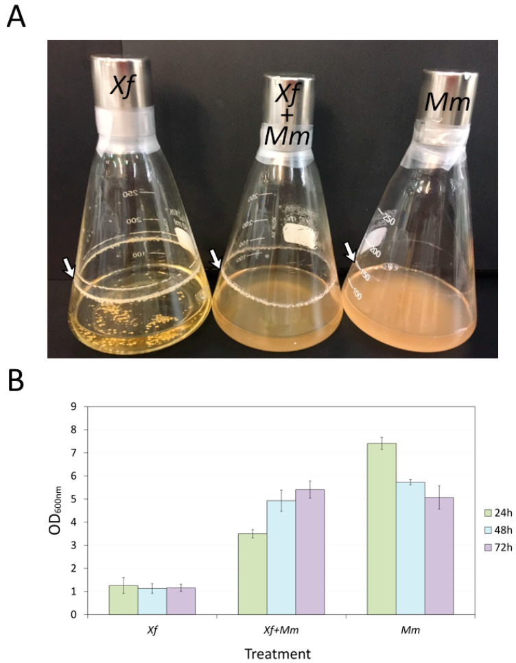
An initial experiment was performed to evaluate endophyte and pathogen growth in monoculture and co-culture for 24 h, 48 h, and 72 h in PWG broth (Figure 1) to investigate the interaction between these bacteria isolated from citrus. Figure 1A shows biofilm formation in the three cultures Xf, Mm, and co-culture, with Xf forming a thicker biofilm. The results also show that Xf monoculture density was stable over the time evaluated, while that of the co-culture (Xf+Mm) increased, and Mm monoculture decreased after 24 h (Figure 1B).
Figure 1.
Phenotypic and growth evaluation of Xf and Mm monocultures and co-culture. (A) Phenotypic growth and biofilm formation (indicated by the white arrow) of Xf, Xf+Mm, and Mm after 72 h of culture. (B) Quantification of bacteria growth. Measurements of OD600nm of each treatment before and after co-culture. Measurements were performed after 24 h, 48 h, and 72 h of culture. Xf: X. fastidiosa 9a5c; Xf+Mm: X. fastidiosa 9a5c and M. mesophilicum SR1.6/6; Mm: M. mesophilicum SR1.6/6.
3.2. Bacteria Quantification by qPCR
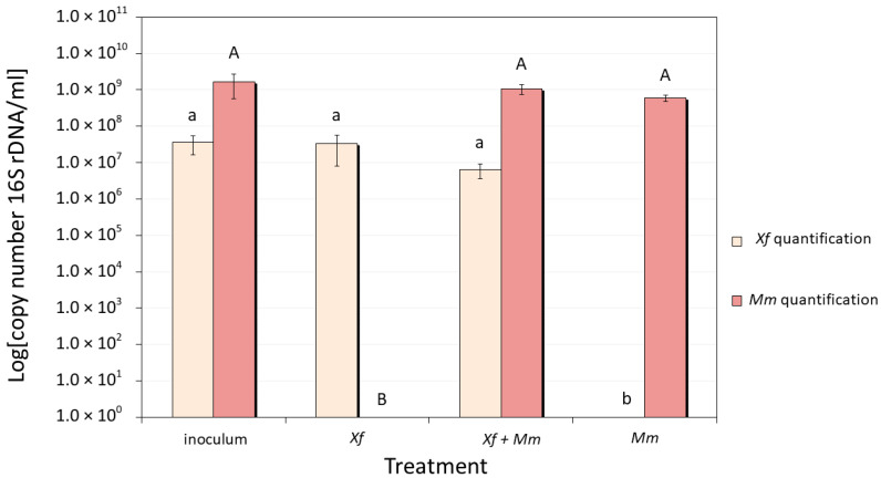
Aliquots of bacterial cultures for RNA-Seq analyses were also used to isolate DNA to quantify Mm and Xf gene copy numbers. The initial inoculum of Xf and Mm present similar growth verified by the OD600nm measurement. After 24 h, in control cultures (Xf or Mm monocultures) and co-culture (Xf and Mm), quantification showed no significant alterations in the number of cells for both Mm and Xf when comparing with the inoculum (Figure 2), contributing to reduced transcriptomic analyses variation.
Figure 2.
Quantification by qPCR of the number of copies of specific genes in aliquots of bacterial cells used in RNA-Seq analyses (after 24 h of co-culture). M. mesophilicum SR1.6/6 quantification using the pair of primers MMC1/MMC2 [31]; and X. fastidiosa 9a5c quantification using the pair of primers CVC-1/272-2-int [51]. Xf: X. fastidiosa 9a5c monoculture; Xf+Mm: X. fastidiosa 9a5c and M. mesophilicum SR1.6/6 co-culture; Mm: M. mesophilicum SR1.6/6 monoculture. Statistical analyses were performed using the Duncan test. Capital letters show statistical differences when comparing Mm quantification and lower-case letters when comparing Xf quantification, different letters indicate statistically significant different mean.
3.3. Transcriptomic Analyses
A total of 4,400,994 reads of Xf in monoculture, 3,981,063 reads of Mm in monoculture, and 4,408,696 reads of Xf+Mm co-culture, all of them after 24 h incubation, were obtained from cDNA library sequencing. The reads were mapped on the reference Xf 9a5c and Mm SR1.6/6 genomes, which present 2,679,305 bp (2708 genes including plasmids) and 6,214,449 bp (5899 genes), respectively. The average percentage of uniquely mapped reads was high in both Xf (80.16%) and Mm (71.45%) monoculture, while in the co-cultures the percentage was lower (51.05% in Xf and 26.41% in Mm) (Table 1 and Table S3).
Table 1.
Average number of mapped reads in Xf and Mm using CLC Genomics Workbench software. Xf: X. fastidiosa; Mm: M. mesophilicum.
| Treatment | Number of Reads | Mapped Reads in Pairs | % Mapped Reads | Unique Fragments |
|---|---|---|---|---|
| Xf monoculture | 4,400,994 | 3,523,819 | 80.16 | 1,759,574 |
| Co-culture mapped in Xf genome | 4,410,865 | 2,284,762 | 51.05 | 1,140,964 |
| Co-culture mapped in Mm genome | 4,410,865 | 1,158,502 | 26.41 | 578,282 |
| Mm monoculture | 3,981,063 | 2,892,542 | 71.45 | 1,444,234 |
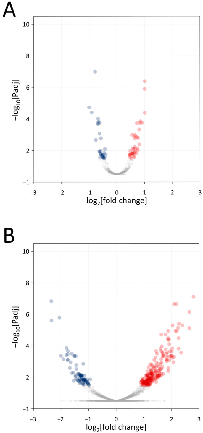
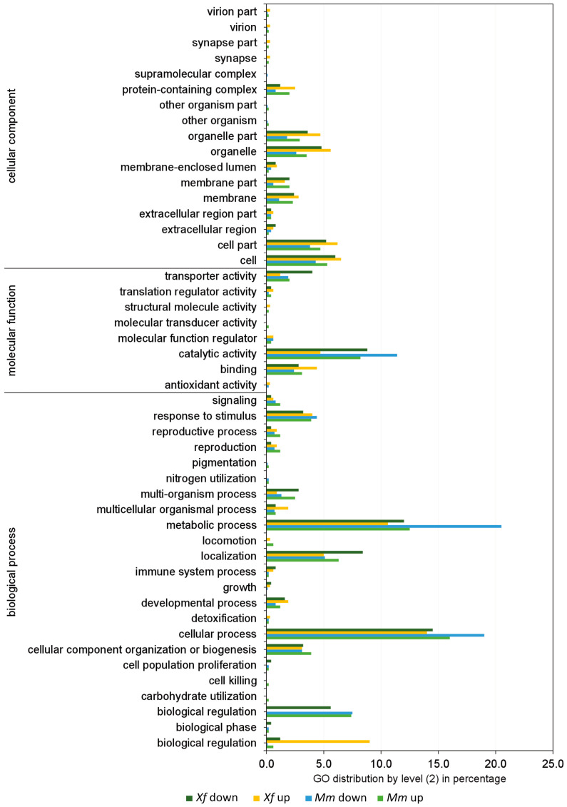
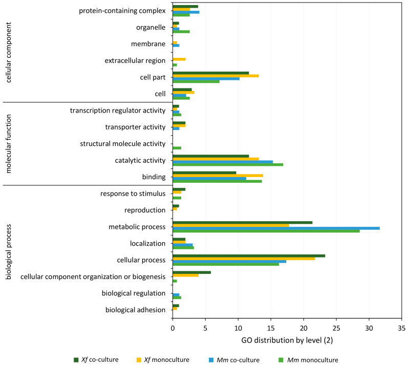
We identified 68 differentially expressed genes (DEGs) in Xf and 288 DEGs in Mm monoculture in comparison with Xf and Mm co-culture (Figure 3). The complete lists of differentially expressed genes for Xf and Mm are shown in Supplementary Tables S4 and S5, respectively. The DEGs identified in monocultures of Xf and Mm and co-culture treatments were annotated according to the Gene Ontology (GO) (Figure 3 and Figure 4). We highlight the presence of DEGs (both up- and down-regulated) related to the categories “molecular function” (catalytic activity, transport activity, and binding), and “biological process” (cellular and metabolic process, biological regulation, and response to stimulus) in both Xf and Mm cultures (Figure 3 and Figure 4).
Figure 3.
Volcano plots of differentially expressed genes. (A). Results of the comparison between the Xf+Mm co-culture with the control (Xf monoculture) (B). Results of the comparison between the Xf+Mm co-culture with the control (Mm monoculture). In the x-axis are the log2fold change values; in the y-axis are the −log10 adjusted p-values (padj). Points in red and blue represent genes with padj < 0.1, and thus, considered statistically differentially expressed; red dots represent genes up-regulated, while blue dots represent genes down-regulated. Xf: X. fastidiosa 9a5c; Xf+Mm: X. fastidiosa 9a5c and M. mesophilicum SR1.6/6; Mm: M. mesophilicum SR1.6/6.
Figure 4.
Frequency (%) of the categories of differentially expressed genes (up- and down-regulated) in co-culture generated by Blast2GO software. Xf down: genes down-regulated in Xf in the comparison between the co-culture (Xf+Mm) and the control (Xf); Xf up: genes up-regulated in Xf in the comparison between the co-culture (Xf+Mm) and the control (Xf monoculture). Mm down: genes down-regulated in Mm in the comparison between the co-culture (Xf+Mm) and the control (Mm monoculture); Mm up: genes up-regulated in Mm in the comparison between the co-culture (Xf+Mm) and the control (Mm monoculture). Amino acid sequences of differentially expressed genes were used for annotation by “biological process”, “molecular function” and “cellular component”. Xf: X. fastidiosa 9a5c; Xf+Mm: X. fastidiosa 9a5c and M. mesophilicum SR1.6/6; Mm: M. mesophilicum SR1.6/6.
3.4. Transcriptome Analysis of Xf+Mm Co-Culture Compared to Xf Monoculture
The following sections describe genes related to selected functional categories that were differentially expressed by comparing Xf monoculture to Xf+Mm co-culture transcriptomes.
3.4.1. Macromolecule Metabolism
Genes related to RNA metabolism were up-regulated during co-culture. All genes of protein metabolism related to tRNA were down-regulated. All transcripts related to cell division were down-regulated, such as predicted integral membrane proteins containing uncharacterized repeats (XF_2349/XF9a_02226), cell division septal protein (XF_0800/XF9a_00732), and a MAF protein (XF_1124/XF9a_01036) (Table S4).
3.4.2. Transport
Overall transport proteins (for sugars, amino acids and ions) were down-regulated: Mg2+ and Co2+ transporters (XF_0900/XF9a_00833), sulfate ABC transporters (XF_1346/XF9a_01231), permeases (XF_0139/XF9a_00121), oligopeptide transporter, OPT Family (XF_2261/XF9a_02149) and transporter, as well as the SSS (solute:sodium symporter) (XF_2251/XF9a_02141). However, a single transporter was up-regulated, the phosphate ABC transporter, phosphate-binding protein (XF_2141/XF9a_02027), which primarily imports nutrients during phosphate starvation (Table S4).
3.4.3. Stress-Related Genes
Several Xf chaperone genes were up-regulated during co-culture: molecular chaperone, HSP90 family (XF_0978/XF9a_00903), chaperonin GroEL (XF_0615/XF9a_00557), cochaperonin GroES (HSP10) (XF_0616/XF9a_00558), chaperone protein DnaK (XF_2340/XF9a_02217), molecular chaperone GrpE (heat shock protein) (XF_2341/XF9a_02218) and Zn-dependent protease with chaperone function (XF_2625/XF9a_02481). Meanwhile, three Xf stress-related genes were down-regulated: ascorbate metabolism—2,3-diketo-L-gulonate reductase (XF_2449/XF9a_02311), glutathione metabolism—lactoylglutathione lyase (XF_1399/XF9a_01278) and stress-induced morphogen (activity unknown) (XF_2450/XF9a_02312) (Table S4).
3.4.4. Pathogenicity
Genes related to membrane lysis were up-regulated in Xf during co-culture: esterase/lipase (XF_0357/XF9a_00323), protease (XF_0185/XF9a_00171) and Zn-dependent oligopeptidase (XF_1944/XF9a_01835). Instead, genes coding for iron storage proteins such as bacterioferritin (XF_0395/XF9a_00357) and copper-binding protein of YfiH family (XF_0940/XF9a_00870), as well as membrane and biofilm-related genes, such as an adhesin (XF_1529/XF9a_01401), a lipoprotein (XF_2185/XF9a_02068) and a lysozyme activity protein (XF_0907/XF9a_00840) were down-regulated in Xf during co-culture (Table S4).
3.5. Transcriptome Analysis of Xf+Mm Co-Culture Compared to Mm Monoculture
The following sections describe genes related to selected functional categories that were differentially expressed by comparing Mm monoculture to Xf+Mm co-culture transcriptomes.
3.5.1. DNA, RNA, and Protein Metabolism
Overall, genes related to DNA replication and DNA repair were down-regulated. Several transcriptional regulators were down-regulated in Mm during co-culture: MucR family transcriptional regulator, related to symbiosis and exopolysaccharide production; MerR family DNA-binding transcriptional regulator (MMSR116_RS14485), related to metal sensing; Crp/Fnr family transcriptional regulator (MMSR116_RS00375) and bifunctional DNA-binding transcriptional regulator/O6-methylguanine-DNA methyltransferase Ada (MMSR116_RS30165), related to the activation of stress-related genes.
A general transcriptional regulator (MMSR116_RS08620), an endonuclease (MMSR116_RS22505), the RNA polymerase sigma factor RpoH (MMSR116_RS11170), a methyltransferase domain-containing protein (MMSR116_RS23260), a DNA-binding response regulator (MMSR116_RS06015) and the nitrogen regulatory protein P-II (MMSR116_RS28615) were also down-regulated. Genes related to translation were up-regulated such as ribosomal proteins. Cell division genes were also up-regulated in Mm during co-culture (Table S5).
3.5.2. Lipid, Sugar, Amino Acid and Nucleotide Metabolism
Other down-regulated genes include those of amino acid biosynthesis functions: the L-homocysteine biosynthesis gene Adenosylhomocysteinase (MMSR116_RS19390); serine biosynthesis genes, such as D-serine ammonia-lyase (MMSR116_RS11755), serine protease (MMSR116_RS01620), serine O-acetyltransferase (MMSR116_RS25615), and PrkA family serine protein kinase (MMSR116_RS24210); besides other amino acids related genes: a hypothetical protein (MMSR116_RS21385), a 3-keto-5-aminohexanoate cleavage protein (MMSR116_RS29585), an aspartate ammonia-lyase (MMSR116_RS09695), an aspartate-semialdehyde dehydrogenase (MMSR116_RS07215), a D-amino acid dehydrogenase (MMSR116_RS01745) and a methylmalonate-semialdehyde dehydrogenase (CoA acylating) (MMSR116_RS25555).
Purine and pyrimidine metabolism genes were also down-regulated: xanthine dehydrogenase family protein (MMSR116_RS22755), hypothetical protein (MMSR116_RS29495), NUDIX domain-containing protein (MMSR116_RS09635), NMT1/THI5-like domain-containing protein (MMSR116_RS15475) and polyphosphate kinase 2 (MMSR116_RS14700) (Table S5).
3.5.3. Transport
Fifteen genes related to transport were down-regulated in Mm during co-culture. Among them are genes for ion transporters: mechanosensitive ion channel family protein (MMSR116_RS24965), oxalate/formate MFS antiporter (MMSR116_RS02620), STAS/SEC14 domain-containing protein (MMSR116_RS21710), citrate transporter (MMSR116_RS16110), SulP family inorganic anion transporter (MMSR116_RS01695) and a putative sulfate exporter family transporter (MMSR116_RS08495). Other transporters were found related to carbohydrate transport uptake, such as the PTS fructose transporter subunit IIA (MMSR116_RS15270); protein transport, such as a nuclear transport factor 2 family protein (MMSR116_RS16730); amino acid transport: ABC transporter substrate-binding protein (MMSR116_RS29515, MMSR116_RS29475), and a dicarboxylate/amino acid:cation symporter (MMSR116_RS20220); and general transporters, such as MFS transporter (MMSR116_RS22290, MMSR116_RS27205, MMSR116_RS21120, MMSR116_RS07435).
On the other hand, siderophore transport genes were up-regulated in Mm: TonB-dependent siderophore receptor (MMSR116_RS23315, MMSR116_RS21805), TonB-dependent receptor (MMSR116_RS13925), iron ABC transporter permease (MMSR116_RS08120), and the porins MMSR116_RS12465, MMSR116_RS12045, MMSR116_RS03325, and MMSR116_RS25895 (Table S5).
3.5.4. Stress-Related Genes
Eighteen stress-related genes were down-regulated in Mm during co-culture. Most of them are related to glutathione, thioredoxin, and the metabolism of iron-sulfur clusters.
Chemotaxis-related genes were up-regulated in Mm: HAMP domain-containing protein (MMSR116_RS22055), chemotaxis phosphatase CheZ (MMSR116_RS26365), and a response regulator (MMSR116_RS26370). Cell adhesion genes were also up-regulated in Mm: the outer membrane protein assembly factors BamA (MMSR116_RS12315) and BamE (MMSR116_RS22310) (Table S5).
3.5.5. Defense Mechanism-Related Genes
Enzymes related to cell wall and membrane cleavage were up-regulated in Mm during co-culture: cell wall hydrolase SleB (MMSR116_RS01255), glycoside hydrolase (MMSR116_RS03180), a hypothetical protein (MMSR116_RS14565) and lytic transglycosylase domain-containing protein (MMSR116_RS03845) (Table S5).
3.6. Secretome Analyses
3.6.1. Total Secretome of Xf and Mm
A total of 112 unique proteins were detected across the secretome samples analyzed by LC-MS/MS and identified by two or more peptides. More Xf proteins were detected in the secretome samples compared to Mm (64 and 48 proteins, respectively). The secretome of Mm and Xf are detailed in Table 2 and Table 3, respectively. Fifty-five proteins were identified in the co-culture, of which 32 were from Xf and 23 were from Mm. None of the proteins detected for Xf in co-culture was exclusive to this treatment. On the other hand, four Mm proteins were detected only in the co-culture as reported below (Table 3).
Table 2.
Proteins identified in the Xf secretome in monoculture (MC) or in co-culture (CC) with Mm. MC: Ranking of proteins in order of abundance of spectra in the monoculture, where the most abundant is number 1. CC: Ranking of proteins in order of abundance of spectra in the co-culture.
| MC | CC | CDS Number IMG Xf * | CDS Number NCBI Xf * | Description and Symbol ** | Predicted Molecular Mass (kDa) | Sequence Coverage (%) | SignalP | SecP Score |
|---|---|---|---|---|---|---|---|---|
| 1 | 3 | XF9a_01687 | XF_1803 | Omp1X | 21 | 26 | 20|21 | |
| 2 | 1 | XF9a_01416 | XF_1547 | Membrane lipoprotein Lpp | 16 | 73 | 17|18 | |
| 3 | 2 | XF9a_00315 | XF_0343 | Outer membrane protein mopB | 42 | 28 | 0.94 | |
| 4 | 11 | XF9a_00326 | XF_0363 | Outer membrane protein OmpA | 26 | 33 | 27|28 | |
| 5 | 4 | XF9a_00948 | XF_1026 | Serine protease PspB | 95 | 18 | 0.95 | |
| 6 | 7 | XF9a_01869 | XF_1981 | Adhesin XadA3 | 118 | 23 | 0.95 | |
| 7 | 12 | XF9a_00900 | XF_0975 | PorinO OprP | 44 | 35 | 21|22 | |
| 8 | 6 | XF9a_00602 | XF_0668 | Hemolysin toxin protein RTX | 128 | 22 | 0.74 | |
| 9 | 13 | XF9a_00831 | XF_0898 | Membrane lipoprotein | 13 | 53 | ||
| 10 | 9 | XF9a_02226 | XF_2349 | Autotransporter beta-domain | 81 | 26 | 0.85 | |
| 11 | 5 | XF9a_01123 | XF_1219 | Protein of Unknown Function | 9 | 23 | ||
| 12 | 10 | XF9a_02129 | XF_2237 | TonB-dependent receptor | 103 | 21 | 31|32 | |
| 13 | - | XF9a_01121 | XF_1217 | Protein of Unknown Function | 9 | 17 | ||
| 14 | 8 | XF9a_00936 | XF_1011 | Hemolysin toxin protein RTX | 173 | 5 | 0.57 | |
| 15 | 14 | XF9a_00892 | XF_0964 | Membrane lipoprotein | 19 | 24 | 0.56 | |
| 16 | 15 | XF9a_01736 | XF_1851 | Serine protease | 105 | 10 | 0.95 | |
| 17 | - | XF9a_01786 | XF_1896 | Outer membrane protein OmpA | 20 | 10 | 27|28 | |
| 18 | 21 | XF9a_00582 | XF_0644 | Peptidylprolyl isomerase Fkbp | 25 | 18 | 19|20 | |
| 19 | 19 | XF9a_00514 | XF_0565 | Lipid-binding SYLF | 32 | 13 | 22|23 | |
| 20 | 16 | XF9a_02272 | XF_2407 | Hemolysin toxin protein RTX | 219 | 5 | 0.69 | |
| 21 | 18 | XF9a_02555 | XF_2713 | Porin CirA | 96 | 15 | 24|25 | |
| 22 | 29 | XF9a_02447 | XF_2586 | Outer membrane export factor TolC | 49 | 23 | 21|22 | |
| 23 | 17 | XF9a_00557 | XF_0615 | Chaperonin GroEL | 58 | 16 | ||
| 24 | 20 | XF9a_01776 | XF_1887 | Cysteine/serine peptidase PS-46 | 79 | 16 | 22|23 | |
| 25 | 26 | XF9a_01787 | XF_1897 | protein TolB | 48 | 17 | 0.78 | |
| 26 | - | XF9a_02412 | XF_2548 | Succinyl-CoA ligase SucD | 30 | 21 | 0.64 | |
| 27 | 30 | XF9a_00323 | XF_0357 | Lipase/Esterase LesA | 42 | 15 | 0.90 | |
| 28 | - | XF9a_01186 | XF_1297 | Gluconolactonase | 37 | 5 | 25|26 | |
| 29 | 23 | XF9a_01725 | XF_1840 | Protein of Unknown Function | 25 | 7 | 0.90 | |
| 30 | - | XF9a_01712 | XF_1827 | Organic hydroperoxide reductase OsmC/OhrA | 15 | 20 | ||
| 31 | - | XF9a_00558 | XF_0616 | Chaperonin GroES | 10 | 39 | ||
| 32 | - | XF9a_01475 | XF_1604 | Glutathione peroxidase | 21 | 16 | 27|28 | |
| 33 | - | XF9a_00607 | XF_0672 | Acyl carrier protein AcpP | 9 | 24 | ||
| 34 | 22 | XF9a_00073 | XF_0082 | Fimbrial chaperone protein PapD | 29 | 15 | 30|31 | |
| 35 | - | XF9a_00777 | XF_0855 | Lipoprotein NlpD | 26 | 8 | 26|27 | |
| 36 | - | XF9a_00319 | XF_0353 | Translation initiation inhibitor | 14 | 18 | ||
| 37 | 25 | XF9a_02217 | XF_2340 | Chaperone protein DnaK | 68 | 15 | ||
| 38 | - | XF9a_01697 | XF_1811 | Outer membrane protein Slp | 18 | 17 | 0.91 | |
| 39 | - | XF9a_01043 | XF_1133 | Tryptophan repressor binding protein WrbA | 20 | 10 | 0.92 | |
| 40 | 24 | XF9a_01514 | XF_1649 | Protein of Unknown Function | 30 | 11 | ||
| 41 | - | XF9a_00742 | XF_0816 | Zn-dependent peptidase | 108 | 4 | ||
| 42 | - | XF9a_00954 | XF_1036 | Porin | 111 | 9 | 33|34 | |
| 43 | 27 | XF9a_00221 | XF_0239 | Polyribonucleotide nucleotidyltransferase Pnp | 76 | 7 | ||
| 44 | 28 | XF9a_00502 | XF_0550 | TonB-dependent receptor | 114 | 4 | 34|35 | |
| 45 | - | XF9a_02478 | XF_2622 | Cold shock protein, CspA | 9 | 21 | 0.89 | |
| 46 | - | XF9a_02496 | XF_2640 | Elongation factor Tu tufA | 43 | 16 | ||
| 47 | - | XF9a_01153 | XF_1253 | Acetyl esterase/lipase | 35 | 6 | 0.59 | |
| 48 | - | XF9a_00069 | XF_0078 | Fimbrial protein MrkD | 37 | 6 | 37|38 | |
| 49 | - | XF9a_02168 | XF_2283 | Beta-lactamase-like | 34 | 8 | 23|24 | |
| 50 | - | XF9a_01476 | XF_1605 | Fkbp-type peptidyl-prolyl cis-trans isomerase FkpA | 32 | 7 | 0.74 | |
| 51 | - | XF9a_00181 | XF_0196 | DUF2059 | 20 | 20 | 26|27 | |
| 52 | - | XF9a_00120 | XF_0138 | Aminopeptidase PepA | 52 | 12 | ||
| 53 | - | XF9a_00760 | XF_0838 | Chaperone SurA | 51 | 5 | 25|26 | |
| 54 | 32 | XF9a_00744 | XF_0820 | Zn-dependent amino- or carboxypeptidase | 58 | 9 | 0.52 | |
| 55 | - | XF9a_01399 | XF_1527 | Type II protein GspD/PulD | 81 | 3 | 0.95 | |
| 56 | - | XF9a_02411 | XF_2547 | Succinate--CoA ligase subunit beta SucC | 41 | 7 | ||
| 57 | - | XF9a_01116 | XF_1211 | Malate dehydrogenase Mdh | 35 | 8 | ||
| 58 | - | XF9a_01785 | XF_1895 | Tol-pal System protein YbgF | 30 | 8 | 23|24 | |
| 59 | - | XF9a_00964 | XF_1046 | Outer membrane protein BamA | 88 | 6 | 26|27 | |
| 60 | - | XF9a_01449 | XF_1577 | DUF2184 | 37 | 6 | 0.53 | |
| 61 | 31 | XF9a_02619 | XF_2773 | Hemagglutinin HxfA | 361 | 1 | 0.96 | |
| 62 | - | XF9a_00008 | XF_0007 | Protein of Unknown Function | 44 | 6 | 0.75 | |
| 63 | - | XF9a_00264 | XF_0290 | Aconitase | 98 | 3 | 0.78 | |
| 64 | - | XF9a_00072 | XF_0081 | Outer membrane FimD | 98 | 2 | 0.91 |
* Number of the locus tag of Xf gene. ** Name assigned to gene and retrieved from UNIPROT and IMG/ER platform.
Table 3.
Proteins identified in the secretome of M. mesophilicum in monoculture (IC) or in co-culture (CC) with X. fastidiosa. MC: Ranking of proteins in order of abundance of spectra in the monoculture, where the most abundant is number 1. CC: Ranking of proteins in order of abundance of spectra in the co-culture.
| MC | CC | CDS Number Mb * | CDS Number “Old NCBI Locustag” Mb * |
Description and Symbol ** | Predicted Molecular Mass kDa |
Sequence Coverage (%) | SignalP | SecP Score |
|---|---|---|---|---|---|---|---|---|
| 1 | - | MMSR116_RS18845 | MMSR116_19085 | Flagellin | 41 | 16 | 0.97 | |
| 2 | 2 | MMSR116_RS23965 | MMSR116_24295 | Cytochrome c class I | 13 | 15 | 22|23 | |
| 3 | - | MMSR116_RS28515 | MMSR116_28855 | Uncharacterized protein | 12 | 10 | 24|25 | |
| 4 | 3 | MMSR116_RS16505 | MMSR116_16705 | Formaldehyde-activating enzyme | 18 | 8 | 0.82 | |
| 5 | 10 | MMSR116_RS16515 | MMSR116_16715 | Chaperonin GroES | 11 | 21 | ||
| 6 | - | MMSR116_RS22410 | MMSR116_22715 | Uncharacterized protein | 17 | 19 | 25|26 | |
| 7 | - | MMSR116_RS23130 | MMSR116_23445 | Peptidyl-prolyl cis-trans isomerase | 19 | 19 | 22|23 | |
| 8 | 9 | MMSR116_RS12040 | MMSR116_12205 | Thioredoxin Trx | 12 | 20 | 0.73 | |
| 9 | 5 | MMSR116_RS00465 | MMSR116_00470 | Porin | 29 | 19 | 20|21 | |
| 10 | - | MMSR116_RS24205 | MMSR116_24535 | Uncharacterized protein | 15 | 14 | 23|24 | |
| 11 | 8 | MMSR116_RS12140 | MMSR116_12305 | Uncharacterized protein | 10 | 12 | 23|24 | |
| 12 | 22 | MMSR116_RS25265 | MMSR116_25605 | Elongation factor Tuf1 | 43 | 6 | ||
| 13 | - | MMSR116_RS13860 | MMSR116_14035 | Signal peptide protein | 19 | 20 | 21|22 | |
| 14 | 6 | MMSR116_RS20185 | MMSR116_20450 | Extracellular solute-binding protein | 33 | 23 | 27|28 | |
| 15 | 12 | MMSR116_RS27490 | MMSR116_27830 | Multiple sugar-binding periplasmic receptor ChvE | 38 | 15 | 27|28 | |
| 16 | 7 | MMSR116_RS07030 | MMSR116_07145 | Glyceraldehyde-3-phosphate dehydrogenase | 36 | 13 | ||
| 17 | 4 | MMSR116_RS11970 | MMSR116_12135 | Cysteine synthase A | 34 | 14 | ||
| 18 | - | MMSR116_RS17175 | MMSR116_17365 | Peptidase PepSY | 20 | 21 | 21|22 | |
| 19 | 13 | MMSR116_RS09940 | MMSR116_10105 | Chaperone protein DnaK | 69 | 10 | ||
| 20 | - | MMSR116_RS16510 | MMSR116_16710 | Chaperone protein GroEL | 58 | 20 | ||
| 21 | 1 | MMSR116_RS07550 | MMSR116_07665 | Extracellular ligand-binding receptor | 39 | 26 | 20|21 | |
| 22 | 14 | MMSR116_RS09540 | MMSR116_09680 | NAD-binding 6-phosphogluconate dehydrogenase | 30 | 14 | ||
| 23 | - | MMSR116_RS26210 | MMSR116_26565 | Catalase-related peroxidase | 35 | 8 | 19|20 | |
| 24 | - | MMSR116_RS09370 | MMSR116_09510 | Superoxide dismutase | 23 | 9 | 0.51 | |
| 25 | - | MMSR116_RS25335 | MMSR116_25675 | Transcription elongation factor GreA | 16 | 13 | ||
| 26 | - | MMSR116_RS18790 | MMSR116_19035 | Uncharacterized protein | 8 | 28 | 23|24 | |
| 27 | - | MMSR116_RS03745 | MMSR116_03785 | Uncharacterized protein | 23 | 7 | 23|24 | |
| 28 | - | MMSR116_RS24405 | MMSR116_24735 | Transcriptional regulator, MucR family | 18 | 10 | 0.96 | |
| 29 | - | MMSR116_RS12930 | MMSR116_13100 | Uncharacterized protein | 22 | 9 | 27|28 | |
| 30 | - | MMSR116_RS09955 | MMSR116_10120 | Redoxin | 17 | 11 | ||
| 31 | - | MMSR116_RS23810 | MMSR116_24135 | Citrate synthase | 48 | 3 | ||
| 32 | - | MMSR116_RS14870 | MMSR116_15050 | Uncharacterized protein | 17 | 16 | 36|37 | |
| 33 | - | MMSR116_RS01755 | MMSR116_01770 | Alanine racemase domain-containing protein | 40 | 12 | ||
| 34 | 15 | MMSR116_RS14980 | MMSR116_15160 | Phosphoenolpyruvate carboxykinase PckA | 59 | 6 | ||
| 35 | 18 | MMSR116_RS20370 | MMSR116_20635 | Malate dehydrogenase Mdh | 34 | 7 | ||
| 36 | - | MMSR116_RS20190 | MMSR116_20455 | branched-chain amino acid ABC transporter substrate-binding protein |
43 | 9 | 25|26 | |
| 37 | 20 | MMSR116_RS00865 | MMSR116_00880 | Methanol/ethanol family PQQ-dependent dehydrogenase | 69 | 3 | 24|25 | |
| 38 | 19 | MMSR116_RS20480 | MMSR116_20745 | Polyribonucleotide nucleotidyltransferase Pnp | 80 | 3 | ||
| 39 | 23 | MMSR116_RS29630 | MMSR116_29970 | Ketol-acid reductoisomerase IlvC | 37 | 5 | ||
| 40 | - | MMSR116_RS11180 | MMSR116_11330 | Adenylosuccinate synthetase PurA | 48 | 6 | ||
| 41 | - | MMSR116_RS21010 | MMSR116_21290 | Methylenetetrahydrofolate dehydrogenase | 29 | 8 | 0.81 | |
| 42 | - | MMSR116_RS05635 | MMSR116_05745 | 30S ribosomal protein S1 RpsA | 63 | 3 | ||
| 43 | - | MMSR116_RS03750 | MMSR116_03790 | Aconitate hydratase | 97 | 3 | 0.60 | |
| - | 11 | MMSR116_RS21075 *** | MMSR116_21360 | Inorganic pyrophosphatase Ppa | 20 | 8 | ||
| 44 | - | MMSR116_RS13495 | MMSR116_13665 | Pyruvate, phosphate dikinase | 97 | 2 | ||
| - | 16 | MMSR116_RS19390 *** | MMSR116_19640 | Adenosylhomocysteinase AhcY | 51 | 5 | ||
| - | 17 | MMSR116_RS19735 *** | MMSR116_19990 | Phosphate-binding protein PstS | 37 | 6 | 23|24 | |
| - | 22 | **** | MMSR116_18145 *** | Porin | 59 | 3 | 0.89 |
* Number of the locus tag of the bacterium M. mesophilicum. ** Name assigned to the gene and retrieved from the UNIPROT platform. *** Proteins only detected in the co-culture. **** There is no equivalent locustag on this genome annotation.
3.6.2. Xf Secretome
There were no exclusive proteins from Xf in its co-culture with Mm, meaning that all those detected in this treatment were also found in the Xf monoculture. However, many proteins detected in the Xf monoculture secretome were not detected in the co-culture secretome (Table 2). Among the proteins absent in co-culture, we highlight those linked to carbon metabolism, defense mechanisms, and translation. The most abundant proteins found in both Xf monoculture and co-culture were membrane proteins: Omp1X (XF_1803/XF9a_01687), membrane lipoprotein Lpp (XF_1547/XF9a_01416), and outer membrane protein mopB (XF_0343/XF9a_00315).
3.6.3. Mm Secretome
In the Mm secretome, the following proteins were detected under monoculture and co-culture: porins, oxidative stress proteins (thioredoxin), and chaperones (GroES and DnaK). Flagellin, a motility-related protein, is the most abundant protein for Mm. There are four exclusive proteins in the co-culture for Mm: the inorganic pyrophosphatase Ppa (MMSR116_RS21075), the adenosylhomocysteinase AhcY (MMSR116_RS19390), the phosphate-binding protein PstS (MMSR116_RS19735) and a porin (MMSR116_18145) (Table 3 and Figure 5). On the other hand, in Mm monoculture, there were 25 exclusive proteins. MMSR116_RS18845, flagellin, is related to cell motility and is also a microbe-associated molecular pattern (MAMP). Several uncharacterized proteins and the majority of metabolism proteins were also found.
Figure 5.
Gene Ontology classifications of secretome proteins. Proteins detected in the secretome analysis were annotated using Blast2GO (version 5.2.5) software in level 2 GO terms for Cellular Component, Molecular Function, and Biological Process ontologies. The percentage of annotated proteins with each indicated GO term level 2 is shown. The classification was applied to the secretomes of the monoculture of X. fastidiosa, X. fastidiosa on co-culture with M. mesophilicum, the monoculture of M. mesophilicum and M. mesophilicum on co-culture with X. fastidiosa.
3.6.4. Secretome Predictions and Functional Analyses
The prediction tools SignalP and SecretomeP analysis revealed 26 out of a total of 55 proteins (~58%) present in the co-culture secretome to have known secretion prediction. Additionally, 20 cytoplasmic or other non-predicted secreted proteins were detected in the Mm secretome, as compared to 16 in the Xf secretome in the co-culture. For Mm and Xf monoculture secretomes, respectively, 37 (67%) and 48 (75%) detected proteins have secretion prediction.
Significant differences were observed between the secretomes of Xf and Mm in the co-culture, as compared to their corresponding monocultures. In the co-culture, the number of detected proteins for Xf and Mm decreased by around half, resulting in 32 and 23 proteins, respectively, compared to their monocultures, which had 64 (Xf) and 44 (Mm) proteins. Furthermore, while the ranking of the top 20 most abundant proteins for Xf and Mm monocultures changed in terms of presence or absence in the co-culture, the majority of these top 20 proteins remained consistent for both Xf and Mm.
Further, we annotated the proteins detected at the secretome of Xf and Mm according to GO, consistent with our previous transcriptomic analyses (Figure 5). Between 1 to 4% of the proteins from the full secretome (monoculture or co-culture, Xf or Mm) submitted returned no GO terms (including hypothetical proteins).
For Xf secretome, 51 out of 64 abundant proteins detected (80%) in the monoculture, and 24 out of 32 abundant proteins detected (75%) in the co-culture were assigned to the GO level 2 categories (Figure 5) using the tool Gene Ontology 2nd Level (https://oseias-r-junior.github.io/Gene_Ontology_2nd_Level/). GO annotation of Mm proteins assigned 28 out of 44 abundant proteins detected (64%) in the monoculture and 20 out of 23 abundant proteins detected (87%) in the co-culture with the GO 2nd level categories (Figure 5). Most of these gene ontology terms for both Xf and Mm were distributed into more specific GO-level subcategories. GO categories distribution follows a similar pattern as in RNA-Seq analyses, with most proteins grouped into cellular process and metabolic process (BP) and catalytic activity (MF).
GO categories “binding” and “catalytic activity” are higher for Xf in the co-culture compared to monoculture, but lower for Mm in the same comparison. “binding” and “catalytic activity” are usually associated with the description of enzymes. “Response to stimulus” was only found on Mm and Xf monocultures and “biological adhesion” was present only on Xf monoculture. Moreover, “biological regulation” was not detected in Xf co-culture.
3.7. Integrating Transcriptome and Secretome Data
Usually omics integration is performed through the comparison between differentially expressed genes (DEGs) which are common among omics data [65]. In this work, although we found DEGs for Xf or Mm transcriptome analyses we did not access differentially expressed proteins for Xf or Mm secretome analysis. Thus, we adopted a strategy to search for correlation and major trends between the two techniques, similar to what have been done before [66].
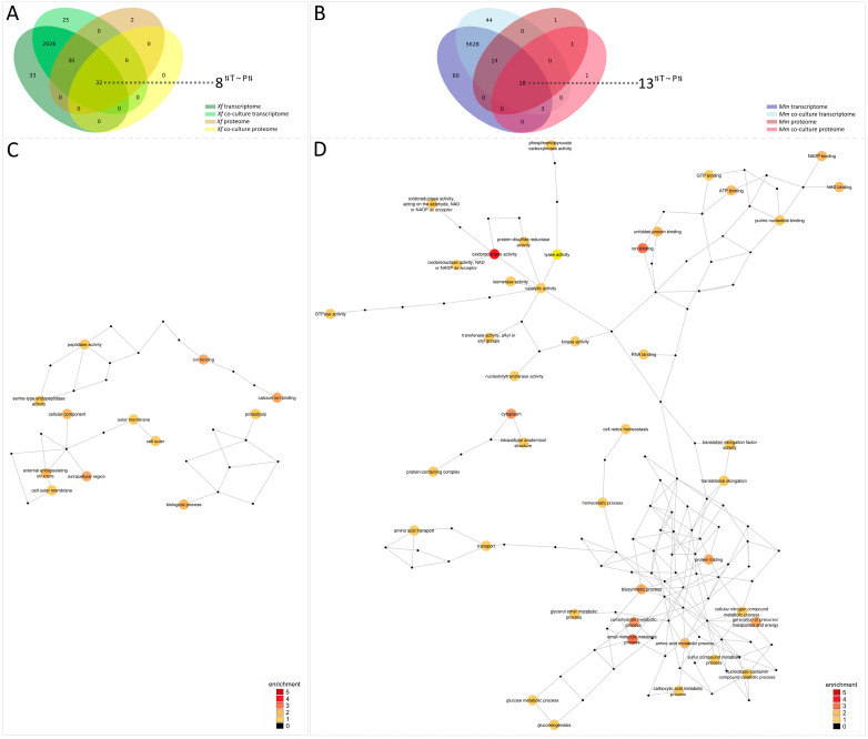
We found 32 common genes between the two analyzed secretomes and the two analyzed transcriptomes of co-culture versus monoculture from Xf. Regarding the comparison of co-culture versus monoculture from Mm, 18 common genes were detected (Figure 6A,B). To establish an appropriate comparison with transcriptome data, the proteome data were normalized to PPT values similarly to TPM values, as described in the Materials and Methods section. Following this, a Mantel test was used to obtain a correlation between the secretome and transcriptome. The intersection genes detected in transcriptome and secretome (32) from Xf have a correlation shown by a p-value of 0.05742, which indicates a medium-to-high correlation. A much lower correlation was obtained in the intersection genes detected in transcriptome and secretome (18) from Mm where a p-value ≅ of 0.8038 was calculated based also on a Mantel test. By the removal of the outlier gene MMSR116_RS20190 (Table S7), we calculated a much smaller p-value of 0.08273, indicating a relative medium-to-high correlation between transcriptome and secretome also for Mm.
Figure 6.
Integration of transcriptome and secretome analyses. Venn diagrams show the intersection between transcriptome and proteome as well as monoculture and co-culture from Xf (A) and Mm (B). Numbers highlighted outside the Venn diagrams indicate the intersection of genes with the same trend, illustrated by the divergent arrows, in the transcriptome (T) and secretome (S). Xf: X. fastidiosa 9a5c; Xf+Mm: X. fastidiosa 9a5c + M. mesophilicum SR1.6/6; Mm: M. mesophilicum SR1.6/6. GO terms networks of Xf (C) and Mm (D) interaction data. Networks representing gene ontology (GO) terms in molecular function, biological process, or cellular component categories enriched among the integration of transcriptome and secretome analyses, showing genes specifically affected by co-culture between Mm and Xf. Enriched GO terms were identified after a pipeline involving a Mantel correlation test and Blast2Go and visualized with Cytoscape software v3.10.1. The GO terms were connected based on their parent-child relationships. The colors of the circles indicate the GO terms filtered after the interactome pipeline.
Following, the intersection genes from both Xf and Mm were further analyzed. The ratios between gene values in the transcriptome (co-culture:monoculture) were compared to the ratios of gene products values in the secretome (co-culture:monoculture), i.e., TPM and PPT, respectively. Gene transcripts that were up- or down-regulated in a given condition do not always result in up- or down-regulation at the protein level [65]. Therefore, we again limited our observations to major trends. Ratios in transcriptome and secretome that simultaneously were more than one or less than one were observed in 8 out of 32 genes for Xf and in 13 out of 18 genes for Mm (Tables S6 and S7). These genes with conserved trends for GO annotations were then used to build networks (Figure 6C,D and Figure S1). The GO annotations network for Xf showed particularly two different functionally enriched annotations: extracellular region, and calcium ion binding/ion binding. On the other hand, Mm had generated a huge GO network, which was particularly enriched in GO annotations associated with oxidoreductase activity, ion binding, and small molecule metabolic process.
4. Discussion
Gene expression profiles can reveal valuable information about the mechanisms related to interactions among bacterial species in a particular environmental niche. Several endophytic bacteria can occupy the same niche/habitat as the phytopathogen Xf in the lumen of xylem vessels [27,28,31,32]. Araujo et al. [28] isolated a high number of Methylobacterium spp. in asymptomatic Xf-infected citrus plants. Moreover, physiological tests were performed showing the growth inhibition of the phytopathogen Xf in the presence of M. mesophilicum, M. extorquens, and Curtobacterium sp. in vitro [29] and in the presence of Mm in planta [31]. Induction of stress-related genes and the repression of growth genes in Xf were detected in co-culture with Mm [30]. However, the DEG of Mm as well as new Xf-expressed genes were not assessed in that initial study. The genome sequences of both species are available, improving transcriptomic and proteomic analyses. Recently, a transcriptomic study of Mm SR1.6/6 interacting with soybean was performed [48], which has analyzed planktonic bacterial cells under the influence of soybean exudates in comparison with control treatments, showing that several stress genes are modulated by plant colonization. Here we investigated the transcriptomic response of both interacting species using RNA-seq and complemented this dataset with the secretome of both bacterial species. We hypothesized that the co-culture of Mm with Xf would generate nutrient deprivation for both bacteria, as previous works in the literature suggested environmental niche competition by Mm towards Xf [29,30].
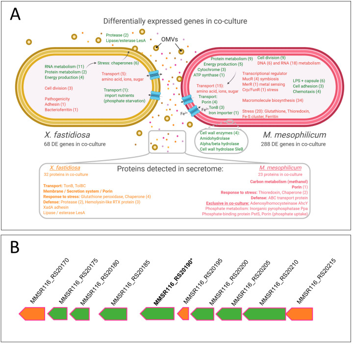
Transcriptomic and growth analyses showed that Xf genes associated with cell division were down-regulated in co-culture, while genes related to energy generation were up-regulated. Similar results were obtained in previous work using microarrays of Xf-Mm [30]. Overall, the present work shows that genes related to metabolism (RNA, protein, and energy production) were up-regulated (Figure 7A).
Figure 7.
(A) Schematic representation of differentially expressed genes and proteins detected in the secretome of Xf and Mm during co-culture. Green represents genes up-regulated during co-culture and red those that are down-regulated during co-culture. The number of differentially expressed genes in the functional categories is indicated in brackets. The bottom box shows the schematic representation of the secretome. Secretome analysis identified four proteins in M. mesophilicum exclusively produced in co-culture with X. fastidiosa, among these, three are related to phosphorous uptake. Orange represents Xf proteins and pink represents Mm proteins detected during co-culture. The number of proteins identified in the functional categories is indicated in brackets. OMVs—Outer membrane vesicles. Xf: X. fastidiosa 9a5c; Xf+Mm: X. fastidiosa 9a5c + M. mesophilicum SR1.6/6; Mm: M. mesophilicum SR1.6/6. (B) Mm ABC transporter operon. Green arrows represent up-regulated genes in Xf+Mm co-culture according to transcriptome analysis, orange arrows represent genes with similar expression in all tested treatments. (*) indicates an up-regulated gene in co-culture which encoded protein was detected in the secretome of Mm monoculture.
On the other hand, Xf showed up-regulation of pathogenesis-related genes, such as oligopeptidases, proteases, and lipase/esterase (LesA). As Xf is under stress and starvation due to competition against Mm, cell wall degrading enzymes are expressed possibly to provide additional nutrient sources. Furthermore, those enzymes were shown to be secreted via OMVs, which were previously shown to be produced in stress conditions to improve bacterial spread inside the host [20,25]. Additionally, mechanisms to secrete Xf proteases and lipases by different systems under diverse situations to target membrane cells of the competitor Mm have been hypothesized in recent articles [67,68,69].
Six different chaperone genes were also up-regulated in response to Mm presence. An increase in stress response was also found in previous microarray analyses [30].
Interestingly, several Xf transport genes were down-regulated during co-culture suggesting reduction of sugar and nutrient intake. However, a single phosphate transport gene (XF_2141/XF9a_02027- phosphate ABC transporter) was up-regulated (Figure 7B). Upregulation of phosphate transporter has been shown to be related to import nutrients when facing phosphate starvation in M. tuberculosis and M. bovis [70]. This expression profile might represent nutrient starvation by Xf following interaction with Mm.
Genes related to cell division were up-regulated in this condition. This may occur due to an increase in Mm metabolism but does not necessarily result in an increase in cell number. Moreover, similar to Xf, genes related to metabolism and energy production were also induced in Mm.
Mm also down-regulated several transport genes upon interaction with Xf, particularly those related to ions, carbohydrate, and protein uptake. However, several other transport genes were up-regulated, including ABC transporter, TonB, as well as porins involved mainly in iron and phosphorus uptake. Additionally, despite the constant agitation in broth culture, four cell adhesion genes were up-regulated in Mm, which may be related to glass adhesion. LPS and capsule genes were also up-regulated, which may play roles in protection and adherence [71]. Four chemotaxis and four hydrolase genes related to bacterial interaction were also up-regulated. These suggest that Mm might be both taking nutrients from Xf (mainly Fe and P) and simultaneously producing enzymes to cleave the Xf cell wall. Twenty Mm stress-related genes were down-regulated, including genes related to oxidative stress, mainly glutathione, thioredoxin, and iron detoxification and storage. Antitoxin genes related to host response were also down-regulated, indicating that Mm may not sense the presence of Xf as a stressful condition.
Analysis of the secretome data reveals that the response of phytopathogen Xf in co-culture shows some overlap when compared to the endophyte Mm in co-culture. Proteins with putative functional similarities have been identified, hinting at potential functional convergences across Xf and Mm. Notably, both organisms appear to possess porin proteins PorinO/OprP (Table 2) and Porin (Table 3) that likely serve as outer membrane channels for the translocation of molecules. Additionally, the presence of chaperone proteins, such as Chaperonin GroEL (Table 2), and Chaperonin GroES, and Chaperone protein DnaK (Table 3), suggests shared roles in assisting protein folding and maintenance. Furthermore, the occurrence of proteins labeled as “uncharacterized” in both datasets emphasizes the need for extensive characterization efforts to elucidate the precise functions of those proteins. Also, several Xf proteins associated with nutrient transport, including membrane proteins (OmpA, Omp1X) and porins (OprP, CirA), along with adhesins (XadA3), lipases (Lpp and LesA), and pathogenicity-related proteins (RTX), were identified, consistent with their presence in previous monoculture studies [14,17,20]. The presence of proteins related to pathogenicity and antibiotic resistance in Xf implies its continuous metabolic activity in the investigated treatments, allowing it to persist within its ecological niche even in the presence of potential competitors. These findings align with the RNA-Seq data, confirming that the pathogen is compatible with environments characterized by limited nutrient availability and competition with other microorganisms. The pathogen’s capacity to endure such conditions appears to involve the production of these defense effectors, even at the expense of its survival [7,72,73].
For instance, the RTX toxicity proteins, which have been reported in previous Xf proteomics work conducted in our group [25], have been shown to be relatively more abundant in the co-cultured Xf secretome (Table 2). RTX toxicity proteins belong to a class with diverse functions described in Gram-negative bacteria. Proteins of the RTX type may have bacteriocin activity, protease activity, or lipase activity, although most of them do not have a known function [73,74].
While the secretome analyses presented in this study are not directly comparable, the protein spectra identified in the co-culture secretome for both Xf and Mm strongly indicate that these proteins may be considerably more abundant than in the respective monoculture treatments. From the detected proteins in the Xf and Mm secretome, several indicate competition for nutrients and resources. Four exclusive proteins of Mm were detected in co-culture: Inorganic pyrophosphatase Ppa, Phosphate-binding protein PstS, Adenosylhomocysteinase AhcY, and Porin (MMSR116_18145). Inorganic pyrophosphatase Ppa and Phosphate-binding protein PstS proteins are involved with phosphate metabolism and linked to lipid metabolism; the protein Adenosylhomocysteinase AhcY participates in the synthesis of the nucleoside adenosine and homocysteine biosynthesis. Two of these proteins, Phosphate-binding protein PstS (MMSR116_RS19505) and Porin (MMSR116_18145) are membrane proteins that facilitate phosphate entry. Porin protein, also present in transcriptomes, can be involved in phosphate acquisition, nutrient uptake, bacteriocin, and phage receptors.
Apparently, Xf synchronizes the expression of certain proteins (e.g., OmpA and porin CirA) to allow nutrient intake from the environment and increase proteins related to catalytic reactions (Glutathione peroxidase), whereas Mm focuses exclusively on expressing proteins to allow nutrient intake (porins). In the GO annotations, notably, “metabolic process” emerges as the predominant one across all treatment conditions, suggesting the vital involvement of these proteins in diverse metabolic pathways. Furthermore, the “cellular process” also exhibits significant enrichment in the secretome, underscoring the importance of cellular-level activities. Intriguingly, specific GO annotations appear to be unique to each bacterial strain. In the case of Xf, annotations like “biological adhesion”, “protein-containing complex”, “cell part,” “cell”, “transporter activity”, “catalytic activity”, and “reproduction” are exclusively associated with its secretome (either mono or co-culture), shedding light on Xf specialized protein secretion in response to the treatments. Conversely, “biological regulation” is an exclusive GO annotation for Mm, implying its distinct roles in regulating biological processes within the co-culture environment. These findings collectively enhance our understanding of the functional roles and adaptations of Xf and Mm in various treatment conditions, contributing to the broader knowledge of their secretome characteristics and their implications in biological interactions.
It is well-established that during the later stages of bacterial growth, cell death can lead to the release of intracellular components, resulting in medium contamination with membrane components and cytosolic proteins, as evidenced in numerous previous studies [75,76,77,78,79]. However, it is important to note that bacterial cell lysis was not specifically evaluated in our experiments. Still, the presence of typical intracellular proteins in the secretome, such as elongation factors (EF-Tu), chaperones (DnaK, GroEL, and GroES), and cold-shock protein CspA, is not a novel finding. GroEL and EF-Tu are among the most abundant proteins within bacterial cells, and their occurrence in secreted proteomes could be attributed to substantial cell lysis. Remarkably, both of these proteins are recognized as moonlighting proteins, capable of performing multiple physiologically relevant biochemical or biophysical functions, which are not the result of gene fusions [79,80,81,82]. EF-Tu, for instance, has been found in the cell walls, membranes, and secretomes of several bacteria [83,84]. GroEL is believed to have an extracellular function in pathogenic processes [85,86].
Moreover, outer membrane vesicles (OMVs) were previously reported in Xf being associated with pathogenesis, such as by trafficking degradative enzymes against competing bacteria [87] and quorum sensing diffusible signaling factors [25], or by acting as an extracellular antiadhesive, blocking surface attachment and modulating plant colonization [23]. A total of 8 outer membrane proteins were detected in Xf monoculture, among those the XadA protein, an adhesin, which is reported to be associated with OMVs. Four outer membrane proteins were also detected in Xf co-culture, which included XadA. Another protein reported to be secreted in OMVs is the lipase/esterase LesA [20], but also secreted through the Type 2 Secretion System [88], which was found to be up-regulated in the Xf transcriptome and present in both mono and co-culture treatment secretomes. LesA is reported as a key virulence factor, we speculate that it can also potentially act on other competing bacteria, such as Mm. Remains to be investigated if OMVs can be used as a defense mechanism of Xf against Mm or other bacteria-sharing niches.
Moreover, our approach to analyzing common differentially expressed genes/proteins in the transcriptome and secretome analyses reveals relevant trends between up- or down-regulation from transcript to protein expression level. The GO annotation networks highlighted these trends as the investigated genes were elucidated through functional information.
5. Conclusions
The present study is the first report of endophyte-phytopathogen interaction using a global approach combining physiological assays with RNA-Seq and secretome analyses. Integrating all obtained results suggests Mm can suppress the phytopathogen Xf mainly by nutrient competition within xylem vessels, and not only by an active killing mechanism.
Acknowledgments
We thank Matthew Rolfe, from LMU BioCenter, Genetics, for fruitful discussions.
Supplementary Materials
The following supporting information can be downloaded at: https://www.mdpi.com/article/10.3390/microorganisms11112755/s1, Table S1: Quantification of cDNA libraries using Nanodrop (absorbance values). Treatments: Xf: X. fastidiosa 9a5c; Xf+Mm: X. fastidiosa 9a5c + M. mesophilicum SR1.6/6; Mm: M. mesophilicum SR1.6/6; Table S2: Amount of reads per run and treatment. Treatments: Xf: X. fastidiosa 9a5c; Xf+Mm: X. fastidiosa 9a5c + M. mesophilicum SR1.6/6; Mm: M. mesophilicum SR1.6/6; Table S3: Number of mapped reads in Xf and Mm using CLC Genomics Workbench software. Xf: X. fastidiosa; Mm: M. mesophilicum; Table S4: Differentially expressed genes in the phytopathogen Xf only and co-culture; Table S5: Differentially expressed genes in the endophyte Mm only and during co-culture; Table S6: Trends in genes detected in transcriptome and secretome of Xf; Table S7: Trends in genes detected in transcriptome and secretome of Mm; Figure S1: Detailed integration of transcriptome and secretome analyses.
Author Contributions
Conceptualization, M.N.D., P.M.P. and O.R.F.-J.; writing—original draft preparation, M.N.D., P.M.P. and O.R.F.-J.; data curation, P.A.Z., D.B., G.U.-C. and A.M.D.; Transcriptomic analysis, M.N.D. and P.M.P.; Secretome analysis, O.R.F.-J. and P.A.Z.; Integration of transcriptomics and proteomics analysis, O.R.F.-J. and D.B.; funding acquisition, supervision, and writing—review and editing, A.M.d.S. and W.L.A. All authors have read and agreed to the published version of the manuscript.
Data Availability Statement
Transcriptome sequencing (RNA-seq) data reported in this work have been deposited in the NCBI SRA database under BioSample accessions SAMN37339366, SAMN37339367, SAMN37339368, SAMN37339369, SAMN37339370, SAMN37339371, SAMN37339372, SAMN37339373 and SAMN37339374.
Conflicts of Interest
The authors declare no conflict of interest.
Funding Statement
Funding for this work was provided by the São Paulo Research Foundation (FAPESP), research grant 08/11703-4, Coordination for the Improvement of Higher Education Personnel (CAPES), research grant 3385/2013, and the Pierce’s Disease Control Program of the California Department of Food and Agriculture. D.B (88887.285065/2018-00 and 88887.318125/2019-00), O.R.F.J. (88887.160901/2017-00 and 88887.313727/2019-00), and P.M.P (88882.160114/2017-01). were supported by fellowships from CAPES. G.U.C. was supported by FAPESP fellowship 21/04062-7. A.M.d.S. was supported in part by Research Fellowship Award 309182/2016-6 from the National Council for Scientific and Technological Development (CNPq).
Footnotes
Disclaimer/Publisher’s Note: The statements, opinions and data contained in all publications are solely those of the individual author(s) and contributor(s) and not of MDPI and/or the editor(s). MDPI and/or the editor(s) disclaim responsibility for any injury to people or property resulting from any ideas, methods, instructions or products referred to in the content.
References
- 1.Wells J.M., Raju B.C., Hung H.-Y., Weisburg W.G., Mandelco-Paul L., Brenner D.J. Xylella fastidiosa gen. nov., sp. nov: Gram-negative, xylem-limited, fastidious plant bacteria related to Xanthomonas spp. Int. J. Syst. Bacteriol. 1987;37:136–143. doi: 10.1099/00207713-37-2-136. [DOI] [Google Scholar]
- 2.Hopkins D.L. Xylella fastidiosa: Xylem-limited bacterial pathogen of plants. Ann. Rev. Phytopathol. 1989;27:271–290. doi: 10.1146/annurev.py.27.090189.001415. [DOI] [Google Scholar]
- 3.Chang C.J., Garnier M., Zreik L., Rossetti V., Bove J.M. Culture and serological detection of the xylem-limited bacterium causing citrus variegated chlorosis and its identification as a strain ofXylella fastidiosa. Curr. Microbiol. 1993;27:137–142. doi: 10.1007/BF01576010. [DOI] [PubMed] [Google Scholar]
- 4.Hopkins D.L., Purcell A.H. Xylella fastidiosa: Cause of Pierce’s disease of grapevine and other emergent diseases. Plant Dis. 2002;86:1056–1066. doi: 10.1094/PDIS.2002.86.10.1056. [DOI] [PubMed] [Google Scholar]
- 5.Cariddi C., Saponari M., Boscia D., De Stradis A., Loconsole G., Nigro F., Porcelli F., Potere O., Martelli G.P. Isolation of a Xylella fastidiosa strain infecting olive and oleander in Apulia, Italy. J. Plant Pathol. 2014;96:425–429. [Google Scholar]
- 6.Sicard A., Zeilinger A.R., Vanhove M., Schartel T.E., Beal D.J., Daugherty M.P., Almeida R.P.P. Xylella fastidiosa: Insights into an Emerging Plant Pathogen. Annu. Rev. Phytopathol. 2018;56:181–202. doi: 10.1146/annurev-phyto-080417-045849. [DOI] [PubMed] [Google Scholar]
- 7.Chatterjee S., Almeida R.P., Lindow S. Living in two worlds: The plant and insect lifestyles of Xylella fastidiosa. Annu. Rev. Phytopathol. 2008;46:243–271. doi: 10.1146/annurev.phyto.45.062806.094342. [DOI] [PubMed] [Google Scholar]
- 8.Janse J.D., Obradovic A. Xylella fastidiosa: Its biology, diagnosis, control and risks. J. Plant Pathol. 2010;92:S1.35–S31.48. [Google Scholar]
- 9.Backus E.A., Morgan D.J. Spatiotemporal colonization of Xylella fastidiosa in its vector supports the role of egestion in the inoculation mechanism of foregut-borne plant pathogens. Phytopathology. 2011;101:912–922. doi: 10.1094/PHYTO-09-10-0231. [DOI] [PubMed] [Google Scholar]
- 10.de Souza J.B., Almeida-Souza H.O., Zaini P.A., Alves M.N., de Souza A.G., Pierry P.M., da Silva A.M., Goulart L.R., Dandekar A.M., Nascimento R. Xylella fastidiosa subsp. pauca Strains Fb7 and 9a5c from Citrus Display Differential Behavior, Secretome, and Plant Virulence. Int. J. Mol. Sci. 2020;21:6769. doi: 10.3390/ijms21186769. [DOI] [PMC free article] [PubMed] [Google Scholar]
- 11.Zaini P.A., Fogaca A.C., Lupo F.G., Nakaya H.I., Vencio R.Z., da Silva A.M. The iron stimulon of Xylella fastidiosa includes genes for type IV pilus and colicin V-like bacteriocins. J. Bacteriol. 2008;190:2368–2378. doi: 10.1128/JB.01495-07. [DOI] [PMC free article] [PubMed] [Google Scholar]
- 12.Ciraulo M.B., Santos D.S., Rodrigues A.C., de Oliveira M.V., Rodrigues T., de Oliveira R.C., Nunes L.R. Transcriptome analysis of the phytobacterium Xylella fastidiosa growing under xylem-based chemical conditions. J. Biomed. Biotechnol. 2010;2010:781365. doi: 10.1155/2010/781365. [DOI] [PMC free article] [PubMed] [Google Scholar]
- 13.Giampetruzzi A., Morelli M., Saponari M., Loconsole G., Chiumenti M., Boscia D., Savino V.N., Martelli G.P., Saldarelli P. Transcriptome profiling of two olive cultivars in response to infection by the CoDiRO strain of Xylella fastidiosa subsp. pauca. BMC Genom. 2016;17:475. doi: 10.1186/s12864-016-2833-9. [DOI] [PMC free article] [PubMed] [Google Scholar]
- 14.Gouran H., Gillespie H., Nascimento R., Chakraborty S., Zaini P.A., Jacobson A., Phinney B.S., Dolan D., Durbin-Johnson B.P., Antonova E.S., et al. The Secreted Protease PrtA Controls Cell Growth, Biofilm Formation and Pathogenicity in Xylella fastidiosa. Sci. Rep. 2016;6:31098. doi: 10.1038/srep31098. [DOI] [PMC free article] [PubMed] [Google Scholar]
- 15.Parker J.K., Chen H., McCarty S.E., Liu L.Y., De La Fuente L. Calcium transcriptionally regulates the biofilm machinery of Xylella fastidiosa to promote continued biofilm development in batch cultures. Env. Microbiol. 2016;18:1620–1634. doi: 10.1111/1462-2920.13242. [DOI] [PubMed] [Google Scholar]
- 16.Chen H., De La Fuente L. Calcium transcriptionally regulates movement, recombination and other functions of Xylella fastidiosa under constant flow inside microfluidic chambers. Microb. Biotechnol. 2020;13:548–561. doi: 10.1111/1751-7915.13512. [DOI] [PMC free article] [PubMed] [Google Scholar]
- 17.Smolka M.B., Martins-de-Souza D., Winck F.V., Santoro C.E., Castellari R.R., Ferrari F., Brum I.J., Galembeck E., Della Coletta Filho H., Machado M.A., et al. Proteome analysis of the plant pathogen Xylella fastidiosa reveals major cellular and extracellular proteins and a peculiar codon bias distribution. Proteomics. 2003;3:224–237. doi: 10.1002/pmic.200390031. [DOI] [PubMed] [Google Scholar]
- 18.Silva M.S., De Souza A.A., Takita M.A., Labate C.A., Machado M.A. Analysis of the biofilm proteome of Xylella fastidiosa. Proteome Sci. 2011;9:58. doi: 10.1186/1477-5956-9-58. [DOI] [PMC free article] [PubMed] [Google Scholar]
- 19.Mendes J.S., Santiago A.S., Toledo M.A., Horta M.A., de Souza A.A., Tasic L., de Souza A.P. In vitro Determination of Extracellular Proteins from Xylella fastidiosa. Front. Microbiol. 2016;7:2090. doi: 10.3389/fmicb.2016.02090. [DOI] [PMC free article] [PubMed] [Google Scholar]
- 20.Nascimento R., Gouran H., Chakraborty S., Gillespie H.W., Almeida-Souza H.O., Tu A., Rao B.J., Feldstein P.A., Bruening G., Goulart L.R., et al. The Type II Secreted Lipase/Esterase LesA is a Key Virulence Factor Required for Xylella fastidiosa Pathogenesis in Grapevines. Sci. Rep. 2016;6:18598. doi: 10.1038/srep18598. [DOI] [PMC free article] [PubMed] [Google Scholar]
- 21.Ryan R.P., Dow J.M. Communication with a growing family: Diffusible signal factor (DSF) signaling in bacteria. Trends Microbiol. 2011;19:145–152. doi: 10.1016/j.tim.2010.12.003. [DOI] [PubMed] [Google Scholar]
- 22.Voegel T.M., Warren J.G., Matsumoto A., Igo M.M., Kirkpatrick B.C. Localization and characterization of Xylella fastidiosa haemagglutinin adhesins. Microbiology. 2010;156:2172–2179. doi: 10.1099/mic.0.037564-0. [DOI] [PubMed] [Google Scholar]
- 23.Ionescu M., Zaini P.A., Baccari C., Tran S., da Silva A.M., Lindow S.E. Xylella fastidiosa outer membrane vesicles modulate plant colonization by blocking attachment to surfaces. Proc. Natl. Acad. Sci. USA. 2014;111:E3910–E3918. doi: 10.1073/pnas.1414944111. [DOI] [PMC free article] [PubMed] [Google Scholar]
- 24.Santiago A.D., Mendes J.S., Dos Santos C.A., de Toledo M.A., Beloti L.L., Crucello A., Horta M.A., Favaro M.T., Munar D.M., de Souza A.A., et al. The Antitoxin Protein of a Toxin-Antitoxin System from Xylella fastidiosa Is Secreted via Outer Membrane Vesicles. Front. Microbiol. 2016;7:2030. doi: 10.3389/fmicb.2016.02030. [DOI] [PMC free article] [PubMed] [Google Scholar]
- 25.Feitosa-Junior O.R., Stefanello E., Zaini P., Nascimento R., Pierry P.M., Dandekar A., Lindow S., da Silva A.M. Proteomic and metabolomic analyses of Xylella fastidiosa OMV-enriched fractions reveal association with virulence factors and signaling molecules of the DSF family. Phytopathology. 2019;109:1344–1353. doi: 10.1094/PHYTO-03-19-0083-R. [DOI] [PubMed] [Google Scholar]
- 26.Merfa M.V., Niza B., Takita M.A., De Souza A.A. The MqsRA Toxin-Antitoxin System from Xylella fastidiosa Plays a Key Role in Bacterial Fitness, Pathogenicity, and Persister Cell Formation. Front. Microbiol. 2016;7:904. doi: 10.3389/fmicb.2016.00904. [DOI] [PMC free article] [PubMed] [Google Scholar]
- 27.Anguita-Maeso M., Ares-Yebra A., Haro C., Roman-Ecija M., Olivares-Garcia C., Costa J., Marco-Noales E., Ferrer A., Navas-Cortes J.A., Landa B.B. Xylella fastidiosa Infection Reshapes Microbial Composition and Network Associations in the Xylem of Almond Trees. Front. Microbiol. 2022;13:866085. doi: 10.3389/fmicb.2022.866085. [DOI] [PMC free article] [PubMed] [Google Scholar]
- 28.Araujo W.L., Marcon J., Maccheroni W., Jr., Van Elsas J.D., Van Vuurde J.W., Azevedo J.L. Diversity of endophytic bacterial populations and their interaction with Xylella fastidiosa in citrus plants. Appl. Env. Microbiol. 2002;68:4906–4914. doi: 10.1128/AEM.68.10.4906-4914.2002. [DOI] [PMC free article] [PubMed] [Google Scholar]
- 29.Lacava P.T., Araujo W.L., Marcon J., Maccheroni W., Jr., Azevedo J.L. Interaction between endophytic bacteria from citrus plants and the phytopathogenic bacteria Xylella fastidiosa, causal agent of citrus-variegated chlorosis. Lett. Appl. Microbiol. 2004;39:55–59. doi: 10.1111/j.1472-765X.2004.01543.x. [DOI] [PubMed] [Google Scholar]
- 30.Dourado M.N., Santos D.S., Nunes L.R., Costa de Oliveira R.L., de Oliveira M.V., Araujo W.L. Differential gene expression in Xylella fastidiosa 9a5c during co-cultivation with the endophytic bacterium Methylobacterium mesophilicum SR1.6/6. J. Basic. Microbiol. 2015;55:1357–1366. doi: 10.1002/jobm.201400916. [DOI] [PubMed] [Google Scholar]
- 31.Lacava P.T., Li W.B., Araujo W.L., Azevedo J.L., Hartung J.S. Rapid, specific and quantitative assays for the detection of the endophytic bacterium Methylobacterium mesophilicum in plants. J. Microbiol. Methods. 2006;65:535–541. doi: 10.1016/j.mimet.2005.09.015. [DOI] [PubMed] [Google Scholar]
- 32.Andreote F.D., Lacava P.T., Gai C.S., Araujo W.L., Maccheroni W., Jr., van Overbeek L.S., van Elsas J.D., Azevedo J.L. Model plants for studying the interaction between Methylobacterium mesophilicum and Xylella fastidiosa. Can. J. Microbiol. 2006;52:419–426. doi: 10.1139/w05-142. [DOI] [PubMed] [Google Scholar]
- 33.Gai C.S., Lacava P.T., Quecine M.C., Auriac M.C., Lopes J.R., Araujo W.L., Miller T.A., Azevedo J.L. Transmission of Methylobacterium mesophilicum by Bucephalogonia xanthophis for paratransgenic control strategy of citrus variegated chlorosis. J. Microbiol. 2009;47:448–454. doi: 10.1007/s12275-008-0303-z. [DOI] [PubMed] [Google Scholar]
- 34.Rogers E.E., Backus E.A. Anterior foregut microbiota of the glassy-winged sharpshooter explored using deep 16S rRNA gene sequencing from individual insects. PLoS ONE. 2014;9:e106215. doi: 10.1371/journal.pone.0106215. [DOI] [PMC free article] [PubMed] [Google Scholar]
- 35.Dourado M.N., Camargo Neves A.A., Santos D.S., Araujo W.L. Biotechnological and agronomic potential of endophytic pink-pigmented methylotrophic Methylobacterium spp. Biomed. Res. Int. 2015;2015:909016. doi: 10.1155/2015/909016. [DOI] [PMC free article] [PubMed] [Google Scholar]
- 36.Bogas A.C., Aguilar-Vildoso C.I., Camargo Neves A.A., Araujo W.L. Effects of growth-promoting endophytic Methylobacterium on development of Citrus rootstocks. Afr. J. Microbiol. Res. 2016;10:646–653. [Google Scholar]
- 37.Madhaiyan M., Suresh Reddy B.V., Anandham R., Senthilkumar M., Poonguzhali S., Sundaram S.P., Sa T. Plant growth-promoting Methylobacterium induces defense responses in groundnut (Arachis hypogaea L.) compared with rot pathogens. Curr. Microbiol. 2006;53:270–276. doi: 10.1007/s00284-005-0452-9. [DOI] [PubMed] [Google Scholar]
- 38.Tani A., Sahin N., Kimbara K. Methylobacterium oxalidis sp. nov., isolated from leaves of Oxalis corniculata. Int. J. Syst. Evol. Microbiol. 2012;62:1647–1652. doi: 10.1099/ijs.0.033019-0. [DOI] [PubMed] [Google Scholar]
- 39.Jirakkakul J., Khoiri A.N., Duangfoo T., Dulsawat S., Sutheeworapong S., Petsong K., Wattanachaisaereekul S., Paenkaew P., Tachaleat A., Cheevadhanarak S., et al. Insights into the genome of sp. NMS14P, a novel bacterium for growth promotion of maize, chili, and sugarcane. PloS ONE. 2023;18:e0281505. doi: 10.1371/journal.pone.0281505. [DOI] [PMC free article] [PubMed] [Google Scholar]
- 40.Vera R.T., García A.J.B., Alvarez F.J.C., Ruiz J.M., Martín F.F. Application and effectiveness of as a biological inoculant in maize and strawberry crops. Folia Microbiol. 2023 doi: 10.1007/s12223-023-01078-4. [DOI] [PMC free article] [PubMed] [Google Scholar]
- 41.Trotsenko Y.A., Ianova E.G., Doronina N.V. Aerobic methylotrophic bacteria as phytosymbionts. Microbiology. 2001;70:623–632. doi: 10.1023/A:1013167612105. [DOI] [Google Scholar]
- 42.Meena K.K., Kumar M., Kalyuzhnaya M.G., Yandigeri M.S., Singh D.P., Saxena A.K., Arora D.K. Epiphytic pink-pigmented methylotrophic bacteria enhance germination and seedling growth of wheat (Triticum aestivum) by producing phytohormone. Antonie Van. Leeuwenhoek. 2012;101:777–786. doi: 10.1007/s10482-011-9692-9. [DOI] [PubMed] [Google Scholar]
- 43.Cervantes-Martínez J., Lopez-Díaz S., Rodriguez-Garay B. Detection of the effects of Methylobacterium in Agave tequilana Weber var. azul by laser-induced fluorescence. Plant Sci. 2004;166:889–892. doi: 10.1016/j.plantsci.2003.11.029. [DOI] [Google Scholar]
- 44.Lee H.S., Madhaiyan C.W.K., Choi S.J., Chung K.Y., Sa T.M. Physiological enhancement of early growth of rice seedlings (Oryza sativa L.) by production of phytohormone of N2-fixing methylotrophic isolates. Biol. Fertil. Soils. 2006;42:402–408. doi: 10.1007/s00374-006-0083-8. [DOI] [Google Scholar]
- 45.Van Dien S.J., Marx C.J., O’brien B.N., Listrom M. Genetic characterization of the carotenoid biosynthetic pathway in Methylobacterium extorquens AM1 and isolation of a colorless mutant. Appl. Environ. Microbiol. 2003;69:7563–7566. doi: 10.1128/AEM.69.12.7563-7566.2003. [DOI] [PMC free article] [PubMed] [Google Scholar]
- 46.Toyama H., Anthony C., Lidstrom M.E. Construction of insertion and deletion mxa mutants of Methylobacterium extorquens AM1 by electroporation. FEMS Microbiol. Lett. 1998;166:1–7. doi: 10.1111/j.1574-6968.1998.tb13175.x. [DOI] [PubMed] [Google Scholar]
- 47.Dourado M.N., Bogas A.C., Pomini A.M., Andreote F.D., Quecine M.C., Marsaioli A.J., Araujo W.L. Methylobacterium-plant interaction genes regulated by plant exudate and quorum sensing molecules. Braz. J. Microbiol. 2013;44:1331–1339. doi: 10.1590/S1517-83822013000400044. [DOI] [PMC free article] [PubMed] [Google Scholar]
- 48.Araujo W.L., Santos D.S., Dini-Andreote F., Salgueiro-Londono J.K., Camargo-Neves A.A., Andreote F.D., Dourado M.N. Genes related to antioxidant metabolism are involved in Methylobacterium mesophilicum-soybean interaction. Antonie Van. Leeuwenhoek. 2015;108:951–963. doi: 10.1007/s10482-015-0548-6. [DOI] [PubMed] [Google Scholar]
- 49.Li W.B., Zreik L., Fernandes N.G., Miranda V.S., Teixeira D.C., Ayres A.J., Garnier M., Bov J.M. A triply cloned strain of xylella fastidiosa multiplies and induces symptoms of citrus variegated chlorosis in sweet orange. Curr. Microbiol. 1999;39:106–108. doi: 10.1007/s002849900428. [DOI] [PubMed] [Google Scholar]
- 50.Davis M.J., French W.J., Schaad N.W. Axenic culture of the bacteria associated with phony disease of peach and plum leaf scald. Curr. Microbiol. 1981;6:309–314. doi: 10.1007/BF01566883. [DOI] [Google Scholar]
- 51.Pooler M.R., Hartung J.S. Specific PCR detection and identification of Xylella fastidiosa strains causing citrus variegated chlorosis. Curr. Microbiol. 1995;31:377–381. doi: 10.1007/BF00294703. [DOI] [PubMed] [Google Scholar]
- 52.Klindworth A., Pruesse E., Schweer T., Peplies J., Quast C., Horn M., Glockner F.O. Evaluation of general 16S ribosomal RNA gene PCR primers for classical and next-generation sequencing-based diversity studies. Nucleic Acids Res. 2013;41:e1. doi: 10.1093/nar/gks808. [DOI] [PMC free article] [PubMed] [Google Scholar]
- 53.Andrews S., Lindenbaum P., Howard B., Ewels P. FastQC High Throughput Sequence QC Report. Babraham Bioinformatics; Cambridge, UK: 2014. 0.11.2. [Google Scholar]
- 54.Mortazavi A., Williams B.A., McCue K., Schaeffer L., Wold B. Mapping and quantifying mammalian transcriptomes by RNA-Seq. Nat. Methods. 2008;5:621–628. doi: 10.1038/nmeth.1226. [DOI] [PubMed] [Google Scholar]
- 55.Love M.I., Huber W., Anders S. Moderated estimation of fold change and dispersion for RNA-Seq data with DESeq2. Genome Biol. 2014;15:550. doi: 10.1186/s13059-014-0550-8. [DOI] [PMC free article] [PubMed] [Google Scholar]
- 56.Conesa A., Gotz S., Garcia-Gomez J.M., Terol J., Talon M., Robles M. Blast2GO: A universal tool for annotation, visualization and analysis in functional genomics research. Bioinformatics. 2005;21:3674–3676. doi: 10.1093/bioinformatics/bti610. [DOI] [PubMed] [Google Scholar]
- 57.Ashburner M., Ball C.A., Blake J.A., Botstein D., Butler H., Cherry J.M., Davis A.P., Dolinski K., Dwight S.S., Eppig J.T., et al. Gene ontology: Tool for the unification of biology. The Gene Ontology Consortium. Nat. Genet. 2000;25:25–29. doi: 10.1038/75556. [DOI] [PMC free article] [PubMed] [Google Scholar]
- 58.Craig R., Beavis R.C. TANDEM: Matching proteins with tandem mass spectra. Bioinformatics. 2004;20:1466–1467. doi: 10.1093/bioinformatics/bth092. [DOI] [PubMed] [Google Scholar]
- 59.Petersen T.N., Brunak S., von Heijne G., Nielsen H. SignalP 4.0: Discriminating signal peptides from transmembrane regions. Nat. Methods. 2011;8:785–786. doi: 10.1038/nmeth.1701. [DOI] [PubMed] [Google Scholar]
- 60.Bendtsen J.D., Kiemer L., Fausboll A., Brunak S. Non-classical protein secretion in bacteria. BMC Microbiol. 2005;5:58. doi: 10.1186/1471-2180-5-58. [DOI] [PMC free article] [PubMed] [Google Scholar]
- 61.Zhao S., Ye Z., Stanton R. Misuse of RPKM or TPM normalization when comparing across samples and sequencing protocols. RNA. 2020;26:903–909. doi: 10.1261/rna.074922.120. [DOI] [PMC free article] [PubMed] [Google Scholar]
- 62.Python-Core-Team . Python: A Dynamic, Open Source Programming Language. Python-Core-Team; Beaverton, OR, USA: 2015. [Google Scholar]
- 63.Mantel N. The detection of disease clustering and a generalized regression approach. Cancer Res. 1967;27:209–220. [PubMed] [Google Scholar]
- 64.Shannon P., Markiel A., Ozier O., Baliga N.S., Wang J.T., Ramage D., Amin N., Schwikowski B., Ideker T. Cytoscape: A software environment for integrated models of biomolecular interaction networks. Genome Res. 2003;13:2498–2504. doi: 10.1101/gr.1239303. [DOI] [PMC free article] [PubMed] [Google Scholar]
- 65.Ludvigsen M., Honoré B. Encyclopedia of Life Sciences. John Wiley; Hoboken, NJ, USA: 2018. Transcriptomics and Proteomics: Integration? pp. 1–7. [Google Scholar]
- 66.Rinschen M.M., Gödel M., Grahammer F., Zschiedrich S., Helmstädter M., Kretz O., Zarei M., Braun D.A., Dittrich S., Pahmeyer C., et al. A Multi-layered Quantitative Expression Atlas of the Podocyte Unravels Kidney Disease Candidate Genes. Cell Rep. 2018;23:2495–2508. doi: 10.1016/j.celrep.2018.04.059. [DOI] [PMC free article] [PubMed] [Google Scholar]
- 67.Stubbendieck R.M., Straight P.D. Multifaceted Interfaces of Bacterial Competition. J. Bacteriol. 2016;198:2145–2155. doi: 10.1128/JB.00275-16. [DOI] [PMC free article] [PubMed] [Google Scholar]
- 68.Alteri C.J., Mobley H.L.T. The Versatile Type VI Secretion System. Microbiol. Spectr. 2016;4:337–356. doi: 10.1128/microbiolspec.VMBF-0026-2015. [DOI] [PMC free article] [PubMed] [Google Scholar]
- 69.Kazmierczak B.I. The Enemy of my Enemy: Bacterial Competition in the Cystic Fibrosis Lung. Cell Host Microbe. 2020;28:502–504. doi: 10.1016/j.chom.2020.09.009. [DOI] [PubMed] [Google Scholar]
- 70.Lefevre P., Braibant M., de Wit L., Kalai M., Roeper D., Grotzinger J., Delville J.P., Peirs P., Ooms J., Huygen K., et al. Three different putative phosphate transport receptors are encoded by the Mycobacterium tuberculosis genome and are present at the surface of Mycobacterium bovis BCG. J. Bacteriol. 1997;179:2900–2906. doi: 10.1128/jb.179.9.2900-2906.1997. [DOI] [PMC free article] [PubMed] [Google Scholar]
- 71.Roberts I.S. The biochemistry and genetics of capsular polysaccharide production in bacteria. Annu. Rev. Microbiol. 1996;50:285–315. doi: 10.1146/annurev.micro.50.1.285. [DOI] [PubMed] [Google Scholar]
- 72.Wang N., Li J.L., Lindow S.E. RpfF-dependent regulon of Xylella fastidiosa. Phytopathology. 2012;102:1045–1053. doi: 10.1094/PHYTO-07-12-0146-R. [DOI] [PubMed] [Google Scholar]
- 73.Gambetta G.A., Matthews M.A., Syvanen M. The Xylella fastidosa RTX operons: Evidence for the evolution of protein mosaics through novel genetic exchanges. BMC Genom. 2018;19:329. doi: 10.1186/s12864-018-4731-9. [DOI] [PMC free article] [PubMed] [Google Scholar]
- 74.Linhartova I., Bumba L., Masin J., Basler M., Osicka R., Kamanova J., Prochazkova K., Adkins I., Hejnova-Holubova J., Sadilkova L., et al. RTX proteins: A highly diverse family secreted by a common mechanism. FEMS Microbiol. Rev. 2010;34:1076–1112. doi: 10.1111/j.1574-6976.2010.00231.x. [DOI] [PMC free article] [PubMed] [Google Scholar]
- 75.Kato S., Kowashi Y., Demuth D.R. Outer membrane-like vesicles secreted by Actinobacillus actinomycetemcomitans are enriched in leukotoxin. Microb. Pathog. 2002;32:1–13. doi: 10.1006/mpat.2001.0474. [DOI] [PubMed] [Google Scholar]
- 76.Kulp A., Kuehn M.J. Biological functions and biogenesis of secreted bacterial outer membrane vesicles. Annu. Rev. Microbiol. 2010;64:163–184. doi: 10.1146/annurev.micro.091208.073413. [DOI] [PMC free article] [PubMed] [Google Scholar]
- 77.Siljamaki P., Varmanen P., Kankainen M., Sukura A., Savijoki K., Nyman T.A. Comparative exoprotein profiling of different Staphylococcus epidermidis strains reveals potential link between nonclassical protein export and virulence. J. Proteome Res. 2014;13:3249–3261. doi: 10.1021/pr500075j. [DOI] [PubMed] [Google Scholar]
- 78.Perez-Cruz C., Delgado L., Lopez-Iglesias C., Mercade E. Outer-inner membrane vesicles naturally secreted by gram-negative pathogenic bacteria. PLoS ONE. 2015;10:e0116896. doi: 10.1371/journal.pone.0116896. [DOI] [PMC free article] [PubMed] [Google Scholar]
- 79.Schwechheimer C., Kuehn M.J. Outer-membrane vesicles from Gram-negative bacteria: Biogenesis and functions. Nat. Rev. Microbiol. 2015;13:605–619. doi: 10.1038/nrmicro3525. [DOI] [PMC free article] [PubMed] [Google Scholar]
- 80.Huberts D.H., van der Klei I.J. Moonlighting proteins: An intriguing mode of multitasking. Biochim. Biophys. Acta. 2010;1803:520–525. doi: 10.1016/j.bbamcr.2010.01.022. [DOI] [PubMed] [Google Scholar]
- 81.Jeffery C.J. Why study moonlighting proteins? Front. Genet. 2015;6:211. doi: 10.3389/fgene.2015.00211. [DOI] [PMC free article] [PubMed] [Google Scholar]
- 82.Wang W., Jeffery C.J. An analysis of surface proteomics results reveals novel candidates for intracellular/surface moonlighting proteins in bacteria. Mol. Biosyst. 2016;12:1420–1431. doi: 10.1039/C5MB00550G. [DOI] [PubMed] [Google Scholar]
- 83.Caldas T.D., El Yaagoubi A., Richarme G. Chaperone properties of bacterial elongation factor EF-Tu. J. Biol. Chem. 1998;273:11478–11482. doi: 10.1074/jbc.273.19.11478. [DOI] [PubMed] [Google Scholar]
- 84.Gupta S., Wardhan V., Kumar A., Rathi D., Pandey A., Chakraborty S., Chakraborty N. Secretome analysis of chickpea reveals dynamic extracellular remodeling and identifies a Bet v1-like protein, CaRRP1 that participates in stress response. Sci. Rep. 2015;5:18427. doi: 10.1038/srep18427. [DOI] [PMC free article] [PubMed] [Google Scholar]
- 85.Neckers L. Molecular Chaperones in Pathogen Virulence: Emerging New Targets for Therapy. Cell Host Microbe. 2009;4:519–527. doi: 10.1016/j.chom.2008.10.011. [DOI] [PMC free article] [PubMed] [Google Scholar]
- 86.Ghazaei C. Molecular chaperones impacts in growth, metabolism, pathogenicity and production of virulence factors in bacterial pathogen. Rev. Med. Microbiol. 2018;29:24–29. doi: 10.1097/MRM.0000000000000123. [DOI] [Google Scholar]
- 87.Evans A.G.L., Davey H.M., Cookson A., Currinn H., Cooke-Fox G., Stanczyk P.J., Whitworth D.E. Predatory activity of Myxococcus xanthus outer-membrane vesicles and properties of their hydrolase cargo. Microbiology. 2012;158:2742–2752. doi: 10.1099/mic.0.060343-0. [DOI] [PubMed] [Google Scholar]
- 88.Ingel B., Castro C., Burbank L., Her N., De Anda N.I., Way H., Wang P., Roper C. Xylella fastidiosa requires the Type II secretion system for pathogenicity and survival in grapevine. Mol. Plant Microbe Interact. 2023;36 doi: 10.1094/MPMI-03-23-0027-R. [DOI] [PubMed] [Google Scholar]
Associated Data
This section collects any data citations, data availability statements, or supplementary materials included in this article.
Supplementary Materials
Data Availability Statement
Transcriptome sequencing (RNA-seq) data reported in this work have been deposited in the NCBI SRA database under BioSample accessions SAMN37339366, SAMN37339367, SAMN37339368, SAMN37339369, SAMN37339370, SAMN37339371, SAMN37339372, SAMN37339373 and SAMN37339374.


