



Abstract
Background and Aims
Colonic bacterial biofilms are frequently present in ulcerative colitis [UC] and may increase dysplasia risk through pathogens expressing oncotraits. This prospective cohort study aimed to determine [1] the association of oncotraits and longitudinal biofilm presence with dysplasia risk in UC, and [2] the relation of bacterial composition with biofilms and dysplasia risk.
Methods
Faeces and left- and right-sided colonic biopsies were collected from 80 UC patients and 35 controls. Oncotraits [FadA of Fusobacterium, BFT of Bacteroides fragilis, colibactin [ClbB] and Intimin [Eae] of Escherichia coli] were assessed in faecal DNA with multiplex quantitative polymerase chain reaction [qPCR]. Biopsies were screened for biofilms [n = 873] with 16S rRNA fluorescent in situ hybridiation. Shotgun metagenomic sequencing [n = 265], and ki67-immunohistochemistry were performed. Associations were determined with a mixed-effects regression model.
Results
Biofilms were highly prevalent in UC patients [90.8%] with a median persistence of 3 years (interquartile range [IQR] 2–5 years). Biofilm-positive biopsies showed increased epithelial hypertrophy [p = 0.025] and a reduced Shannon diversity independent of disease status [p = 0.015], but were not significantly associated with dysplasia in UC: adjusted odds ratio [aOR] 1.45, 95% confidence interval [CI] 0.63–3.40. In contrast, ClbB independently associated with dysplasia [aOR 7.16, 95% CI 1.75–29.28], and FadA and Fusobacteriales were associated with a decreased dysplasia risk in UC [aOR 0.23, 95% CI 0.06–0.83, p <0.01].
Conclusions
Biofilms are a hallmark of UC; however, because of their high prevalence are a poor biomarker for dysplasia. In contrast, colibactin presence and FadA absence independently associate with dysplasia in UC and might therefore be valuable biomarkers for future risk stratification and intervention strategies.
Keywords: Microbiology, biomarkers, pathology
Graphical abstract
Graphical Abstract.

1. Introduction
Ulcerative colitis [UC] is characterised by chronic inflammation of the colonic mucosa and is an increasing burden worldwide.1 In UC, chronic inflammation may lead to the development of mucosal dysplasia that can progress to colorectal cancer [CRC].2,3 Detection of dysplasia and risk stratification are dependent on invasive techniques including surveillance colonoscopies every 1–5 years. Cancer prevention in UC is limited by our knowledge on accurate predictors of dysplasia and human errors in dysplasia detection, with 30–55% of CRC diagnosed as interval lesions.4–6 Current non-invasive tests for CRC detection are based on faecal occult blood or haemoglobin detection, which is unreliable in UC because of high false-positive rates due to inflammation-related bleeding. Investigation of novel biomarkers might provide new leads for less invasive risk stratification. The colonic microbiota and its interaction with the [impaired] mucosal barrier may play a role in colitis-associated cancer development, and could potentially provide novel biomarkers for dysplasia in UC.
The inner mucosal layer of UC patients is frequently covered with spatially organised bacteria in an adherent polymeric matrix, so-called biofilm.7,8 Cross-sectional studies have shown endoscopic visibility of biofilms in 34% of UC patients7 and microscopic detection in 70% of UC patients.8 Bacteria frequently detected in mucosal biofilms in UC include Bacteroides fragilis, Escherichia coli, Klebsiella sp., Fusobacterium peridonticum, and Ruminococcus gnavus.7,8 Biofilms provide a microenvironment for bacteria to thrive on the mucosal surface. Such bacteria may be pathogenic by carrying or producing pro-oncogenic bacterial products, so called oncotraits, as recently discovered in familial adenomatous polyposis.9 Dejea et al. demonstrated that two oncotraits, colibactin from Escherichia coli and Bacteroides fragilis toxin [BFT], co-occur in biofilms and combined cause tumour formation in mice. Besides BFT and Colibactin, intimin [Eae] of E. coli and Fusobacterium adhesin protein A [FadA] have also been linked to CRC development.10–14 These oncotraits are detectable in faeces and bear potential as biomarker for an increased dysplasia risk or presence of dysplasia.15 However, the exact role of oncotraits and bacterial biofilms and their potential as biomarkers of carcinogenesis in UC is unknown.
In this study we analysed longitudinal colonic biopsies and faecal samples from UC patients undergoing surveillance, to determine [1] the association of oncotraits and longitudinal biofilm presence with dysplasia risk in UC, and [2] bacterial composition and its relation with biofilms and dysplasia risk.
2. Materials and Methods
2.1. Study design
We performed a prospective cohort study and employed an additional retrospective longitudinal arm for in-depth biofilm characterisation of the included UC patients. For the additional longitudinal biofilm assessment, only high-quality data were included, that is [1] patients with at least three colonoscopies including the study procedure, and [2] colonoscopies withto at least left- and right-sided biopsies. Longitudinal data of the UC patients were gathered up the moment of the study colonoscopy.
2.2. Study population
Patients were included at the Gastroenterology Department [Radboud University Medical Centre, The Netherlands] in a consecutive manner from December 2016 to September 2018. Cases with UC were included if they had [1] disease duration longer than 8 years, left-sided colitis, or extensive disease [Montreal score E2 or E3], and [2] before colonoscopic surveillance in clinical remission [Montreal score S0] for patients with UC. Exclusion criteria were antibiotics within 3 months before colonoscopy. Control group patients [n = 35] were included based on an indication for a colonoscopy for unexplained complaints of the intestine, such as changed bowel habits [34.3%], unclarified abdominal pain [34.3%], iron deficiency anaemia [28.6%], faecal blood loss [34.3%], or other indications [22.9%], of whom 11 patients had two indications and four patients had three indications. Exclusion criteria for this group were an inflammatory bowel disease [IBD] diagnosis, a history of CRC, colonic surgery, or antibiotics within 3 months before colonoscopy, and abnormal findings during study colonoscopy [Figure 1]. The study [NL55930.091.16] was approved by the Internal Revenue Board CMO-Arnhem Nijmegen [CMO 2016-2616] and the board of the Radboudumc. All participants gave written informed consent.
Figure 1.

Study workflow from inclusion to collection of data and samples to analysis.
2.3. Tissue and faecal sample collection
Patients were asked to bring a cooled home-collected faecal sample to the appointment, with the instruction to collect it shortly before bowel cleansing. Faecal samples were homogenised, aliquoted, and stored at −80°C, the day after collection. From all patients, biopsies were taken from the ascending and descending colon and, when present, at the site of a suspected dysplastic lesion. All biopsies were collected according to three different methods: 1] fixed in methacarn [60% methanol, 10% acetic acid, and 30% chloroform]; 2] formalin; and 3] snap-frozen. Snap-frozen biopsies were stored at −80°C until use. Endoscopic assessment of the severity of inflammation was performed for each biopsy location using the endoscopic Mayo score. In addition, retrospective data from prior colonoscopies were collected through patient files.
2.4. Pathology
Formalin- and methacarn-fixed biopsies were paraffin embedded, and pathology diagnoses were retrieved from patient files. Pathology diagnoses were revised by a gastrointestinal [GI] pathologist on haematoxylin and eosin [H&E]-stained slides. Retrospective, formalin-fixed, paraffin-embedded tissue biopsies from UC patients were gathered from the Radboudumc archives up to the first registered surveillance colonoscopy, resulting in 402 colonoscopies of which we analysed 873 biopsies from the 1240 available biopsies. Pathology data, such as inflammation score [mild, moderate, severe], and dysplasia grade [low-grade, high-grade] were extracted from pseudonymised pathology reports, and additional data were collected through questionnaires, such as patient demographics, CRC risk factors, [IBD-related] medication, Simple Clinical Colitis Activity Index [SCCAI], and Bristol stool chart. Methods for tissue processing for periodic acid Schiff [PAS], immunohistochemistry [IHC], and fluorescent in situ hybridisation [FISH], and corresponding analysis are provided in the Supplementary Data 1.
2.5. Biofilm scoring
Mucosa-associated bacteria were visualised with a universal 16S rRNA probe [Cy3-EUB338-Cy3] and fluorescence microscopy [Leica DMRA] [Supplementary Data 1]. The tissue was scored based on bacterial abundance and biofilm formation. Tissue was scored in four tiers: no bacteria [0], low [1], moderate [2], or high [3] bacterial abundance on the mucosa, which is a semi-quantitative measurement that associated with number of bacterial reads attained through shotgun metagenomic sequencing.16 Biofilms were defined as a continuous plaque of bacteria covering at least 100 μm of the epithelial surface and were measured with Fiji [version 1.51n]. All biopsies from the study colonoscopy were scored by two independent observers, and a third observer was consulted in case of disagreement to reach consensus. For the longitudinal retrospective biopsies, a random selection of 174 of the 873 biopsies was scored by a second observer. The agreement was 86.8%, resulting in a kappa of 0.63 with substantial agreement (95% confidence interval [CI] 0.48–0.77); Supplementary Table 1].
2.6. Oncotrait prevalence
To detect the bacterial oncotraits, BFT from Bacteroides fragilis, colibactin [ClbB] and intimin [Eae] of Escherichia coli, and FadA of Fusobacterium sp., a multiplex polymerase chain reaction [PCR] was performed on faecal DNA [Supplementary Data 1].
2.7. Metagenomics analysis
Bacterial DNA isolation was performed with a bacterial DNA enrichment step, was sequenced using shotgun metagenomic sequencing, and was analysed as previously described in Bruggeling et al., 2021,16 in which study control and UC patients contributed to a subset of the presented sequencing data [Supplementary Data 1 and Supplementary Table 2].
2.8. Statistics
The number of patients needed for the study was based on previously observed biofilm rates in UC patients [70%]8 versus controls [15%]17 and on the rate of BFT in controls [51.3%] and CRC patients [88.5%].18 Based on these rates, 27 patients per group are needed for chi square statistics. To allow correction for at least one confounder in binary logistic regression, we aimed to include 40 controls, 40 high-risk, and 40 low-risk UC patients. High-risk UC patients were considered those with current or previous dysplasia, or concomitant primary sclerosing cholangitis [PSC]. All other patients were considered low-risk UC. Statistical tests were performed using Graphpad Prism v9.0.0 [GraphPad Software LLC, USA] and IBM SPSS Statistics v25 [Armonk, NY, USA]. Descriptive statistics were assessed, chi square tests were performed, and odds ratios are displayed with 95% confidence intervals. Correlations were assessed with Pearson’s or Spearman’s tests. A binary logistic regression model was used to: [1] assess univariable associations with dysplasia, [2] adjust for confounders, and [3] independence of found associations.
A regression framework was made using Daggity [Supplementary Figure 1].19 Factors were selected for the multivariable model based on expert opinion and clinical relevance. In addition, to adjust for repeated measures, a binary logistic mixed model was used with subjects as random effects to assess longitudinal biofilm data. Two-sided p-values <0.05 were considered statistically significant. For the cross-sectional data, we employed dysplasia at the study colonoscopy or in the prior 5 years as a composite endpoint. The following definitions were used for comparison of the longitudinal data: right-sided colon included caecum and ascending colon; left-sided colon included descending colon and sigmoid. Rectal biopsies were excluded because of sparse data.
3. Results
3.1. Patient demographics
3.1.1. Baseline characteristics
After exclusion of 11 controls with abnormal colonoscopy findings [low-grade dysplasia, inflammation, microscopic colitis, and spirochetosis], 115 patients [n = 80 UC patients and n = 35 controls] were included. UC patients were more often male compared with controls [58.8% vs 31.4%, p <0.01], had a diagnosis of pancolitis [77.2%], PSC [16.3%], and a median disease duration of 21 years (interquartile range [IQR] 12.5–29.5). Of these, 68.8% used aminosalicylates and 17.5% were on biologic therapy. The median retrospective follow-up time was 7.5 years [IQR 4.5–11.0 years]. Details of the study selection and baseline characteristics are displayed in Figure 1 and Tables 1 and 2.
Table 1.
Baseline characteristics for ulcerative colitis patients and controls*.
| Characteristics | Ulcerative colitis [n = 80] | Controls [n = 35] |
Total sample [n = 115] |
|---|---|---|---|
| Female sex, n [%] | 33 [41.3] | 24 [68.6] | 57 [49.6] |
| Age, mean [+-SD] | 52.0 [13.0] | 55.1 [14.8] | 53.5 [13.5] |
| Family history of CRC, FDR, n [%] | 8 [10.0] | 4 [11.4] | 12 [10.4] |
| Smoking, n [%] | |||
| Current | 7 [8.8] | 4 [11.4] | 11 [9.6] |
| Past | 35 [43.8] | 12 [34.3] | 47 [40.9] |
| None | 38 [47.5] | 19 [54.3] | 57 [49.6] |
| History of colon surgery, n [%] | 3 [3.8] | 0 [0.0] | 3 [2.6] |
| Antibiotic use in past year, n [%] | 13 [16.3] | 11 [31.4] | 24 [20.9] |
| History of neoplasms, n [%] | |||
| LGD | 22 [27.5] | 2 [5.7] | 24 [34.8] |
| HGD | 1 [1.3] | 0 [0.0] | 1 [0.9] |
| CRC | 1 [1.3] | 0 [0.0] | 1 [0.9] |
| At inclusion with neoplasms, n [%] | |||
| LGD | 11a [13.8] | 0 [0.0] | 11 [9.6] |
| PARIS grading, n neoplasms [%] | 12 | NA | NA |
| Type lp | 1 [8.3] | ||
| Type Is | 7 [58.3] | ||
| Type IIa | 3 [25.0] | ||
| Type IIb | 1 [8.3] | ||
| Boston Bowel Preparation Score [BPPS],b median [IQR] | 9 [2] | 9 [1] | 9 [1] |
| Endoscopic Mayo scoreb [total], median [IQR] | 0 [1] | 0 [2] | 0 [1] |
| SSCAI, total score, median [IQR] | 1 [2] | 3 [3] | 2 [2] |
SD, standard deviation; FDR , first-degree relative; LGD , low-grade dysplasia; HGD , high-grade dysplasia; CRC , colorectal cancer; IQR , interquartile range; SSCAI , Simple Clinical Colitis Activity Index; NA , not applicable.
aOnepatient with two neoplasms with LGD.
bOne missing.
Table 2.
Ulcerative colitis-specific baseline characteristics.
| UC-specific baseline characteristics | |
|---|---|
| Age at UC diagnosis, mean [±SD] | 31.2 [12.5] |
| PSC, n [%] | 13 [16.3] |
| History of post inflammatory polyps, n [%] | 19 [23.8] |
| <100 | 14 [66.6] |
| >100 | 5 [6.3] |
| Number of colonoscopies,a median [IQR] | 4 [4] |
| Follow-up after UC diagnosis until index colonoscopy, median years [IQR] | 21 [17] |
| Maximal endoscopic extent [Montreal],an [%] | |
| E2 | 18 [22.8] |
| E3 | 51 [77.2] |
| Maximal histological extent [Montreal],bn [%] | |
| E2 | 19 [24.4] |
| E3 | 49 [62.8] |
| Unknown | 1 [1.3] |
| Maximal endoscopic inflammation severity, n [%] | |
| Mild | 18 [22.5] |
| Moderate | 17 [21.3] |
| Severe | 25 [31.3] |
| Unknown | 20 [25.0] |
| IBD medication,cn [%] | |
| Aminosalicylates | 55 [68.8] |
| Immunomodulator | 17 [23.0] |
| Biological | 13 [17.5] |
| Prednisone | 8 [13.5] |
UC, ulcerative colitis; IQR , interquartile range; IBD , inflammatory bowel disease; PSC , primary sclerosing cholangitis; SD , standard deviation, .
aOne missing.
bTwo missing.
cSix missing.
3.1.2. Dysplasia
Dysplastic lesions identified in UC patients [n = 11] during the study colonoscopy were all classified as tubular adenomas with low-grade dysplasia [58.3% right colon, 25% left colon, and 16.6% rectum] [Supplementary Tables 3 and 4]. Of the 11 patients with dysplastic lesions during the study colonoscopy, eight patients [72.7%] had a history of one or more dysplastic lesions. In addition, 16 patients had a history of dysplasia, of which nine were within 5 years before but not during the study colonoscopy. Disease duration (odds ratio [OR] 1.06, 95% confidence interval [CI]: 1.01–1.11), and pseudopolyp presence [OR 1.80, 95% CI: 1.00–3.23] were significantly associated with dysplasia development [p <0.05]. Family history of CRC showed a non-significant increased odds [OR 3.50, 95% CI: 0.79–15.60; p = 0.10]. All univariable associations with dysplasia are displayed in Table 3.
Table 3.
Univariable regression analysis for associations with dysplasia at the study colonoscopy or in the prior 5 years.
| Risk factor | OR [95% CI] | p-value |
|---|---|---|
| Age [per year] | 1.02 [0.98–1.06] | 0.35 |
| Smoking [history or present] | 1.41 [0.61–3.22] | 0.42 |
| Medication | ||
| 5-ASA | 2.14 [0.63–7.35] | 0.23 |
| Thiopurine + MTX | 0.58 [0.17–1.99] | 0.39 |
| Anti-TNF | 0.56 [0.11–2.80] | 0.48 |
| NSAIDs | 1.56 [0.26–9.21] | 0.63 |
| Corticosteroids | 0.45 [0.05–3.91] | 0.47 |
| PPI | 1.49 [0.45–4.96] | 0.52 |
| Opioids | 3.50 [0.21–59.13] | 0.39 |
| Bile acid binders | 0.38 [0.04–3.30] | 0.38 |
| Biofilm presence | ||
| Any | 1.59 [0.50–5.10] | 0.43 |
| Left-sided | 0.88 [0.29–2.70] | 0.82 |
| Right-sided | 1.32 [0.47–3.71] | 0.60 |
| Extensive disease | 0.80 [0.27–2.33] | 0.68 |
| Familial history of CRC | 3.50 [0.79–15.60] | 0.10 |
| Pseudopolyp presence | 1.80 [1.00–-3.23] | 0.049 |
| PSC | 0.50 [0.10–2.45] | 0.39 |
| Disease duration [cont., for each year] | 1.06 [1.01–1.11] | 0.03 |
| Oncotraits | ||
| Eae | 2.59 [0.73–9.2] | 0.14 |
| ClbB | 4.77 [1.36–16.71] | 0.02 |
| BFT | 1.06 [0.32–3.51] | 0.93 |
| FadA | 0.23 [0.06–0.84] | 0.03 |
OR, odds ratio; CI, confidence interval; 5-ASA, 5-aminosalicylates, MTX, methotrexate; TNF, tumour necrosis factor; NSAIDs, non-steroidal anti-inflammatory drugs; PPI, proton pump inhibitor; CRC, colorectal cancer; PSC, primary sclerosing cholangitis.
3.2. Oncotraits
Oncotraits were assessed in faecal DNA of 59 UC patients [73.8%] and 25 controls [71.6%] who provided a faecal sample. One or more oncotraits were detected in 45 UC patients [76.3%] and 16 controls [64.0%]. FadA was most frequently present in UC patients [52.5%] and controls [44.0%; p = 0.47], followed by BFT [39% UC vs 52.0% in controls; p = 0.27] and ClbB [39.0% in UC vs 48.0% in controls; p = 0.44] [Figure 2A and B]. Thus, no difference in prevalence of oncotraits was observed when comparing UC patients with controls. Whereas FadA and ClbB occur as single oncotrait in UC patients [33% and 26%, respectively], Eae did not occur without ClbB, and BFT did not occur alone. In UC patients, FadA and BFT were the most frequently observed combination. In general, oncotraits rarely occurred alone [35.6% single vs 64.4% multiple oncotraits in UC, and 18.8% vs 81.2% in controls; p < 0.05].
Figure 2.
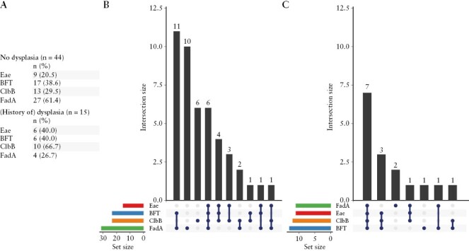
[A] Frequency of oncotraits FadA [green], ClbB [orange], BFT [blue], and Eae [red] in 59 UC patients [B] and 25 controls [C]. Set size represents n patients detected with each oncotrait. Intersection size depicts the number of patients with the combinations of oncotraits [dark blue circles] on the y-axis.
In UC, ClbB was positively associated with dysplasia at the study colonoscopy or in the prior 5 years [OR 4.77, 95% CI 1.36–16.7], whereas FadA was negatively associated [OR 0.23, 95% CI 0.06–0.84; Table 3]. After adjusting for disease duration, an aOR of 7.97 [95% CI 1.77–35.9] was observed for ClbB and aOR 0.15 [95% CI 0.03–0.67; p = 0.01] for FadA. BFT and Eae were not associated with dysplasia in UC. To assess the independence of found associations, an explorative model including both FadA and ClbB was used. The association of ClbB with dysplasia remained significant [aOR 4.0, 1.09–14.7]. ClbB had a positive predictive value [PPV] for dysplasia within the past 5 years of 42.93% [CI: 29.64%–57.32%] and a negative predictive value [NPV] of 86.11% [CI: 74.72%–92.86%]. FadA had a PPV of 12.90% [5.84%–26.15%] and NPV of 61.25% [49.41%–71.89%] for dysplasia in the past 5 years [See Supplementary Figure 2 for NPV/PPV dependence on dysplasia prevalence].20
3.3. Biofilms
3.3.1. Biofilms at study colonoscopy
Biofilms [Figure 3A] were present in 50.0% of the controls at any location [left and/or right colon; Figure 3B] [see Supplementary Figure 3 for images of biofilms]. In total, nine [27.3%] control patients with a right-sided biofilm had a left-sided biofilm as well. Biofilm prevalence was higher in high-risk [n = 38] UC patients [72.2%], PSC [84.6%], dysplasia during endoscopy [70.0%], and any history of dysplasia [69.6%], compared with patients without [a history of] dysplasia or PSC (low-risk [n = 42]; 57.5%), but this was not significant [Figure 3B B; Supplementary Table 5]. Biofilms occurred in 72.2% of patients with dysplasia at the study colonoscopy or in the prior five years versus 62.1% in the remaining group [p = 0.43]. Biofilms occurred numerically more frequently in the ascending versus descending colon for both UC patients and controls [47 vs 36% in UC and 45 vs 32% in controls; p = 0.08] [Supplementary Table 5].
Figure 3.

Biofilms and cell proliferation in ascending and descending colon biopsies. [A] Example of a bacterial biofilm spanning two colonic crypts in an ulcerative colitis [UC] patient. Cyan = nuclear staining [DAPI], yellow = Eubacteria staining [eub338]. [B] Prevalence of biofilms in ascending and descending colon biopsies at study colonoscopy in controls [n = 34 any, n = 33 ascending, and n = 34 descending biopsies], low-risk[n = 40 any, n = 41 ascending, and n = 40 descending biopsies], and high-risk UC patients [n = 36 any, 37 ascending, and 36 descending biopsies]. [C] Average biofilm thickness [µm] and biofilm length [µm] separated by left- and right-sided colon biopsies in UC patients [left: n = 26; right: n = 34] and controls [left: n = 11; right: n = 15]. Right-sided colon = green, left-sided colon = blue. [D] Average mucus depth measured on PAS-stained right- [n = 16 control, n = 59 UC; green] and left-sided [n = 17 control, n = 51 UC; blue] colon biopsies.[E] Number of cells per crypt and number of ki67-positive cells per crypt in biofilm-positive [dotted boxplots] and biofilm-negative [plain boxplots] in UC patients separated by right- [green][n = 19] and left-sided [n = 22] colon [blue]. N = no biofilm, B = biofilm. [F] Representative pictures of ki-67 staining [brown] in biofilm-negative right colon and biofilm-positive right colon of one UC patient. Mann-Whitney U-test was performed for biofilm thickness and length, and mucus thickness; Independent Students t test was performed for number of cells per crypt and number of ki67-positive cells per crypt of biofilm vs no biofilm.
Right-sided biofilms were not associated with age [p = 0.48], smoking [p = 0.13], extensive disease [p = 0.44], or SSCAI score [p = 0.07] [Supplementary Table 6]. Biofilms in the right-sided colon were significantly thicker compared with left-sided biofilms [median 13.8 µm vs 9.2 µm; p = 0.031] [Figure 3C], whereas there was no difference in average biofilm length. There were no significant differences in biofilm thickness, length, and area between UC patients and controls [Supplementary Figure 4]. No correlation was observed between average biofilm and mucus thickness in right- [Spearman R 0.103] and left-sided colon biopsies [Spearman R 0.069] [Figure 3D and Supplementary Figure 5]. Biofilms were associated with epithelial hypertrophy in UC patients measured by the increased number of epithelial cells per crypt [median number of cells per crypt 42.5 vs 51.1; p = 0.025], and a slight non-significant increase in Ki67-positive cells per crypt [22.0 vs 26.1; p = 0.11] [Figure 3E and F].
3.3.2. Longitudinal biofilm characteristics in UC and associations with dysplasia
In 65 UC patients, 264 colonoscopies with left and right biopsies were performed, including 27 patients who developed dysplasia. Left- or right-sided biofilms were present at 141 colonoscopies [53.4%]. Presence [at least on one occasion] of biofilms was observed in 59/65 [90.8%] of patients [Figure 4A and B]. The rate of biofilm-positive surveillance was 0.38 [IQR 0.0–0.6] and 0.43 [IQR 0.29–0.67] for left- and right-sided biopsies in UC patients with dysplasia compared with 0.24 [IQR 0.02–0.5] and 0.25 [IQR 0.02–0.75] for patients without dysplasia [Kolmogrov-Smirnof, not significant, Supplementary Figure 6]. In patients with dysplasia, biofilms were present before dysplasia development in 73% of the cases [n = 19], whereas for eight cases no biopsies were available to assess biofilms prior to dysplasia development [Figure 4B]. The median duration of biofilm persistence in between biofilm-negative colonoscopies was 3 years [IQR 2–5 years] in patients with more than one biofilm-positive colonoscopy. Median number of colonoscopies in biofilm-positive intervals was 2.5 years [IQR 2–3.3 years]. Biofilms were neither associated with endoscopically visible active inflammation in left- and/or right-sided biopsies [p = 0.43–0.92], nor with dysplasia [aOR after confounder correction for disease duration: 1.45, 95% CI 0.63–3.40].
Figure 4.

Longitudinal biofilms in patients with and without dysplasia. [A] Ulcerative colitis [UC] patients without dysplasia [n = 38], and [B] UC patients with dysplasia [n = 27]. For each patient left [L] and right-sided [R] colonoscopies are displayed with disease duration on the x-axis. Data are sorted based on disease duration. Green bars represent biofilm-positive episodes, black bars represent biofilm-negative episodes, red squares indicate episodes with dysplasia present.
3.3.3. Bacterial composition in UC patients and biofilms
Metagenomic shotgun analysis of left- and right-sided biopsies revealed that the bacterial composition in UC patients differed from those in the control population, characterised by a lower Shannon diversity index [4.80 vs 6.01 in controls; p = 0.0009] [Figure 5A; Supplementary Table 7]. Only in UC patients, right-sided biopsies displayed a significantly lower Shannon diversity as opposed to left-sided biopsies [Figure 5B], but was similar between UC patients with a low and high dysplasia risk [Figure 5C].
Figure 5.

Metagenomic shotgun sequencing of healthy-appearing colon ascendens [right] and descendens [left] in ulcerative colitis [UC] and control patients. Bacterial composition displayed at the order level with Shannon diversity [S. diversity]. Samples were sorted by their correlation to the group’s mean composition. [A] UC [n = 80] vs control [n = 32] patients [three controls failed metagenomics sequencing due to low DNA quality]. [B] No biofilm vs biofilm of UC and control patients. [C] Left vs right colon of UC patients. [D] Biofilms in UC vs control patients. [E] High- [1] vs low- [0] risk UC patients in general. [F] Biofilms in high-risk vs low-risk UC patients.
Biopsies with biofilms displayed a lower Shannon diversity [p = 0.0152; Figure 5D], but were not different between control and UC patients [Figure 5E], or between UC patients with low or high dysplasia risk [Figure 5F]. ANCOM analysis showed that biofilm presence could be predicted by Clostridiales, Bacteroidales, and Veillonellales [adj-p = 0.012] in metagenomes of controls and Selenomonadales and Synergistales in metagenomes of UC patients [adj-p = 0.011]. More specifically, presence of Eggerthellales and absence of Leptospirales could predict biofilms in right-sided biopsies of high-risk UC patients [adj-p = 0.023; Supplementary Table 8]. Noteworthy is the observation that high risk for dysplasia in UC patients could be predicted by the absence of Fusobacteriales [p = 0.00892; Supplementary Figure 7], similar to the inverse relationship of FadA with high-risk UC patients.
4. Discussion
Our study provides unique evidence that bacterial oncogenic factors show potential for dysplasia prediction and risk stratification in UC patients. The oncotrait colibactin [ClbB] was independently associated with dysplasia in multivariate analysis and supports a role for colibactin in early carcinogenesis in UC patients. Contrarily, the commonly CRC-associated Fusobacteriales, along with its associated adhesin FadA, was negatively associated with dysplasia in UC patients. Bacterial biofilms were studied longitudinally, spanning median 10 years in >800 biopsies of UC patients undergoing surveillance colonoscopies, and were intermittently present in the majority of UC patients [90.8%]. The high longitudinal prevalence of these biofilms impairs its usage as risk stratification tool in UC patients, and biofilms were not associated with dysplasia. Presence of biofilms associated with a lower Shannon diversity and epithelial hypertrophy. Together our data suggest that specific bacterial factors, such as presence of ClbB and absence of FadA, associate with dysplasia in UC patients, and show potential for faecal oncotrait screening as risk stratification tool.
An altered microbiome composition and function have been associated with the development and progression of UC.21 Currently, UC prediction algorithms based on microbial markers are rapidly developing,22 but these microbial markers have unknown roles in development and prediction of [early] carcinogenesis.23 ClbB is located on the pks + island that encodes colibactin,24 which directly damages DNA and results in a mutational signature in human intestinal organoids which can be found in some CRC patients.10 Thus far, the direct link between colibactin and an increased CRC risk in human patients has been lacking.25 Here, we present ClbB as independent factor associated with recent or prevalent dysplasia [aOR 7.97]. The prevalence of ClbB in our study was similar between controls and UC patients, as opposed to a previous report showing significantly higher colibactin levels in IBD patients compared with controls.26 However, most of those patients had active IBD, whereas all patients in our study were in endoscopic remission. The high prevalence of ClbB in control patients, but also other populations like FAP,9 suggests ClbB may be relevant for dysplasia development in multiple populations and warrants further research.
FadA can bind to E-cadherin on epithelial cells and can promote inflammation and oncogenic processes.27 FadA and Fusobacteriales presence were associated with an absence of [prior] dysplasia, in contrast to literature indicating a relation between CRC and increased FadA presence.28 Our contrasting results underline the growing concept that this bacterium is associated to later carcinogenic stages, possibly by being attracted to tumour metabolites,29 and is not actually an inducer of CRC, in contrast to colibactin.30,31 Another study showed that FadA promotes CRC growth in mice but proposed that FadA only affects cancerous cells.31 Moreover, Fusobacteria have variations in presence and expression of FadA,32 resulting in strain- rather than species-dependent oncogenic potential.33 Recent literature even suggests that Fusobacteria supernatants induce expression of immunomodulatory TGFβ1 in vitro, possibly by its production of butyrate.34 Our results support that Fusobacteriales do not associate with early dysplasia and we even observed an inverse relationship of Fusobacteriales and FadA in early carcinogenesis in UC.
Two other toxin-encoding genes, Eae and BFT, were not associated with dysplasia in UC patients. Eae, which has been shown to induce loss of DNA mismatch repair genes,35–37 did not occur without ClbB in our cohort, possibly because both genes are found in E. coli. The effect of Eae was not significant and therefore is likely to play a minor role in dysplasia risk, as opposed to ClbB. B. fragilis and BFT were reported to be over-expressed in tissue biopsies of UC patients with active disease and to be frequently present in the mucosa of CRC patients and controls.8,18,38 Although compiling evidence shows the potential contribution of BFT to CRC development,30 our prospective study does not associate BFT with dysplasia in the UC population. In our study, BFT was frequently present in UC and control patients, and frequently occurred together with FadA, which shows an inverse relationship. Further longitudinal studies in humans are warranted to investigate combinations of oncotraits on CRC risk in UC patients.39
Colonic biofilms have been associated with gastrointestinal symptoms in patients with UC and other IBDs,7,8 as well as sporadic CRC and FAP.35,40,41 A study from Baumgartner et al. detected visible biofilms in 34% and microscopic biofilms in 79% of UC patients, comparable to our microscopic biofilm rate of 72.2% in high-risk UC patients at study colonoscopy. These authors found a low biofilm prevalence of 6% in controls compared with the higher prevalence in our study [50%]. Importantly, this might be explained by the different selection of controls in our study, also allowing patients with symptoms to be included. Moreover, biofilms were numerically more frequent and thicker in the right-sided colon compared with the more distal colon, were frequently persistent over time, and showed an association with epithelial hypertrophy in UC patients. Samples with biofilms were associated with a higher bacterial abundance, but a lower Shannon diversity, than samples without biofilms,16 indicating the outgrowth of specific bacteria; Clostridiales, Bacteroidales, and Veillonellales in metagenomes of controls, and Selenomonadales and Synergistales in metagenomes of UC patients. In a mouse model, biofilms from both CRC and control patients were carcinogenic,42 However, our longitudinal data did not show a significant association between biofilm presence and dysplasia risk in UC patients. The near uniform longitudinal biofilm presence complicates the investigation of biofilms as a possible risk factor for dysplasia, because only few patients present without biofilms. Moreover, we were not able to show that biofilms associated with high dysplasia risk are different from biofilms in low-risk patients. We speculate that this is the consequence of biofilms harbouring a multitude of bacterial species, not all associated with pro-inflammatory or oncogenic characteristics.
4.1. Strengths and limitations
We extensively assessed biofilm presence over time as well as cross-sectionally in a prospective fashion, providing a link with clinical and histological presence of inflammation and dysplasia. The UC population used for this study may present with a higher disease burden compared with the general UC population, due the recruitment in a tertiary IBD referral centre that includes patients with PSC, a longer disease duration and therapy-refractory disease. The majority of the UC patients presented with low-grade dysplasia [LGD]. Although the risk of advanced neoplasia is elevated after development of LGD, it is uncertain how and for whom progression will take place. Furthermore, our colonoscopy controls were carefully selected and had no abnormal findings during colonoscopy such as dysplasia and inflammation. In addition, this is the first study that assessed oncotrait presence in UC in relation with dysplasia. Although we prospectively collected data from our patients, the additional longitudinal analysis of biopsies that were collected prior to the study colonoscopy has a retrospective nature and involved formalin-fixed rather than methacarn-fixed tissues, which may affect sensitivity of biofilm detection. Finally, from our controls we do not have a longitudinal follow-up, and we are unable to investigate whether the presence of oncotraits in controls is associated with dysplasia. The potential additive or synergistic effects of bacterial oncogenes are subject of our ongoing investigations, and our results should be validated in larger longitudinal cohorts.
4.2. Conclusion
With an incidence of 90.8%, longitudinal biofilms cannot serve as biomarkers to predict dysplasia development in UC patients. Although no association between biofilms and dysplasia was confirmed, it is likely that these biofilms and pathogenic inhabitants can increase dysplasia risk, considering recent literature.40–42 Our data on ClbB show for the first time that its presence in stool was significantly and independently associated with dysplasia in UC patients. On the contrary, FadA showed an inverse significant association, therefore, both ClbB presence and FadA absence are useful biomarkers for UC-risk stratification. Since these markers are detectable in faeces, they could potentially be used to predict low-risk patients and thereby reduce the number of invasive endoscopic procedures for CRC prevention. More research is needed to investigate whether more oncotraits could be incorporated into faecal screening and whether such screening could aid beneficial effects of faecal transfers. Indeed, incorporation of ClbB faecal testing in a CRC detection model for the general population has shown promise and resulted in an improved sensitivity.43,44 This emphasises the potential for ClbB in future CRC risk stratification in UC and in improving the discriminatory value for use in clinical practice.
Supplementary Data
Supplementary data are available at ECCO-JCC online.
Contributor Information
Carlijn E Bruggeling, Department of Pathology, Radboud Institute for Molecular Life Sciences [RIMLS], Radboud University Medical Center, Nijmegen, The Netherlands.
Maarten te Groen, Inflammatory Bowel Disease Center, Department of Gastroenterology, Radboud University Medical Center, Nijmegen, The Netherlands.
Daniel R Garza, Center for Molecular and Biomolecular Informatics [CMBI], Radboud Institute for Molecular Life Sciences [RIMLS], Nijmegen, The Netherlands; KU Leuven, Department of Microbiology, Immunology and Transplantation, Rega Institute for Medical Research, Laboratory of Molecular Bacteriology, Leuven, Belgium.
Famke van Heeckeren tot Overlaer, Department of Pathology, Radboud Institute for Molecular Life Sciences [RIMLS], Radboud University Medical Center, Nijmegen, The Netherlands.
Joyce P M Krekels, Department of Pathology, Radboud Institute for Molecular Life Sciences [RIMLS], Radboud University Medical Center, Nijmegen, The Netherlands.
Basma-Chick Sulaiman, Department of Pathology, Radboud Institute for Molecular Life Sciences [RIMLS], Radboud University Medical Center, Nijmegen, The Netherlands.
Davy Karel, Department of Pathology, Radboud Institute for Molecular Life Sciences [RIMLS], Radboud University Medical Center, Nijmegen, The Netherlands.
Athreyu Rulof, Department of Pathology, Radboud Institute for Molecular Life Sciences [RIMLS], Radboud University Medical Center, Nijmegen, The Netherlands.
Anne R Schaaphok, Department of Pathology, Radboud Institute for Molecular Life Sciences [RIMLS], Radboud University Medical Center, Nijmegen, The Netherlands.
Daniel L A H Hornikx, Department of Pathology, Radboud Institute for Molecular Life Sciences [RIMLS], Radboud University Medical Center, Nijmegen, The Netherlands.
Iris D Nagtegaal, Department of Pathology, Radboud Institute for Molecular Life Sciences [RIMLS], Radboud University Medical Center, Nijmegen, The Netherlands.
Bas E Dutilh, Center for Molecular and Biomolecular Informatics [CMBI], Radboud Institute for Molecular Life Sciences [RIMLS], Nijmegen, The Netherlands; Institute of Biodiversity, Faculty of Biological Sciences, Cluster of Excellence Balance of the Microverse, Friedrich Schiller University Jena, Jena, Germany; Theoretical Biology and Bioinformatics, Science for Life, Utrecht University, Utrecht, The Netherlands.
Frank Hoentjen, Inflammatory Bowel Disease Center, Department of Gastroenterology, Radboud University Medical Center, Nijmegen, The Netherlands; Division of Gastroenterology, Department of Medicine, University of Alberta, Edmonton, AB, Canada.
Annemarie Boleij, Department of Pathology, Radboud Institute for Molecular Life Sciences [RIMLS], Radboud University Medical Center, Nijmegen, The Netherlands.
Funding
This work was supported by the Netherlands Organization for Scientific Research ‘Veni grant 016.166.089’ to A.B. and ‘Vidi grant 864.14.004’ to B.E.D., the Dutch cancer society ‘KWF-kankerberstrijding grant KUN 2015-7739’, the European Research Council ‘Consolidator grant 865694: DiversiPHI’ to B.E.D., the Netherlands Organization for Gastroenterology ‘Gastrostart PROJECT 19-2021’, and the Deutsche Forschungsgemeinschaft ‘German Research Foundation’ under Germany´s Excellence Strategy ‘EXC 2051 – Project-ID 390713860’ to B.E.D.
Conflict of Interest
The authors declare no conflicts of interest.
Author Contributions
Study design [A.B., F.H.], patient recruitment and sample processing [C.E.B., J.K., A.B., F.H.], clinical data acquisition and analyses [A.B., C.E.B., M.tG., J.K., I.D.N., A.R.S.], metagenomics [C.E.B., D.R.G., B.E.D]., biofilm assessment [C.E.B., F.H.O., J.K., A.R., A.B], oncotrait analyses [B.S., D.K., A.B.], mucus and ki67 assessment [C.E.B., A.B., D.L.A.H.], pathology [I.D.N.], supervision [A.B., B.E.D., F.H., I.D.N.], article writing [all authors].
This study was presented as oral presentation during the Digestive Disease Week 2022 in San Diego and the Dutch Digestive Disease Days 2022.
Data Availability
Raw sequencing data with human reads will not be publicly available because of General Data Protection Regulation [GDPR]. Processed sequencing data are available upon request. Patient and research data are anonymised and openly available in Supplementary Data: [https://www.medrxiv.org/content/10.1101/2022.09.09.22279675v1.supplementary-material].
References
- 1. Alatab S, Sepanlou SG, Ikuta K, et al. The global, regional, and national burden of inflammatory bowel disease in 195 countries and territories, 1990–2017: A systematic analysis for the global burden of disease study 2017. Lancet Gastroenterol Hepatol 2020;5:17–30. [DOI] [PMC free article] [PubMed] [Google Scholar]
- 2. Jess T, Rungoe C, Peyrin-Biroulet L.. Risk of colorectal cancer in patients with ulcerative colitis: A meta-analysis of population-based cohort studies. Clin Gastroenterol Hepatol 2012;10:639–45. [DOI] [PubMed] [Google Scholar]
- 3. Lutgens MW, van Oijen MG, van der Heijden GJ, Vleggaar FP, Siersema PD, Oldenburg B.. Declining risk of colorectal cancer in inflammatory bowel disease: An updated meta-analysis of population-based cohort studies. Inflamm Bowel Dis 2013;19:789–99. [DOI] [PubMed] [Google Scholar]
- 4. Burke KE, Nayor J, Campbell EJ, Ananthakrishnan AN, Khalili H, Richter JM.. Interval colorectal cancer in inflammatory bowel disease: The role of guideline adherence. Dig Dis Sci 2020;65:111–8. [DOI] [PMC free article] [PubMed] [Google Scholar]
- 5. Wintjens DSJ, Bogie RMM, van den Heuvel TRA, et al. Incidence and classification of postcolonoscopy colorectal cancers in inflammatory bowel disease: A Dutch population-based cohort study. J Crohns Colitis 2018;12:777–83. [DOI] [PubMed] [Google Scholar]
- 6. Mooiweer E, van der Meulen-de Jong AE, Ponsioen CY, et al.; Dutch Initiative on Crohn’s and Colitis. Incidence of interval colorectal cancer among inflammatory bowel disease patients undergoing regular colonoscopic surveillance. Clin Gastroenterol Hepatol 2015;13:1656–61. [DOI] [PubMed] [Google Scholar]
- 7. Baumgartner M, Lang M, Holley H, et al. Mucosal biofilms are an endoscopic feature of irritable bowel syndrome and ulcerative colitis. Gastroenterology 2021;161:1245–56.e20. [DOI] [PMC free article] [PubMed] [Google Scholar]
- 8. Swidsinski A, Weber J, Loening-Baucke V, Hale LP, Lochs H.. Spatial organization and composition of the mucosal flora in patients with inflammatory bowel disease. J Clin Microbiol 2005;43:3380–9. [DOI] [PMC free article] [PubMed] [Google Scholar]
- 9. Dejea CM, Fathi P, Craig JM, et al. Patients with familial adenomatous polyposis harbor colonic biofilms containing tumorigenic bacteria. Science 2018;359:592–7. [DOI] [PMC free article] [PubMed] [Google Scholar]
- 10. Pleguezuelos-Manzano C, Puschhof J, Rosendahl Huber A, et al.; Genomics England Research Consortium. Mutational signature in colorectal cancer caused by genotoxic pks+ e. Coli. Nature 2020;580:269–73. [DOI] [PMC free article] [PubMed] [Google Scholar]
- 11. Dziubańska-Kusibab PJ, Berger H, Battistini F, et al. Colibactin DNA-damage signature indicates mutational impact in colorectal cancer. Nat Med 2020;26:1063–9. [DOI] [PubMed] [Google Scholar]
- 12. Strakova N, Korena K, Karpiskova R.. Klebsiella pneumoniae producing bacterial toxin colibactin as a risk of colorectal cancer development: a systematic review. Toxicon 2021;197:126–35. [DOI] [PubMed] [Google Scholar]
- 13. Rubinstein MR, Wang X, Liu W, Hao Y, Cai G, Han YW.. Fusobacterium nucleatum promotes colorectal carcinogenesis by modulating e-cadherin/beta-catenin signaling via its fada adhesin. Cell Host Microbe 2013;14:195–206. [DOI] [PMC free article] [PubMed] [Google Scholar]
- 14. Wu S, Rhee KJ, Albesiano E, et al. A human colonic commensal promotes colon tumorigenesis via activation of t helper type 17 t cell responses. Nat Med 2009;15:1016–22. [DOI] [PMC free article] [PubMed] [Google Scholar]
- 15. Pratt M, Forbes JD, Knox NC, Van Domselaar G, Bernstein CN.. Colorectal cancer screening in IBD: can characterization of GI microbiome signatures enhance neoplasia detection? Gastroenterology 2022;162:1409–23. [DOI] [PubMed] [Google Scholar]
- 16. Bruggeling CE, Garza DR, Achouiti S, Mes W, Dutilh BE, Boleij A.. Optimized bacterial DNA isolation method for microbiome analysis of human tissues. Microbiologyopen 2021;10:e1191. [DOI] [PMC free article] [PubMed] [Google Scholar]
- 17. Dejea CM, Wick EC, Hechenbleikner EM, et al. Microbiota organization is a distinct feature of proximal colorectal cancers. Proc Natl Acad Sci U S A 2014;111:18321–6. [DOI] [PMC free article] [PubMed] [Google Scholar]
- 18. Boleij A, Hechenbleikner EM, Goodwin AC, et al. The bacteroides fragilis toxin gene is prevalent in the colon mucosa of colorectal cancer patients. Clin Infect Dis 2015;60:208–15. [DOI] [PMC free article] [PubMed] [Google Scholar]
- 19. Textor J, van der Zander B, Gilthorpe MS, Liśkiewicz M, Ellison GT.. Robust causal inference using directed acyclic graphs: The R package ‘dagitty’. Int J Epidemiol 2017;45:1887–94. [DOI] [PubMed] [Google Scholar]
- 20. Medcalc Software Ltd. Diagnostic Test Evaluation Calculator. https://www.medcalc.org/calc/diagnostic_test.php. Version 20.215 edn. Accessed January 25, 2023
- 21. Sartor RB, Wu GD.. Roles for intestinal bacteria, viruses, and fungi in pathogenesis of inflammatory bowel diseases and therapeutic approaches. Gastroenterology 2017;152:327–39.e4. [DOI] [PMC free article] [PubMed] [Google Scholar]
- 22. Wingfield B, Coleman S, McGinnity TM, Bjourson AJ.. Robust microbial markers for non-invasive inflammatory bowel disease identification. IEEE/ACM Trans Comput Biol Bioinform 2019;16:2078–88. [DOI] [PubMed] [Google Scholar]
- 23. Yalchin M, Baker AM, Graham TA, Hart A.. Predicting colorectal cancer occurrence in IBD. Cancers [Basel] 2021;13:2908. [DOI] [PMC free article] [PubMed] [Google Scholar]
- 24. Faïs T, Delmas J, Barnich N, Bonnet R, Dalmasso GC.. More than a new bacterial toxin. Toxins [Basel] 2018;10:151. [DOI] [PMC free article] [PubMed] [Google Scholar]
- 25. Dubinsky V, Dotan I, Gophna U.. Carriage of colibactin-producing bacteria and colorectal cancer risk. Trends Microbiol 2020;28:874–6. [DOI] [PubMed] [Google Scholar]
- 26. Arthur JC, Perez-Chanona E, Mühlbauer M, et al. Intestinal inflammation targets cancer-inducing activity of the microbiota. Science 2012;338:120–3. [DOI] [PMC free article] [PubMed] [Google Scholar]
- 27. Rubinstein MR, Wang X, Liu W, Hao Y, Cai G, Han YW.. Fusobacterium nucleatum promotes colorectal carcinogenesis by modulating e-cadherin/β-catenin signaling via its fada adhesin. Cell Host Microbe 2013;14:195–206. [DOI] [PMC free article] [PubMed] [Google Scholar]
- 28. Zorron Cheng Tao Pu L, Yamamoto K, Honda T, et al. Microbiota profile is different for early and invasive colorectal cancer and is consistent throughout the colon. J Gastroenterol Hepatol 2020;35:433–7. [DOI] [PubMed] [Google Scholar]
- 29. Garza DR, Taddese R, Wirbel J, et al. Metabolic models predict bacterial passengers in colorectal cancer. Cancer & Metabolism 2020;8:1–3. [DOI] [PMC free article] [PubMed] [Google Scholar]
- 30. Tomkovich S, Yang Y, Winglee K, et al. Locoregional effects of microbiota in a preclinical model of colon carcinogenesis. Cancer Res 2017;77:2620–32. [DOI] [PMC free article] [PubMed] [Google Scholar]
- 31. Rubinstein MR, Baik JE, Lagana SM, et al. Fusobacterium nucleatum promotes colorectal cancer by inducing wnt/β-catenin modulator annexin a1. EMBO Rep 2019;20:e47638. [DOI] [PMC free article] [PubMed] [Google Scholar]
- 32. Kashani N, Bezmin Abadi AT, Rahimi F, Forootan M.. Fada-positive fusobacterium nucleatum is prevalent in biopsy specimens of Iranian patients with colorectal cancer. New Microbes New Infect 2020;34:100651. [DOI] [PMC free article] [PubMed] [Google Scholar]
- 33. Taddese R, Garza DR, Ruiter LN, et al. Growth rate alterations of human colorectal cancer cells by 157 gut bacteria. Gut Microbes 2020;12:1–20. [DOI] [PMC free article] [PubMed] [Google Scholar]
- 34. Martin-Gallausiaux C, Béguet-Crespel F, Marinelli L, et al. Butyrate produced by gut commensal bacteria activates tgf-beta1 expression through the transcription factor sp1 in human intestinal epithelial cells. Sci Rep 2018;8:9742. [DOI] [PMC free article] [PubMed] [Google Scholar]
- 35. Abdulamir AS, Hafidh RR, Bakar FA.. Molecular detection, quantification, and isolation of streptococcus gallolyticus bacteria colonizing colorectal tumors: Inflammation-driven potential of carcinogenesis via il-1, cox-2, and il-8. Mol Cancer 2010;9:249. [DOI] [PMC free article] [PubMed] [Google Scholar]
- 36. Maddocks OD, Scanlon KM, Donnenberg MS.. An escherichia coli effector protein promotes host mutation via depletion of DNA mismatch repair proteins. mBio 2013;4:e00152–13. [DOI] [PMC free article] [PubMed] [Google Scholar]
- 37. Maddocks OD, Short AJ, Donnenberg MS, Bader S, Harrison DJ.. Attaching and effacing escherichia coli downregulate DNA mismatch repair protein in vitro and are associated with colorectal adenocarcinomas in humans. PLoS One 2009;4:e5517. [DOI] [PMC free article] [PubMed] [Google Scholar]
- 38. Zamani S, Hesam Shariati S, Zali MR, et al. Detection of enterotoxigenic bacteroides fragilis in patients with ulcerative colitis. Gut Pathogens 2017;9:1–7. [DOI] [PMC free article] [PubMed] [Google Scholar]
- 39. Scott N, Whittle E, Jeraldo P, Chia N.. A systemic review of the role of enterotoxic bacteroides fragilis in colorectal cancer. Neoplasia 2022;29:100797. [DOI] [PMC free article] [PubMed] [Google Scholar]
- 40. Dejea CM, Fathi P, Craig JM, et al. Patients with familial adenomatous polyposis harbor colonic biofilms containing tumorigenic bacteria. Science 2018;359:592–7. [DOI] [PMC free article] [PubMed] [Google Scholar]
- 41. Dejea CM, Wick EC, Hechenbleikner EM, et al. Microbiota organization is a distinct feature of proximal colorectal cancers. Proc Natl Acad Sci U S A 2014;111:18321–6. [DOI] [PMC free article] [PubMed] [Google Scholar]
- 42. Tomkovich S, Dejea CM, Winglee K, et al. Human colon mucosal biofilms from healthy or colon cancer hosts are carcinogenic. J Clin Invest 2019;129:1699–712. [DOI] [PMC free article] [PubMed] [Google Scholar]
- 43. Eklöf V, Löfgren-Burström A, Zingmark C, et al. Cancer-associated faecal microbial markers in colorectal cancer detection. Int J Cancer 2017;141:2528–36. [DOI] [PMC free article] [PubMed] [Google Scholar]
- 44. Löwenmark T, Löfgren-Burström A, Zingmark C, et al. Parvimonas micra as a putative non-invasive faecal biomarker for colorectal cancer. Sci Rep 2020;10:15250. [DOI] [PMC free article] [PubMed] [Google Scholar]
Associated Data
This section collects any data citations, data availability statements, or supplementary materials included in this article.
Supplementary Materials
Data Availability Statement
Raw sequencing data with human reads will not be publicly available because of General Data Protection Regulation [GDPR]. Processed sequencing data are available upon request. Patient and research data are anonymised and openly available in Supplementary Data: [https://www.medrxiv.org/content/10.1101/2022.09.09.22279675v1.supplementary-material].