

Key Clinical Message
Fetus in fetu (FIF) is a rare congenital anomaly that originates from various sites of the host twin's body. The clinical manifestations of FIF are diverse and the location and size of FIF indicate the degree of threat, which may directly affect the prognosis. A 33‐year‐old woman presented at the hospital with an abdominal mass in her fetus. Prenatal ultrasound observed that mass included soft tissue, bone‐like structures, and fluid. Immature cartilage, nerve tissue, muscle tissue, and glands in the parasitic fetus without signs of neoplastic lesions were reported by histological examination. CNV (copy number variation) and WES (whole exome sequencing) did not detect any abnormal mutations. FIF can continue to grow with gestational age or host infant growth. So complete resection is essential for improving the outcome of the host twin. It is also important that long‐term follow‐up is recommended to monitor any residual or recurrent cysts or malignancies.
Keywords: fetal physiologic changes in pregnancy, fetus in fetu, general genetics, second trimester anomalies

Ultrasonographic images.
1. INTRODUCTION
Fetus in fetu (FIF) is a rare congenital anomaly that occurs when a well‐differentiated fetus with an organ or limb is enclosed within the body of its monozygotic twin. This phenomenon results from the abnormal division of the inner cell mass during early embryonic development. 1 FIF can be found in various locations of the host twin's body, ranging from the cranial cavity to the sacrococcygeal region. The diagnosis of FIF usually relies on prenatal ultrasound and MRI, but histopathological examination is still necessary for definitive diagnosis. 2 The prognosis of FIF depends on the degree of threat that the parasitic twin poses to the host twin's development and survival. Surgical removal of the parasitic twin is usually recommended as soon as possible. 3 In this paper, we report a rare case of FIF that occurred on the abdominal cavity and evidence by pathology.
2. CASE PRESENTATION
The patient was a 33‐year‐old female, G1P0, who conceived spontaneously with no remarkable family history or medical conditions. At 14 weeks of gestation, an ultrasound scan revealed an abnormal mass in the sacrococcygeal region of the fetus. The mass had heterogeneous echogenicity and measured 30 × 27 × 18 mm. It contained hypoplastic lower and upper extremities that were connected to a vertebral column (Figure 1). One week later, reexamination by color Doppler ultrasound confirmed the possibility of a parasitic fetus, and the doctor informed that surgical treatment for FIF is curative after birth. Considering the cost of operation and burden on the family, the pregnant woman requested for induced abortion and for the products of conception to be sent for genetic testing.
FIGURE 1.

Ultrasonographic images. (A) Color Doppler flow imaging (CDFI) showed that a parasitic fetus was attached to the lower abdomen of the host twin. (B) Ultrasound image showed a heterogeneous echoic mass in the sacrococcygeal region of the fetus.
2.1. Gross specimen
Two fetuses were fused at their lower abdomens and lacked external genitalia and anus. They had separate limbs and shared a common umbilical cord and placenta. The host twin measured 40 × 20 × 10 mm while the parasitic twin was approximately 30 × 20 × 10 mm with a flat abdomen (Figure 2).
FIGURE 2.
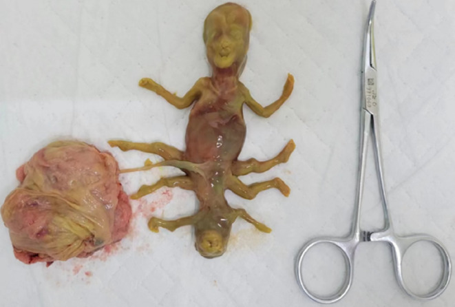
Gross specimen. Two fetuses were fused with each other from the lower abdomen and shared the same umbilical cord and placenta.
2.2. Pathological findings
Microscope examination showed immature cartilage, nerve tissue, muscle tissue, and glands in the parasitic fetus without any sign of neoplastic lesions (Figure 3).
FIGURE 3.

Pathology of the mass. (A) Digestive glandular epithelial tissue (40×); (B) immature cartilage tissue (40×).
2.3. Genetic testing
Copy number variation (CNV) and whole exome sequencing (WES) did not report any abnormal mutations.
3. DISCUSSION
In this case, we reported a rare occurrence of FIF on the abdominal cavity that was diagnosed by prenatal ultrasound and confirmed by histopathology. FIF is a rare congenital anomaly with an estimated incidence of 1 in 500,000 births. 4 It was first described by Meckel in 1808 as an organoid mass protruding from the mouth of a newborn. 5 FIF can originate from various sites of the host twin's body, but it is most commonly found in the abdominal cavity and retroperitoneum. Other reported locations include the intracranial structures, mouth, back, sacrococcygeal region, and scrotum. 6 FIF can grow progressively and compromise the host twin's survival and development. Therefore, early diagnosis and timely surgical removal of the parasitic twin are crucial for improving the prognosis. 7
The pathogenesis of FIF is still controversial. Two main theories have been proposed to explain its origin. One is the monozygotic twin theory, which suggests that FIF results from the asymmetric division of the inner cell mass during blastocyst development. The larger twin envelops the smaller one, leading to its incomplete differentiation. 8 This theory can be supported by genetic testing that shows identical chromosomal patterns between the host and parasitic twins. 9 The other is the teratoma theory, which argues that FIF is a highly differentiated form of teratoma tissue. However, teratomas usually lack functional organs and have malignant potential. There are also cases where both teratoma and FIF coexist in the same host. 10 In our case, pathological examination suggested that immature cartilage, nerve tissue, muscle tissue, and glands in the parasitic fetus without any signs of neoplastic lesions. Long‐term follow‐up is needed to confirm the potential for malignancy and more studies are needed to elucidate the mechanism of this disease.
Prenatal ultrasound is always the first modality to detect the abnormal cyst. Often, it is observed that there is inclusion of soft tissue, bone‐like structure and fluid within the mass. As gestation progresses, the spinal features appear as beaded pattern, and umbilical vascular composition become visible. 11 These features can help differentiate FIF from other ectopic cysts, such as teratomas and meconium pseudocyst. Teratomas are much more common and usually originate from the aggregation of pluripotent cells in the lower abdomen. They show mixed echogenicity with clear border and intact capsule on imaging modality, and they may have calcifications. However, they cannot form organized organs and vertebral column, and they have a risk of malignancy. 12 Meconium pseudocyst mostly present as abdominal calcifications, accompanied by ascites and pneumoperitoneum. Postnatal computed tomography (CT), magnetic resonance imaging (MRI), and x‐ray could be performed to identify contents and to assist in surgical planning. 13
The location and size of FIF may directly affect the prognosis of the host twin. According to previous reports, retroperitoneum is the most frequent site of parasitism (75% of cases), and fortunately, 97% of those had a good prognosis after surgery. 14 FIF can continue to grow with gestational age or host growth, so timely surgery and complete resection are essential for improving the outcome of the host infant. If the resection is incomplete, residual cyst may still degenerate into malignancy. 15 Lu reported a case of FIF in the sacrococcygeal region with incomplete development and anencephaly that was successfully excised with good recovery. 13 In contrast, intracranial occurrences are rare but have poor prognosis due to compression of surrounding structures and the difficulty of surgery. Kai had reported a case of FIF in the brain where the neonate died after birth due to severe intracranial space occupation and tissue edema before any treatment could be administered. 6 Our case belonged to the external type, and pathologic testing discovered that the parasitic fetus was connected to host twin by vertebra. Ultimately, the pregnant mother opted to terminate her pregnancy after considering the burden of surgery and uncertain prognosis.
4. CONCLUSION
FIF is a rare congenital anomaly that can be effectively identified by imaging modalities to show the presence of vertebral column and umbilical vessels in the parasitic mass. The site of parasitism is crucial for the outcome of the host twin, as FIF can grow progressively and compromise the host twin's survival and development by interfering with vital structures. Surgical removal of the parasitic twin should be performed as soon as possible to improve prognosis. The recovery of the host twin is generally good, but long‐term follow‐up is recommended to monitor any residual or recurrent cysts or malignancies. The pathogenesis and associated genetic mutation of FIF are still unclear and will need further investigation. This study adds some basis to pathological diagnosis of FIF, and provides insights for future research on this rare disease.
AUTHOR CONTRIBUTIONS
Ying Zhu: Conceptualization; data curation; writing – original draft. Le Xu: Data curation; resources. Rajluxmee Beejadhursing: Writing – review and editing. Suhua Chen: Conceptualization; supervision.
FUNDING INFORMATION
None.
CONFLICT OF INTEREST STATEMENT
The authors report no conflicts of interest.
ETHICS STATEMENT AND CONSENT TO PARTICIPATE
This report was approved by the ethics committee on human research at Tongji Hospital of Tongji Medical College of Huazhong University of Science and Technology.
CONSENT STATEMENT
Written informed consent was obtained from the patient to publish this report in accordance with the journal's patient consent policy.
Zhu Y, Xu Le, Beejadhursing R, Chen S. External fetus in fetu presenting in the second trimester: A case report and literature review. Clin Case Rep. 2023;11:e8057. doi: 10.1002/ccr3.8057
DATA AVAILABILITY STATEMENT
The data that support the findings of this study are available from the corresponding author upon reasonable request.
REFERENCES
- 1. Matsubara N, Akasaka Y, Kanagaki M, Okamoto S. A case report of fetus in fetu with an aorta‐like structure visualized by contrast‐enhanced CT. Radiol Case Rep. 2020;15(12):2645‐2648. [DOI] [PMC free article] [PubMed] [Google Scholar]
- 2. Ha TN, Nguyen TN, Huynh C, Nguyen HC, Le QT, Dao TH. Intra‐abdominal fetus in fetu presenting at 31 weeks gestational age. Radiol Case Rep. 2021;16(4):863‐866. [DOI] [PMC free article] [PubMed] [Google Scholar]
- 3. Joshi M, Parelkar S, Shah H, Agrawal A, Mishra P. Foetus in fetu with common bile duct injury: a case report and review of literature. ANZ J Surg. 2009;79(9):651‐652. [DOI] [PubMed] [Google Scholar]
- 4. Goyal RB, Gupta R, Prabhakar G, Dagla R. Fetus in fetu: report of two cases. APSP J Case Rep. 2014;5(3):28. [PMC free article] [PubMed] [Google Scholar]
- 5. Narayanasamy JN, Nallusamy MA, Baharuddin ND. Fetus‐in‐fetu: a pediatric rarity. J Surg Case Rep. 2014;2014(2):rju001. [DOI] [PMC free article] [PubMed] [Google Scholar]
- 6. Zhu K. Prenatal and postnatal MRI imaging findings of intracranial parasitic fetus: a case report. Childs Nerv Syst. 2021;37(5):1803‐1806. [DOI] [PubMed] [Google Scholar]
- 7. Harigovind D, Babu Sp H, Nair SV, Sangram N. Fetus in fetu ‐ a rare developmental anomaly. Radiol Case Rep. 2019;14(3):333‐336. [DOI] [PMC free article] [PubMed] [Google Scholar]
- 8. Issa M. Fetus in fetu: a rare case of intra‐abdominal mass. Radiol Case Rep. 2019;14(9):1171‐1174. [DOI] [PMC free article] [PubMed] [Google Scholar]
- 9. Brand A, Alves MC, Saraiva C, et al. Fetus in fetu—diagnostic criteria and differential diagnosis—a case report and literature review. J Pediatr Surg. 2004;39(4):616‐618. [DOI] [PubMed] [Google Scholar]
- 10. Kim YJ, Sohn SH, Lee JY, et al. Misdiagnosis of fetus‐in‐fetu as meconium peritonitis. Korean J Pediatr. 2011;54(3):133‐136. [DOI] [PMC free article] [PubMed] [Google Scholar]
- 11. Hong SS, Goo HW, Jung MR, et al. Fetus in fetu: three‐dimensional imaging using multidetector CT. AJR Am J Roentgenol. 2002;179(6):1481‐1483. [DOI] [PubMed] [Google Scholar]
- 12. Faizi FR, Rasouly N. Lumbar fetiform teratoma; a case report. Radiol Case Rep. 2020;15(7):837‐840. [DOI] [PMC free article] [PubMed] [Google Scholar]
- 13. Lu T, Ma J, Yang X. A rare case of fetus in fetu in the sacrococcygeal region: CT and MRI findings. BMC Pediatr. 2021;21(1):575. [DOI] [PMC free article] [PubMed] [Google Scholar]
- 14. Ji Y, Song B, Chen S, et al. Fetus in fetu in the scrotal sac: case report and literature review. Medicine (Baltimore). 2015;94(32):e1322. [DOI] [PMC free article] [PubMed] [Google Scholar]
- 15. Yaacob R, Zainal Mokhtar A, Abang Jamari DZH, Jaafar N. The entrapped twin: a case of fetus‐in‐fetu. BMJ Case Rep. 2017;2017:bcr2017220801. [DOI] [PMC free article] [PubMed] [Google Scholar]
Associated Data
This section collects any data citations, data availability statements, or supplementary materials included in this article.
Data Availability Statement
The data that support the findings of this study are available from the corresponding author upon reasonable request.