Abstract
PURPOSE
The purpose of this study was to analyze and compare the outcomes of percutaneous microwave ablation (MWA) when used as a primary vs. secondary treatment for hepatocellular carcinoma (HCC).
METHODS
The clinical data of 192 patients with HCC treated with MWA between January 2012 and July 2021 were reviewed retrospectively, with 152 patients being treatment naïve (primary treatment) vs. 40 who had residual or recurrent disease following previous trans-arterial chemoembolization or trans-arterial radioembolization (secondary treatment). The primary outcomes were primary technical efficacy, 1- and 3-year local recurrence-free survival (RFS) and overall survival (OS), local recurrence rates, and adverse events. Pre- and post-intervention liver function tests were compared using a Wilcoxon signed rank test. Univariate and multivariate analyses were also performed, looking at prognostic factors associated with OS and local RFS.
RESULTS
There was no significant difference in 1-year local RFS (primary 93.6% vs. secondary 93.7; P = 0.97) and 3-year local RFS (primary 80.6% vs. secondary 86.5%; P = 0.37) rates. There was no significant difference in 1-year OS (primary 82.4% vs. secondary 86.6%; P = 0.51) and 3-year OS (primary 68.3% vs. secondary 77.4%; P = 0.25) between the two groups. The local recurrence rate (primary 9.8% vs. secondary 14.6%; P = 0.37), primary technical efficacy (primary 96.2% vs. secondary 95%; P = 0.73), and adverse events (primary 8.0% vs. secondary 11.6%; P = 0.45) were also similar between the two groups.
CONCLUSION
Microwave ablation is safe and effective as a secondary treatment for patients with HCC in a clinical salvage scenario and should be utilized more frequently.
Keywords: Hepatocellular carcinoma, microwave ablation, primary treatment, secondary treatment, recurrent HCC
Main points
• Recurrent or residual hepatocellular carcinoma (HCC) following locoregional therapy is still relatively common.
• There are currently no guidelines for the treatment of residual tumors following locoregional therapy.
• Microwave ablation appears safe and effective as a secondary treatment of HCC, with similar local recurrence rates compared with its use as a primary treatment.
The incidence of hepatocellular carcinoma (HCC) recurrence following surgical resection and locoregional therapy (LRT) performed with curative intent is high. Three-year local progression rates after radiofrequency ablation (RFA) vary widely between institutions, ranging from 3.2% to 21.9%.3 Recurrence rates of 50% to 70% (of which 80% to 90% are intrahepatic) have been reported five years after hepatectomy.4 Such recurrences constitute a significant cause of late mortality.
Trans-arterial therapies have traditionally been recommended in those with large tumors or multifocal disease (intermediate stage). Since some patients’ treatment goals are palliative, it is common for patients to undergo more than one treatment, sometimes using combination therapies to achieve disease control.5 The reported rates of residual disease post-trans-arterial chemoembolization (TACE) vary by study but range from 40% to 70%.6,7,8,9 The failure of TACE or a patient being refractory to treatment has been defined as the development of untreatable progression of HCC despite repeated embolizations.10,11,12 Guidelines from international scientific societies recommend switching to systemic therapy in certain patients.13,14,15
While first-line treatment failures are frequently treated with LRT, there is no consensus on when or how these LRTs should be applied. Thermal ablation is an established first-line treatment for small-sized HCCs; however, data evaluating the efficacy of ablation when used as salvage therapy is lacking. Many studies have shown similar outcomes between first-line RFA and microwave ablation (MWA), especially with lesions of less than 3 cm.16,17 One meta-analysis looked at five original studies comparing RFA and MWA outcomes in 431 patients.18 In a patient population of very early- or early-stage HCC [Barcelona Clinic Liver Cancer (BCLC)] (stage 0 or A), they found no differences in complete ablation rates, recurrence-free survival (RFS), overall survival (OS), or complication rates. The average ablation time for MWA was significantly shorter than for RFA.16,17,18,19,20,21,22,23,24,25 In most centers in the USA, MWA has replaced RFA for treating HCC. The purpose of this study was to compare treatment outcomes of MWA in naïve tumors with those with residual or recurrent disease following TACE or transarterial radioembolization (TARE).
Methods
Patient population
This protocol was approved by the Institutional Review Board of Thomas Jefferson University under protocol no: 21E-073 on February 17th, 2021. The requirement for informed consent was waived due to the retrospective nature of the study. In this review, the clinical data of 192 patients (mean age 66.8 ± 8.5 years) with HCC treated with MWA between January 2012 and July 2021 were reviewed retrospectively.
Ablation was performed under general anesthesia. Probe placement was performed under ultrasound guidance, with secondary computed tomography (CT) utilized in hard-to-visualize tumors, such as those within the dome of the liver. The vast majority of cases involved the treatment of a sole lesion, and no patient had more than two tumors treated in any given session. The standard of care was same-day discharge, with some high-risk patients or those with logistical challenges staying overnight. Of the 192 patients, 152 were treatment naïve (primary treatment), and 40 had received prior TACE or TARE (secondary treatment). Within this patient population, a total of 231 HCC lesions were evaluated, of which, 188 were part of the primary treatment group, and 43 were part of the secondary treatment group. Additional relevant data obtained from electronic medical records included patient demographics, tumor characteristics, primary treatment information, MWA treatment parameters, pre- and post-intervention cross-sectional imaging, and pre- and post-intervention liver function tests.
Outcome measures
Treatment response was classified according to the 2018 Liver Imaging Reporting and Data System Treatment Response Algorithm.26 All cases were reviewed at a multidisciplinary liver-tumor board conference. Primary technical efficacy was defined as the percentage of tumors that were reported non-viable on initial 1- to 2-month follow-up imaging. Residual disease was defined as a viable tumor in the ablation cavity on the first follow-up imaging on contrast-enhanced multiphase magnetic resonance imaging or CT. Local recurrence was described as a tumor at or immediately adjacent to the ablation cavity following a previously documented successful treatment. Local RFS was calculated from the date of MWA to either the date of recurrence, liver transplant, or the date of the last imaging follow-up. The OS rate was calculated from the date of MWA to either the date of death or censorship at the last follow-up. Adverse events were classified based on criteria developed by the Society of Interventional Radiology Standards of Practice Committee.27
Statistical analysis
Demographic and clinical data of the primary and secondary treatment groups were compared. Local RFS and OS were compared between the two groups, utilizing a Kaplan–Meier survival analysis with a corresponding log–rank test, with P < 0.05 considered significant. Additionally, 1- and 3-year local RFS and OS were calculated. The rates of local recurrence, primary technical efficacy, and major and minor adverse events were compared using chi-squared tests. Pre- and post-intervention liver function tests were compared using a Wilcoxon signed rank test.
A univariate prognostic factor analysis was performed for both local RFS and OS, analyzing 18 variables. Independent-sample t-tests and chi-squared tests were used for continuous and categorical variables, respectively. All variables found to be significant in the univariate analysis were placed into a binomial logistic regression model for multivariate analysis. All statistical analyses were performed using Statistics for Windows (SPSS) v. 28 statistical software (IBM Corp., Armonk, NY, USA).
Results
Patient demographics, tumor information, and clinical characteristics are summarized in Table 1. Example cases of a treatment-naïve tumor and secondary treatment are provided in Figures 1 and 2, respectively. The mean follow-up time was 18.2 months (range: 0.3–98.6 months). In the secondary treatment group, 28 lesions were previously treated with TACE, 12 with prior TARE, and three with combination TACE/TARE. The BCLC class and Easter Cooperative Oncology Group (ECOG) performance status were significantly different between the groups. Sixty-two percent (117/188) of primary treatment patients were BCLC A vs. 42% (18/43) in the secondary treatment group, 16% (30/188) of primary treatment patients were BCLC B vs. 14% (6/43) in the secondary treatment group, 21% (39/188) of primary treatment patients were BCLC C vs. 44% (19/43) in the secondary treatment group, and 1% (2/188) of primary treatment patients were BCLC D vs. none in the secondary treatment group (P = 0.014). Seventy-eight percent (147/188) of primary treatment patients were ECOG 0 vs. 56% (24/43) in the secondary treatment group, 14% (27/188) of primary treatment patients were ECOG 1 vs. 21% (9/43) in the secondary treatment group, 6% (12/188) of primary treatment patients were ECOG 2 vs. 23% (10/43) in the secondary treatment group, and 1% (2/188) of primary treatment patients were ECOG 3 vs. none in the secondary treatment group (P = 0.003). The likelihood of an overnight stay also significantly varied between the two groups, with 24% (42/175) of primary patients requiring at least an overnight stay vs. 44% (16/36) of secondary patients (P = 0.012). Otherwise, the groups were similarly matched across age, gender, target tumor size, Child–Pugh score, Model for End-Stage Liver Disease (MELD) score, and baseline liver function tests.
Table 1. Patient demographics and clinical characteristics. Continuous variables are reported as mean ± standard deviation, and categorical variables are reported as frequency (%).
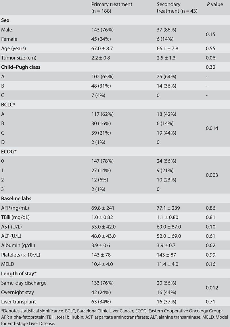
Figure 1.
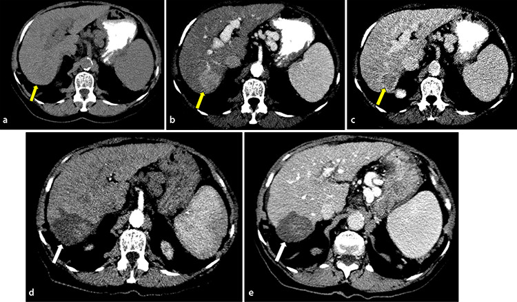
Comparison of pre- and post-treatment imaging in a 78-year-old female with a history of hepatocellular carcinoma (HCC) in the context of cirrhosis due to nonalcoholic steatohepatitis after microwave ablation of a segment 6/7 HCC. Pre-treatment computed tomography (CT) with (a) non-contrast, (b) arterial, and (c) portal venous phases show a 3.7 cm segment 6/7 mass demonstrating early-phase arterial hyperenhancement, capsular appearance, and central washout consistent with an Liver Imaging Reporting and Data System 5 HCC (yellow arrow). Post-treatment CT with (d) arterial and (e) portal venous phases demonstrates no enhancement in the treatment cavity consistent with Liver Imaging Reporting and Data System Treatment Response non-viable (white arrow).
Figure 2.
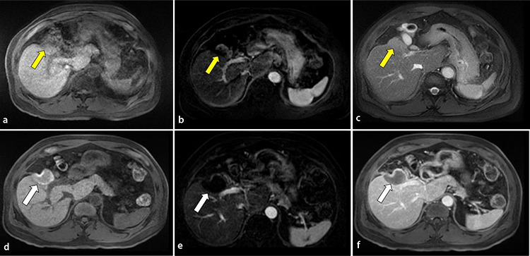
Comparison of pre- and post-treatment imaging in a 70-year-old male with a history of hepatocellular carcinoma (HCC) in the context of chronic hepatitis B after microwave ablation of residual disease in segment 5 sixteen months after trans-arterial chemoembolization. Pre-treatment magnetic resonance imaging (MRI) T1-weighted sequences with (a) non-contrast, (b) arterial, and (c) portal venous phases show a residual 3.5 cm nodular enhancing segment-5 lesion with peripheral washout at the anterosuperior and left lateral borders of the treatment cavity consistent with Liver Imaging Reporting and Data System Treatment Response viable (yellow arrow). Post-treatment MRI T1-weighted sequences with (d) non-contrast, (e) arterial, and (f) portal venous phases demonstrate a new ablation cavity with no evidence of arterial phase hyperenhancement or washout consistent with LIRADS-TR non-viable (white arrow).
Primary technical efficacy was achieved in 96.8% (152/157) of patients in the primary treatment group and in 95% (38/40) of patients in the secondary treatment group (P = 0.58) (Table 2). It should be noted that all MWA was performed on the same system by two treating interventionalists, resulting in a relatively standardized treatment approach. Five of the seven total incomplete ablations were successfully retreated with either MWA, TACE, or TARE. One of the two remaining patients underwent a transplant shortly afterward, and the other was lost to follow-up. No additional treatments for the study tumor were provided until recurrence was detected on cross-sectional imaging. The percentage of patients in the primary (63/188: 34.0%) and secondary groups (16/43: 37.0%) who went on to liver transplant was equal (P = 0.71). The proportion of patients who developed progressive disease at other sites in the liver was similar at 50% (62/125) in the primary treatment group vs. 62% (16/26) in the secondary treatment group (P = 0.42). Although the proportion of patients who developed extra-hepatic metastasis was greater in the secondary treatment group [19% (5/26)] compared with the primary treatment group [7% (8/123)], the difference did not reach statistical significance (P = 0.06).
Table 2. Summary of outcomes in primary vs. secondary treatment groups.
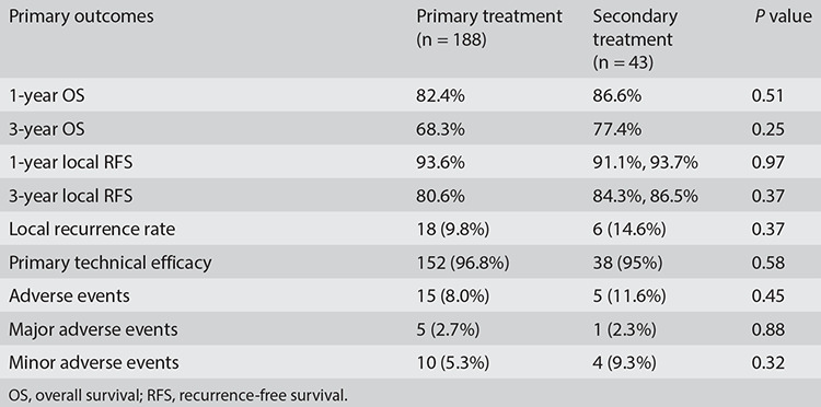
There was no significant difference in OS (P = 0.36) or local RFS (P = 0.68) between the two groups as measured by a Kaplan–Meier survival analysis with corresponding log–rank tests (Figure 3). A survival analysis was performed using a sample size of 182/188 for primary treatment and 41/43 for secondary treatment after excluding cases with failed primary technical efficacy. The 1-year local RFS in the primary group was 93.6% vs. 93.75% in the secondary group (P = 0.97), and the 3-year local RFS in the primary group was 80.58% vs. 86.53% in the secondary group (P = 0.37). The 1-year OS in the primary group was 82.39% vs. 86.65% in the secondary group (P = 0.51), and the 3-year OS in the primary group was 68.34% vs. 77.45% in the secondary group (P = 0.25) (Table 2). The median progression-free survival (PFS) for the primary treatment group was 75.5 months vs. 69.8 months for the secondary treatment group [ratio of 1.08 with a 95% confidence interval (CI) of 0.40 to 2.9]. Similarly, the median OS for the primary treatment group was 95.4 months, while it was undefined in the secondary treatment group.
Figure 3.
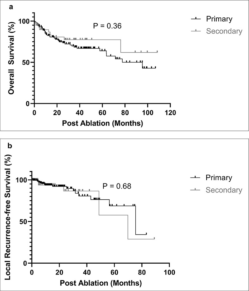
(a) Overall survival curves and (b) local recurrence-free survival curves of patients with hepatocellular carcinoma treated with primary vs. secondary microwave ablation.
The rate of adverse events was 8.0% (15/188) in the primary treatment group and 11.6% (5/43) in the secondary treatment group (P = 0.45). Events were further subdivided into major and minor adverse events. The rate of major adverse events was 2.7% (5/188) in the primary treatment group vs. 2.3% (1/43) in the secondary treatment group (P = 0.88). The rate of minor adverse events was 5.3% (10/188) in the primary treatment group vs. 9.3% (4/43) in the secondary treatment group (P = 0.32) (Table 2). Minor adverse events included pain, abdominal wall hematoma, and biliary stricture, all of which were managed conservatively. Major adverse events included the formation of a hepatic abscess that required percutaneous drainage, hepatorenal syndrome, readmission to an outside hospital for fever attributed to post-ablation syndrome, and hepatic encephalopathy necessitating hospital admission, which was treated successfully with medication. One ablation-related death from sepsis occurred in the secondary treatment group.
The local recurrence rates were calculated after excluding cases with failed primary technical efficacy (6/188 for the primary treatment group and 2/43 for the secondary treatment group). The local recurrence rate for the primary treatment group was 9.8% (18/182) vs. 14.6% (6/41) for the secondary treatment group (P = 0.37) (Table 2). The mean follow-up time for the primary group was 18.2 months (range: 0.3–98.6 months) vs. 18.1 months (range: 0.6–89.0 months) in the secondary group. Twelve recurrent lesions were retreated with either TACE, TARE, or MWA. The remainder of the patients were treated with systemic therapy or radiation therapy, or they were lost to follow-up.
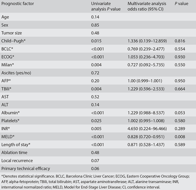
Pre- and post-intervention tumor marker and liver function tests were compared, with alpha-fetoprotein (AFP) (P = < 0.001), total bilirubin (TBili) (P = 0.043), and albumin (P < 0.001) found to be significant (Table 3). Age, Milan criteria, and ablation time were all found to be associated with local RFS by univariate analysis (Table 4). The Milan criteria were an independent predictor of RFS by multivariate analysis, with an odds ratio (OR) of 0.11 (95% CI: 0.018–0.730, P = 0.022). Ablation time was an independent predictor of RFS, with an OR of 1.44 (95% CI: 1.053–1.994, P = 0.023). The Child–Pugh score, BCLC stage, ECOG status, Milan criteria, AFP, TBili, albumin, platelets, international normalized ratio (INR), MELD score, and length of hospital stay were all found to be associated with OS by univariate analysis (Table 5). The MELD score was an independent predictor of OS by multivariate analysis, with an OR of 0.82 (95% CI: 0.720–0.951, P = 0.008).
Table 3. Summary of Wilcoxon signed rank test comparing pre- and post-intervention tumor marker and liver function tests.
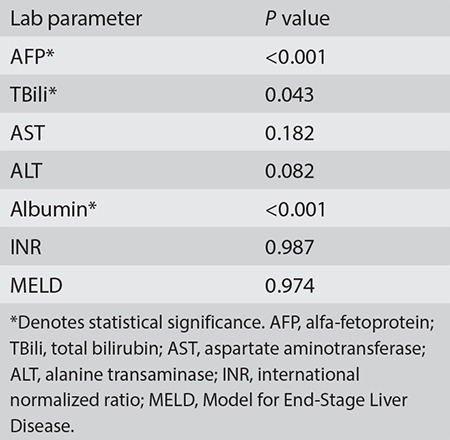
Table 4. Univariate and multivariate prognostic factor analyses for local recurrence-free survival.
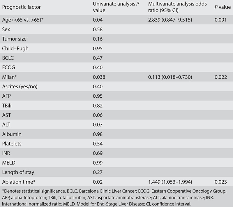
Table 5. Univariate and multivariate prognostic factor analyses for overall survival.
Discussion
In the present study, compared with the primary treatment group, the secondary treatment group had a slightly worse ECOG performance status, presented at a more advanced BCLC stage, and had more patients requiring at least an overnight stay post-intervention. They were otherwise similarly matched in terms of age, sex, target tumor size, Child–Pugh score, MELD score, and liver function tests. Despite having a worse baseline ECOG performance status and a more advanced BCLC stage, we found no difference in the 1- and 3-year local RFS and OS rates, local recurrence rates, primary technical efficacy rates, or rate of adverse events between patients with HCC treated with MWA as their primary vs. secondary treatment. Across both treatment groups, these primary outcomes were also in line with what has been observed in other studies.16,18,28,29 However, it should be noted that overall follow-up was limited, with a mean of 18.2 months (range: 0.3–98.6 months), which may have influenced the OS data.
Comparing the baseline and post-ablation lab test results, we found that AFP, TBili, and albumin were significantly different between the primary and secondary treatment groups”, instead of “Comparing the baseline and post-ablation lab test results, we found that AFP, TBili, and albumin were significantly different. However, the MELD score, which uses a composite of lab values to predict short-term survival, was unchanged. This supports the idea that while there can be short-term fluctuations in liver function tests, MWA is generally well tolerated, and there was no evidence of any acute decompensation among the patients in this study.
The univariate prognostic factor analysis found that age, Milan criteria, and ablation time were related to local RFS. In the multivariate analysis, the Milan criteria and ablation time were independent predictors, indicating that the stage of the disease is an important factor in patient outcomes. The univariate prognostic factor analysis showed that the Child–Pugh score, BCLC stage, ECOG status, AFP, TBili, albumin, platelets, INR, MELD score, and length of hospital stay were all associated with OS. The MELD score was found to be an independent prognostic factor in the multivariate analysis, which is unsurprising given that this clinical characteristic is known to be closely associated with prognosis.
In this study, approximately 19% (43/231) of patients received MWA as a secondary treatment for residual or recurrent disease following TACE or TARE. Of these, 72.1% (31/43) received TACE, with the remaining receiving TARE or combination TACE/TARE therapy. There is currently no consensus on the optimal use of salvage therapy in such patients. However, TACE/TARE is generally favored in patients with large or invasive tumors as well as in those with evidence of portal vein invasion. MWA is usually preferred in patients with smaller tumors in favorable locations away from major anatomical structures, such as the dome of the liver, gallbladder, or biliary system.17 The choice of therapy is also essentially operator and institution dependent. A recent study by Chen et al.30 studied patients with intermediate-stage HCC who were refractory to TACE and compared the OS and RFS of MWA vs. sorafenib as salvage therapy. They found that the MWA group had longer median OS and RFS compared with those of the sorafenib group, suggesting that ablation is a viable option for salvage therapy in such patients.30
Recent studies have evaluated the efficacy and safety of new systemic antineoplastic therapies (atezolizumab/bevacizumab) compared with conventional first-line treatment with sorafenib in patients with unresectable HCC, demonstrating significantly increased OS and PFS rates.31,32 However, to our knowledge, no studies have been conducted comparing the clinical outcomes and disease prognosis associated with the use of local ablative treatments and these newer chemotherapeutic agents in HCC.
In the cohort of patients with a small tumor size (mean: 2.26 ± 0.93 cm), mostly good performance status (ECOG 0 and 1), and primarily early- or intermediate-stage liver disease (BCLC A and B), MWA was equally effective in treating lesions that failed to respond completely to TACE/TARE as it was in treating naïve lesions. We recognize that the population sample size in the secondary treatment group was small and may have contributed to the statistically non-significant results of the primary outcomes. The study group was heterogeneous in terms of disease burden and performance status at presentation. In terms of care goals, treatment intent was curative for some, downstaged to within the Milan criteria for transplant for others, with palliation for those who were not transplant candidates. The analysis did not distinguish between those who had residual disease vs. those with recurrence or between those treated primarily with TACE vs. TARE. While this study showed MWA to be safe and technically effective in a salvage setting, future studies should narrow the focus to include a more specific patient population.
In conclusion, tumor recurrence and its potential impact on survival remain a challenge in patients with HCC treated with hepatectomy and LRTs. Data to guide second-line treatments are very limited. In this retrospective study on clinical experience with MWA as a salvage therapy in patients with residual/recurrent HCC, we showed that despite more advanced tumor stages and poorer performance status at presentation, the survival outcomes and recurrence rates were similar in patients with residual/recurrent HCC compared with those in patients who received MWA as a first-line treatment. When used as a second-line treatment, MWA was technically effective and well tolerated.
Footnotes
Conflict of interest disclosure
The authors declared no conflicts of interest.
Funding
Grant support from Johnson and Johnson. The publication of this article was made possible in part by support from the Thomas Jefferson University Open Access Fund.
References
- 1.Shiina S, Tateishi R, Arano T, et al. Radiofrequency ablation for hepatocellular carcinoma: 10-year outcome and prognostic factors. Am J Gastroenterol. 2012;107(4):569–577. doi: 10.1038/ajg.2011.425. [DOI] [PMC free article] [PubMed] [Google Scholar]
- 2.Kono M, Inoue T, Kudo M, et al. Radiofrequency ablation for hepatocellular carcinoma measuring 2 cm or smaller: results and risk factors for local recurrence. Dig Dis. 2014;32(6):670–677. doi: 10.1159/000367999. [DOI] [PubMed] [Google Scholar]
- 3.Hao Y, Numata K, Ishii T, et al. Rate of local tumor progression following radiofrequency ablation of pathologically early hepatocellular carcinoma. World J Gastroenterol. 2017;23(17):3111–3121. doi: 10.3748/wjg.v23.i17.3111. [DOI] [PMC free article] [PubMed] [Google Scholar]
- 4.Minagawa M, Makuuchi M, Takayama T, Kokudo N. Selection criteria for repeat hepatectomy in patients with recurrent hepatocellular carcinoma. Ann Surg. 2003;238(5):703–710. doi: 10.1097/01.sla.0000094549.11754.e6. [DOI] [PMC free article] [PubMed] [Google Scholar]
- 5.Vogl TJ, Trapp M, Schroeder H, et al. Transarterial chemoembolization for hepatocellular carcinoma: volumetric and morphologic CT criteria for assessment of prognosis and therapeutic success-results from a liver transplantation center. Radiology. 2000;214(2):349–357. doi: 10.1148/radiology.214.2.r00fe06349. [DOI] [PubMed] [Google Scholar]
- 6.Ippolito D, Bonaffini PA, Ratti L, et al. Hepatocellular carcinoma treated with transarterial chemoembolization: dynamic perfusion-CT in the assessment of residual tumor. World J Gastroenterol. 2010;16(47):5993–6000. doi: 10.3748/wjg.v16.i47.5993. [DOI] [PMC free article] [PubMed] [Google Scholar]
- 7.Kono Y, Lucidarme O, Choi SH, et al. Contrast-enhanced ultrasound as a predictor of treatment efficacy within 2 weeks after transarterial chemoembolization of hepatocellular carcinoma. J Vasc Interv Radiol. 2007;18(1 Pt 1):57–65. doi: 10.1016/j.jvir.2006.10.016. [DOI] [PubMed] [Google Scholar]
- 8.Kwan SW, Fidelman N, Ma E, et al. Imaging predictors of the response to transarterial chemoembolization in patients with hepatocellular carcinoma: a radiological-pathological correlation. Liver Transpl. 2012;18(6):727–736. doi: 10.1002/lt.23413. [DOI] [PMC free article] [PubMed] [Google Scholar]
- 9.Shaw CM, Eisenbrey JR, Lyshchik A, et al. Contrast-enhanced ultrasound evaluation of residual blood flow to hepatocellular carcinoma after treatment with transarterial chemoembolization using drug-eluting beads: a prospective study. J Ultrasound Med. 2015;34(5):859–867. doi: 10.7863/ultra.34.5.859. [DOI] [PubMed] [Google Scholar]
- 10.Lencioni R. Loco-regional treatment of hepatocellular carcinoma. Hepatology. 2010;52(2):762–773. doi: 10.1002/hep.23725. [DOI] [PubMed] [Google Scholar]
- 11.Park JW, Amarapurkar D, Chao Y, et al. Consensus recommendations and review by an International Expert Panel on Interventions in Hepatocellular Carcinoma (EPOIHCC) Liver Int. 2013;33(3):327–337. doi: 10.1111/liv.12083. [DOI] [PubMed] [Google Scholar]
- 12.Yamanaka K, Hatano E, Kitamura K, et al. Early evaluation of transcatheter arterial chemoembolization-refractory hepatocellular carcinoma. J Gastroenterol. 2012;47(3):343–346. doi: 10.1007/s00535-011-0511-x. [DOI] [PubMed] [Google Scholar]
- 13.European Association for the Study of the Liver. EASL Clinical Practice Guidelines: management of hepatocellular carcinoma. J Hepatol. 2018;69(1):182–236. doi: 10.1016/j.jhep.2018.03.019. [DOI] [PubMed] [Google Scholar]
- 14.Heimbach JK, Kulik LM, Finn RS, et al. AASLD guidelines for the treatment of hepatocellular carcinoma. Hepatology. 2018;67(1):358–380. doi: 10.1002/hep.29086. [DOI] [PubMed] [Google Scholar]
- 15.Vogel A, Cervantes A, Chau I, et al. Hepatocellular carcinoma: ESMO Clinical Practice Guidelines for diagnosis, treatment and follow-up. Ann Oncol. 2019;30(5):871–873. doi: 10.1093/annonc/mdy510. [DOI] [PubMed] [Google Scholar]
- 16.Izzo F, Granata V, Grassi R, et al. Radiofrequency ablation and microwave ablation in liver tumors: an update. Oncologit. 2019;24(10):990–1005. doi: 10.1634/theoncologist.2018-0337. [DOI] [PMC free article] [PubMed] [Google Scholar]
- 17.Ponniah SA, Zori AG, Cabrera R. Locoregional therapies for bridging and downstaging hepatocellular carcinoma prior to liver transplant. In: Sergi CM, ed. Brisbane, Liver Cancer. 2021. [PubMed] [Google Scholar]
- 18.Yu Q, Liu C, Navuluri R, Ahmed O. Percutaneous microwave ablation versus radiofrequency ablation of hepatocellular carcinoma: a meta-analysis of randomized controlled trials. Abdom Radiol (NY) 2021;46(9):4467–4475. doi: 10.1007/s00261-021-03080-1. [DOI] [PubMed] [Google Scholar]
- 19.Abdelaziz A, Elbaz T, Shousha HI, et al. Efficacy and survival analysis of percutaneous radiofrequency versus microwave ablation for hepatocellular carcinoma: an Egyptian multidisciplinary clinic experience. Surg Endosc. 2014;28(12):3429–3434. doi: 10.1007/s00464-014-3617-4. [DOI] [PubMed] [Google Scholar]
- 20.An C, Li WZ, Huang ZM, et al. Small single perivascular hepatocellular carcinoma: comparisons of radiofrequency ablation and microwave ablation by using propensity score analysis. Eur Radiol. 2021;31(7):4764–4773. doi: 10.1007/s00330-020-07571-5. [DOI] [PMC free article] [PubMed] [Google Scholar]
- 21.Kamal A, Elmoety AAA, Rostom YAM, et al. Percutaneous radiofrequency versus microwave ablation for management of hepatocellular carcinoma: a randomized controlled trial. J Gastrointest Oncol. 2019;10(3):562–571. doi: 10.21037/jgo.2019.01.34. [DOI] [PMC free article] [PubMed] [Google Scholar]
- 22.Nault JC, Sutter O, Nahon P, et al. Percutaneous treatment of hepatocellular carcinoma: state of the art and innovations. J Hepatol. 2018;68(4):783–797. doi: 10.1016/j.jhep.2017.10.004. [DOI] [PubMed] [Google Scholar]
- 23.Shibata T, Iimuro Y, Yamamoto Y, et al. Small hepatocellular carcinoma: comparison of radio-frequency ablation and percutaneous microwave coagulation therapy. Radiology. 2002;223(2):331–337. doi: 10.1148/radiol.2232010775. [DOI] [PubMed] [Google Scholar]
- 24.Som A, Reid NJ, DiCapua J, et al. Microwave ablation as bridging therapy for patients with hepatocellular carcinoma awaiting liver transplant: a single center experience. Cardiovasc Intervent Radiol. 2021;44(11):1749–1754. doi: 10.1007/s00270-021-02873-7. [DOI] [PubMed] [Google Scholar]
- 25.Vietti Violi N, Duran R, Guiu B, et al. Efficacy of microwave ablation versus radiofrequency ablation for the treatment of hepatocellular carcinoma in patients with chronic liver disease: a randomised controlled phase 2 trial. Lancet Gastroenterol Hepatol. 2018;3(5):317–325. doi: 10.1016/S2468-1253(18)30029-3. [DOI] [PubMed] [Google Scholar]
- 26.Chernyak V, Fowler KJ, Kamaya A, et al. Liver imaging reporting and data system (LI-RADS) version 2018: imaging of hepatocellular carcinoma in at-risk patients. Radiology. 2018;289(3):816–830. doi: 10.1148/radiol.2018181494. [DOI] [PMC free article] [PubMed] [Google Scholar]
- 27.Khalilzadeh O, Baerlocher MO, Shyn PB, et al. Proposal of a new adverse event classification by the society of interventional radiology standards of practice committee. J Vasc Interv Radiol. 2017;28(10):1432–1437. doi: 10.1016/j.jvir.2017.06.019. [DOI] [PubMed] [Google Scholar]
- 28.Xu Y, Shen Q, Wang N, et al. Microwave ablation is as effective as radiofrequency ablation for very-early-stage hepatocellular carcinoma. Chin J Cancer. 2017;36(1):14. doi: 10.1186/s40880-017-0183-x. [DOI] [PMC free article] [PubMed] [Google Scholar]
- 29.Swan RZ, Sindram D, Martinie JB, Iannitti DA. Operative microwave ablation for hepatocellular carcinoma: complications, recurrence, and long-term outcomes. J Gastrointest Surg. 2013;17(4):719–729. doi: 10.1007/s11605-013-2164-y. [DOI] [PubMed] [Google Scholar]
- 30.Chen S, Shi M, Shen L, et al. Microwave ablation versus sorafenib for intermediate-stage hepatocellular carcinoma with transcatheter arterial chemoembolization refractoriness: a propensity score matching analysis. Int J Hyperthermia. 2020;37(1):384–391. doi: 10.1080/02656736.2020.1752400. [DOI] [PubMed] [Google Scholar]
- 31.Cheng AL, Qin S, Ikeda M, et al. Updated efficacy and safety data from IMbrave150: Atezolizumab plus bevacizumab vs. sorafenib for unresectable hepatocellular carcinoma. J Hepatol. 2022;76(4):862–873. doi: 10.1016/j.jhep.2021.11.030. [DOI] [PubMed] [Google Scholar]
- 32.Ahmed F, Onwumeh-Okwundu J, Yukselen Z, et al. Atezolizumab plus bevacizumab versus sorafenib or atezolizumab alone for unresectable hepatocellular carcinoma: a systematic review. World J Gastrointest Oncol. 2021;13(11):1813–1832. doi: 10.4251/wjgo.v13.i11.1813. [DOI] [PMC free article] [PubMed] [Google Scholar]


