Abstract
PURPOSE
To identify the optimum strength of advanced modeled iterative reconstruction (ADMIRE) to achieve the best subjective and objective image quality when combining three-dose reduction strategies, ultra-high-pitch computed tomography coronary angiography (FLASH CTCA; with single-dose ivabradine to lower heart rate), low tube voltage, and ADMIRE.
METHODS
Sixty consecutive patients underwent FLASH CTCA at 100 kVp in this single-center prospective study. Single-dose ivabradine was administered to patients whose heart rate was above 75 bpm. Images were reconstructed using the three highest strengths of ADMIRE (A3, A4, and A5). Objective and subjective image quality (using a Likert scale) were evaluated in the three datasets.
RESULTS
The signal strength remained unchanged but mean noise significantly reduced across the increasing strengths of ADMIRE [signal: 513.78 ± 101.7 Hounsfield units (HU) at A3, 515.6 ± 100.5 HU at A4, and 519.7 ± 107.9 HU at A5; noise: 23.4 ± 4.5 HU at A3, 20.2 ± 3.6 HU at A4, and 17.2 ± 3.3 HU at A5]. Signal-to-noise and contrast-to-noise ratios were the highest at A5, and A5 offered significantly higher Likert scores in image noise, vessel sharpness, and overall image quality than A3 or A4. Additionally, A5 did not interfere with image interpretation in any patient.
CONCLUSION
Using all three dose reduction strategies during FLASH CTCA along with single-dose ivabradine administration ensures minimal radiation exposure in daily practice. In this study, A5 datasets had the best overall subjective and objective image quality despite their “plastic appearance”. In the future, enhanced dose reduction can be obtained by further lowering tube voltages.
Keywords: Advanced modeled iterative reconstruction, ultra-high-pitch computed tomography coronary angiography, computed tomography angiography, cardiac, dose reduction strategies
Main points
• Signal strength remains unchanged but mean noise significantly decreases with increasing advanced modeled iterative reconstruction (ADMIRE) strengths (A3 to A5).
• Signal-to-noise and contrast-to-noise ratios are the highest at A5.
• The A5 datasets have the best overall subjective and objective image quality despite producing a “plastic appearance” when combining three-dose reduction strategies: ultra-high-pitch computed tomography coronary angiography (with single-dose ivabradine to lower heart rate), low tube voltage, and ADMIRE.
The main disadvantage of computed tomography coronary angiography (CTCA), despite its non-invasive nature, is radiation exposure. With increasing concerns regarding the accurate estimation of the “true” radiation burden of CTCA caused by the challenges in determining the cardiac-specific conversion factor (k factor), the need to devise new strategies and incorporate multiple dose reduction techniques is of paramount importance. With the newly proposed k factors, even the lower radiation burden of prospectively gated CTCA can no longer be considered “low dose” per se, indicating an urgent need to use all available advanced techniques for every patient to undergo “true” submillisievert CTCAs.1
Lowering the tube potential as a standalone measure leads to a decrease in image quality. The current guidelines recommend 100 kVp for patients with a body mass index (BMI) <30 kg/m2, making an approximate reduction of 50%–60% in radiation dose possible using these strategies compared with strategies using 120 kVp.2 Additional dose reduction may be possible by further reducing the tube voltage and using iterative reconstruction (IR), which permits the decoupling of spatial resolution and noise to offset the noisier image quality obtained with lower tube voltages. IR has rapidly replaced the more archaic filtered back projection (FBP) by enabling the drastic reduction in image noise inherent in the use of low tube voltage.3,4 As a result of advances in computer processing technologies, current generation IR, such as model-based IR, is now available on all vendor platforms and is no longer limited by the absence and expense of the faster computational requirements that IR requires. Previous generations of IR have been shown to reduce image noise by 70%–80% compared with FBP in CT.5,6 The latest generation advanced modeled IR (ADMIRE) is a hybrid technique that removes noise more rapidly by using statistical modeling in both the raw and image domain. Although different strengths of ADMIRE have been shown to deliver a better signal-to-noise ratio (SNR) and contrast-to-noise ratio (CNR) even for CTCA, the best strength of iteration that is optimal remains undetermined. This is because higher strengths of iteration can lead to an excessive smoothing of the image, which degrades subjective image quality.7
Another dose reduction strategy that can be used is high-pitch (>3) prospective electrocardiogram (ECG)-triggered helical data acquisition (FLASH CTCA), which can acquire the complete volumetric dataset within a fraction of a cardiac cycle. The high pitch leads to very low radiation dose (0.5–1 mSv) as long as the patient has low, regular heart rates (HRs).
Prospective ECG-gated CTCA, which is the default “lower-dose CTCA”, is a well-utilized technique. By using forward-looking ECG prediction and step/shoot non-spiral acquisition, it requires the X-ray beam to be turned on for only a small portion of the cardiac cycle. In FLASH CTCA, by using a higher pitch and faster table movement, the beam is switched on for even shorter periods, enabling it to be genuinely “low dose” or “ultra-low dose”.
Combining the three techniques (low tube voltage, FLASH CTCA, and IR) could lead to a significant reduction in radiation exposure and still deliver diagnostic image quality. The objective of this study was to identify the optimum strength of ADMIRE to achieve the best SNR and CNR when using FLASH CTCA at a tube voltage of 100 kVp because the strength of ADMIRE that provides the optimum subjective image quality is still contested. This study also evaluated the role of a single dose of the novel HR-lowering drug ivabradine in decreasing and stabilizing the HR to rates required for FLASH CTCA.
Methods
This study was a prospective single-center study involving 66 consecutive adult patients who underwent CTCA for suspected coronary artery disease. The study was approved by the Institutional Ethics Committee (IECPG-96/21.03.2018), and informed consent was obtained. All studies were conducted after documenting baseline HR and blood pressure and assessing left ventricular ejection fraction through echocardiography. A 384 -slice (192*2) dual-source scanner (Siemens Somatom Force, Erlangen, Germany) was used to perform FLASH CTCA. The acquisition parameters were 2 × 192 × 0.6 mm for detector collimation (Siemens Healthcare Sector, Forcheim, Germany), rotation time of 0.25 s, temporal resolution of 66 ms, pitch of 3.2 mm, and tube voltage of 100 kVp. In cases where the HR was greater than 75 bpm, a single 10 mg dose of ivabradine (in patients without any contraindications for the drug) was administered to the patient, and CTCA was acquired if the HR was less than 75 bpm after 2 h. Ivabradine was not administered to any of the patients already on beta-blockers. Subsequently, 50–60 mL of iodinated contrast media (350 mg I/mL) was injected, followed by a 30 mL saline chaser at a flow rate of 3.6–3.8 mL/s through the antecubital vein. Scans were automatically triggered after an attenuation threshold of 100 Hounsfield units (HU) was reached in the descending thoracic aorta using bolus tracking. Automatic exposure control (CARE kV, Siemens) and attenuation-based tube current modulation (CAREDose, Siemens) was enabled. A dual FLASH protocol was performed using one set of images acquired in the strongest systolic phase (35% of RR interval) and one set of images acquired in the strongest diastolic phase (65% of RR interval). All patients, including those who were given ivabradine, were observed carefully for adverse effects before, during, and after the CTCA. Patients with HRs above 75 bpm, despite being on oral beta-blockers, and patients with hypersensitivity to iodinated contrast were excluded from the study.
Computed tomography image reconstruction
CTCA images were reconstructed using ADMIRE (Siemens, Germany). Both the systolic and diastolic phases were reconstructed using three increasing strengths of noise reduction: ADMIRE 3 (A3), ADMIRE 4 (A4), and ADMIRE 5 (A5). The parameters for reconstruction for both the systolic and diastolic datasets were a 0.75 mm slice thickness with 0.5 mm increments and a 512 × 512 matrix using a medium soft tissue reconstruction kernel (Bv-40).
The resulting six series of images were archived and then evaluated using a commercially available software (Siemens Syngo.via VB10B).
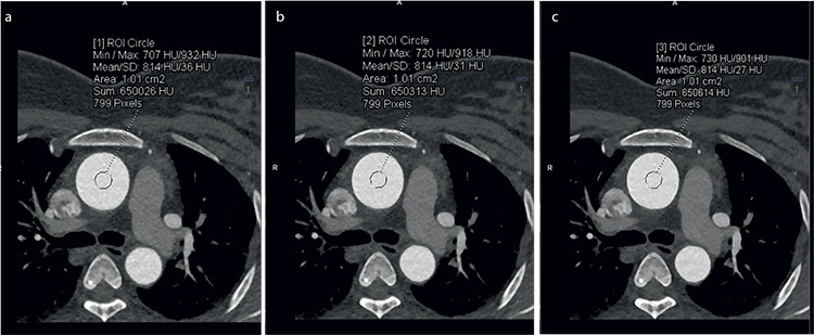
Regions of interest (ROIs) of 1 cm2 drawn in the aortic root at the level of the left main coronary artery on an axial image were used to measure signal strength (mean HU value). The standard deviation (SD) obtained from the same ROI represented the objective image noise (Figure 1). Similar ROIs (as large as possible) were drawn on the right coronary, proximal left main, left anterior descending, and left circumflex arteries (within 1 cm of their origins on axial images).
Figure 1.

(a-c) Axial computed tomography images showing image noise measurements in the aortic root in a 45-year-old female patient (62 kg, 150 cm) at advanced modeled iterative reconstruction (ADMIRE) 3 [image noise: 36 Hounsfield units (HU)], ADMIRE 4 (image noise: 31 HU), and ADMIRE 5 (image noise: 27 HU). ROI, Regions of interest; SD, standard deviation.
Only one phase, either the highest quality diastolic or systolic images, were chosen for the assessment of subjective and objective image analysis based on which had the least cardiac motion in the preliminary analysis of both datasets.
Quantitative analysis
An automated copy/paste tool ensured that all measurements were performed in an identical location for all three datasets (A3, A4, and A5), which were viewed side by side simultaneously by NK (Figures 2, 3, 4).
Figure 2.

(a-c) Curved multiplanar computed tomography images of the right coronary artery of a 45-year-old female patient (62 kg, 150 cm, heart rate: 69 bpm) obtained through electrocardiogram-triggered high-pitch acquisition at advanced modeled iterative reconstruction (ADMIRE) 3 (a), ADMIRE 4 (b), and ADMIRE 5 (c).
Figure 3.

(a-c) Curved multiplanar computed tomography images of the left anterior descending artery of a 45-year-old male patient (88 kg, 170 cm, heart rate: 66 bpm) obtained through electrocardiogram-triggered high-pitch acquisition at advanced modeled iterative reconstruction (ADMIRE) 3 (a), ADMIRE 4 (b), and ADMIRE 5 (c).
Figure 4.

(a-c) Curved multiplanar computed tomography images of the left main coronary artery of a 45-year-old male patient (88 kg, 170 cm, heart rate: 66 bpm) obtained through electrocardiogram-triggered high-pitch acquisition at advanced modeled iterative reconstruction (ADMIRE) 3 (a), ADMIRE 4 (b), and ADMIRE 5 (c).
Mean signal strength and noise were calculated by averaging the values obtained from the five vessels mentioned above.
In addition, SNR was defined as the ratio of the mean HU value of the coronary artery and its SD (noise), and CNR was calculated by dividing the contrast attenuation by image noise.
Qualitative analysis
Two independent and blinded cardiothoracic radiologists (GG and KG, with 20 and 6 years of experience, respectively, in cardiac imaging) performed the qualitative image analysis at a different time from the quantitative measurements. The ADMIRE strengths used on each dataset were hidden from the readers to prevent bias.
Maximum intensity projections, curved multiplanar images, and routine axial datasets were used at the discretion of the individual readers. A Likert score was assigned for each dataset with respect to 1) subjective image noise, 2) coronary wall definition (sharpness), 3) beam-hardening artifacts, and 4) overall subjective image quality. The readers ignored artifacts resulting from motion and poor gating that were not caused by the reconstruction algorithm.
These four parameters were assessed using a single score on a 4-point Likert scale (range 1–4) based on the segment of the coronary artery with the poorest appearance. The Likert scale was defined as follows: image noise and beam-hardening artifacts were assessed as (1) present and not acceptable, (2) present and interfering with assessment, (3) present but not interfering with assessment, and (4) minimal or absent; vessel sharpness was defined as (1) blurry, (2) poorer than average, (3) better than average, and (4) the sharpest; overall image quality was graded as (1) unacceptable (inadequate to evaluate coronary stenosis), (2) fair (sufficient for evaluating coronary stenosis), (3) good, and (4) excellent quality. The Likert scores from both readers were averaged and used for statistical analysis.
Estimation of radiation dose
The CT volume dose index and dose–length product (DLP) were obtained from the scanner. The effective dose was derived from the product of DLP and the conversion coefficient (k) for the chest.8 To make our study more comparable with previous studies, a coefficient of 0.014 mSv mGy-1 cm-1 was used.
Statistical analysis
Data were analyzed using SPSS version 23.0 (SPSS, Chicago, IL, USA). Descriptive statistics are presented in frequencies with percentages, mean+/− SD, or median (min–max), depending on the distribution of the data. For the assessment of image quality score and quantitative parameters between the three reconstruction algorithms, a One-Way analysis of variance (for normal data) and Kruskal–Wallis tests (for non-normal data) were used. For pairwise comparisons, a post hoc test (Dunn’s test) was applied. Student’s test (paired) was used for comparing HRs in patients who had been administered ivabradine (before or after ivabradine administration). Interobserver reliability was assessed using the Kappa coefficient. A P value < 0.05 was considered statistically significant.
Results
In total, 66 patients were enrolled, with 6 patients excluded because of an elevated preprocedural HR above 75 bpm despite being on routine beta-blocker therapy. The mean age of the 33 men and 27 women was 49.9 ± 9.6 years (range: 30–75). The mean BMI and average HR during CTCA were 27.4 ± 4.1 kg/m2 (range: 19.5–40.9) and 69 ± 5.7 bpm (range: 50–75), respectively. Ivabradine was administered to 31 patients, after which the average HR was 67.9 ± 6.2 bpm compared with 87.3 ± 7.8 bpm (P = 0.038) prior to administration. No adverse effect was identified in any patient.
Quantitative analysis
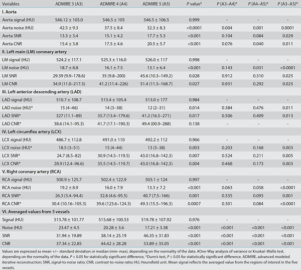
Mean signal strength (averaged value of the ROIs in the five vessels) remained unchanged across the three datasets reconstructed at the different strengths of ADMIRE (513.78 ± 101.7 HU in A3, 515.6 ± 100.5 HU in A4, and 519.7 ± 107.9 HU in A5) (P = 0.976).
Mean image noise (averaged value of the ROIs in the five vessels) significantly decreased with increasing strengths of ADMIRE (23.4 ± 4.5 HU in A3, 20.2 ± 3.6 HU in A4, and 17.2 ± 3.3 HU in A5) (P = 0.033), although pairwise comparison produced varying results in different vessels (Table 1). Mean noise was reduced by 13.66% at A4 compared with A3, 14.83% at A5 compared with A4, and 26.49% at A5 compared with A3 (Table 1). Thus, a linear improvement in the mean SNR and CNR was observed with increasing iterations, with an improvement of 39.91% and 37.12%, respectively, at A5 compared with A3 (P < 0.0001) (Table 1).
Table 1. Objective image quality parameters in the study population.

Qualitative analysis
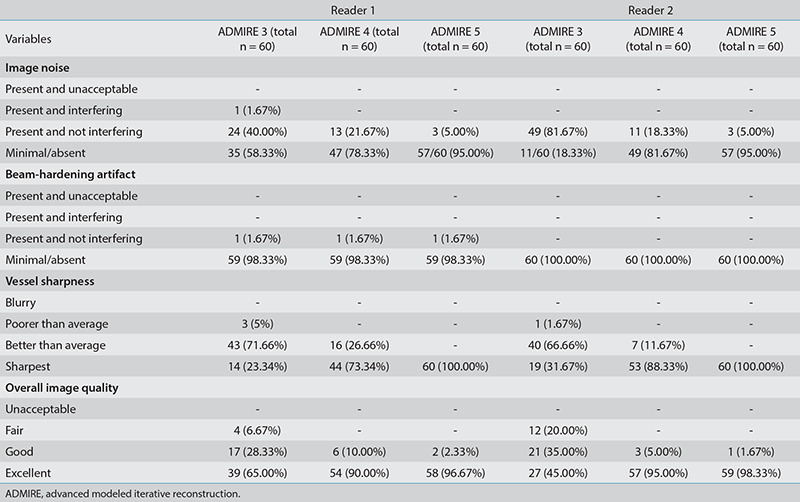
The optimal overall image quality was obtained at A5, with readers 1 and 2 rating 96.67% (58/60) and 98.33% (59/60), respectively, of the A5 images as excellent (Table 2).
Table 2. Subjective comparison between different strengths of ADMIRE.

Subjective image noise was at its maximum at A3, with readers 1 and 2 grading it as minimal or absent only in 58.33% and 18% of the A3 datasets. Subjective image noise was at a minimum at A5, with both readers grading it as minimal or absent in 95% of the A5 datasets.
Beam-hardening artifacts were minimal or absent in almost all the datasets for both readers at all three ADMIRE strengths and did not interfere in the interpretation in any of the datasets for either reader.
Vessel sharpness in all 60 patients was lowest at A3 [14/60 (23.33%) for reader 1, 19/60 (31.67%) for reader 2] and highest in those reconstructed at A5 [60/60 (100%) for both readers]. Interobserver reliability was excellent for subjective image noise, vessel sharpness, and overall image quality at A5 (Table 3). Poor agreement for subjective image noise was determined at A3. This may be because the more experienced reader 1 (GG) was more accepting of image noise. However, image noise did not interfere with image interpretation for either reader.
Table 3. Interobserver variability between readers 1 and 2.

Pairwise comparisons revealed that A4 and A5 offered significantly higher Likert scores for image noise, vessel sharpness, and overall image quality compared with A3 (Table 4), and A5 was associated with a significant increase in all three of the aforementioned parameters according to the Likert scale (all P < 0.0001). Likert scores for beam-hardening artifacts were not significantly different between the groups (P = 0.999).
Table 4. Likert scores for different ADMIRE strengths.

The average effective radiation dose was 1.6 ± 0.29 mSv (range 1.2–2.5 mSv; dual FLASH protocol). The radiation dose with a single FLASH scan was 0.60 ± 0.09 mSv [an average of the radiation dose from the strongest diastolic dataset that was most frequently (59/60) used for interpretation].
Discussion
High CTCA accuracy is desirable when using conventional angiography only in patients requiring revascularization therapy. FBP, which is usually used for CTCA, is fast, requires less computing power, and is more easily implemented, but it leads to noisier images with artifacts at reduced doses because of its inherent mathematical assumptions of the CT system. FBP ignores essential information such as the Poisson distribution of photons and CT hardware details (focal spot size, active detector area, and image voxel shape). The more complex IR has been reintroduced for CT imaging as advances in computer hardware and processing have overcome its traditional time and processing-intensive drawbacks. It has since become routine9 and can compensate for a lower tube potential or even tube current, which are associated with noisier images, by producing images of higher quality in the setting of a very low SNR, as demonstrated by multiple authors.10 To further lower the patient’s radiation burden, CTCA acquisition is, in the current era, primarily performed using the prospective “step-and-shoot” acquisition and by lowering the tube current and using tube current modulation. The resulting incremental increase in noise and decrease in spatial reduction is negated by shifting from FBR to IR. Further advances in IR with the development of hybrid-IR algorithms (e.g., ASIR, AIDR 3D, and iDose) and model-based IR algorithms (e.g., ADMIRE, IMR, VEO, and FIRST) have demonstrated that a “virtually noise-free” image with a higher CNR can be obtained.11 Second-generation dual-source prospective FLASH CTCA provides an additional opportunity to combine all three techniques to obtain a maximum reduction in radiation dose with excellent image quality, mainly by lowering the tube voltage/current and using IR. The low radiation exposure results from the absent slice overlap of the FLASH CTCA protocol compared with the conventional prospectively triggered protocols. Nearly all unnecessary radiation exposure is eliminated when FLASH CTCA is used only at the beginning and end of the scanned volume. With a pitch of 3.4, time resolution of 75 ms, and full heart coverage in <0.25 s, FLASH CTCA has a reported success rate of 100% in the delineation of coronary arteries up to 1.5 mm.12 FLASH CTCA is, therefore, an acceptable angiographic method for reducing radiation dose without compromising image quality. The challenge with using FLASH CTCA is that it requires a stable and low HR. HR control medications such as beta-blockers have long attempted to achieve this objective, but they have numerous drawbacks and contraindications. However, newer drugs such as ivabradine, a funny channel blocker, have recently been successfully used and have proved in studies more efficacious at lowering and stabilizing the HR.13,14,15 We therefore incorporated this into the protocol to establish the utility of a single dose of ivabradine in lowering the HR.
Studies have also demonstrated that it is possible to perform CTCA with ultra-low-dose radiation exposure in the order of 0.1–0.5 mSv.16,17 What remains unanswered is if the higher iteration strength images interfere with interpretation because of their extreme smoothing effect and the possibility of achieving the low HR required for these studies in daily routine clinical practice. The apparent subjective degradation is likely caused by the reader’s preference for obtaining images that have been used to receive FBP, indicating the need for a more conscious and concerted shift toward higher IR strengths despite the “apparent” degradation, which is an opinion shared by other authors.18
In our study, the image noise was lowest at A5 among the three IR datasets (23.4 ± 4.5 HU at A3, 20.2 ± 3.6 HU at A4, and 17.2 ± 3.3 HU at A5). The SNR was 21.3 ± 9.4 at A3, 25.9 ± 5.6 at A4, and 30.6 ± 6.9 at A5, and CNR was 26.1 ± 5.1 at A3, 30.2 ± 5.9 at A4, and 36.5 ± 7.36 at A5. Both SNR and CNR were highest at A5. The subjective image noise was highest at A3 and lowest at A5. Vessel sharpness and overall subjective image quality increased with the higher ADMIRE strengths and were highest at A5. These results are similar to those of the studies by Gordic et al.16, who found that both the objective as well as subjective image noise decreased with higher ADMIRE strengths. Although A4 (selected in 84%) was the preferred dataset in their study for making the diagnosis, our readers preferred A5 (selected in >96%). Images with excessive smoothing and a plastic appearance at A5, which was cited as a drawback in the previous studies, was not a disadvantage in this study. This in consistent with another study by Scholtz et al.19, who opined that despite an artificially smoothed image appearance in CT angiographies of the neck, CNR and overall quality were rated better at A5 than at A3; however, the difference was non-significant. The conclusion of both the previous and present studies is that higher strengths of iteration do not degrade image quality nor interfere with coronary assessment despite the images having a more plastic appearance. The dual FLASH protocol, consisting of two high-pitch CTCA scans performed sequentially, achieves good image quality despite an unexpectedly high HR (caused by anxiety or pain during contrast injection) and occasional premature ventricular contractions. In this study, however, we concluded that a single FLASH is sufficient for an evaluation of all the coronary vessels because, in our sample, only one patient (1/60, 1.67%) required an evaluation of both systolic and diastolic datasets for the depiction of all coronary arteries. This was likely possible because we followed a strict protocol of performing the FLASH technique only after ensuring a stable or regular HR of <75 bpm. If the patient’s HR was >75 bpm, a single 10 mg dose of ivabradine was administered 2 h before examination. In the small subset of patients receiving ivabradine, a significant reduction in the HR was noted. To the best of our knowledge, this is the first study to suggest that a single 10 mg dose of ivabradine may be efficacious in lowering the HR. A recently published abstract by Baig et al.20 revealed similar results with the administration of a 15 mg dose of ivabradine. However, larger randomized studies are required to verify that this is a valid method and not confounded by the 2 h interval period prior to CTCA.
The mean radiation dose per patient was 1.6 and 0.6 mSv when only one scan (systole or diastole) was considered, which demonstrates that FLASH CTCA, when used with a low tube voltage and IR, can lead to submillisievert scans and still obtain diagnostic image quality. Earlier studies have demonstrated that diagnostic image quality can also be achieved with 80 kVp using similar high-pitch techniques with exposure levels as low as 0.1–0.3 mSv despite using lower levels of IR, such as A3 or A4.19,20,23 Based on the findings of our study, we hypothesize that these remarkably low exposure levels can become routinely feasible and be applied to larger subsets of the general population if adequate attention is paid to HR control (with novel and safe drugs such as ivabradine), using FLASH CTCA at 70–80 kVp, and using higher strengths of iteration to compensate for the noisy image. In routine daily practice, coronary artery evaluation is feasible with good-to-excellent image quality while simultaneously reducing radiation exposure significantly.
This study has limitations. First, this was a single vendor study from a single center only; thus, the replication of our results to other scanners and centers may be limited. Second, although anonymized images were used, the differences in image quality between reconstruction methods were at times apparent and may have impacted observer blinding. Third, quantitative noise assessment in IR can be inaccurate when using the SD from ROIs because noise is unequally distributed in the images; however, this is still an accepted criterion for noise estimation. Fourth, the effect of higher grades of iteration on the quantitative assessment of the degree of coronary artery stenosis was not assessed in this study. However, other studies have conclusively proved that stenosis assessment is not hampered with the higher iteration grade of images.19,20,23 Finally, the role played by a single dose of ivabradine needs further validation through trials. The small study group is also a limitation of our study.
In conclusion, we conclude that combining FLASH CTCA, lowering tube voltage, and using IR leads to a significant reduction in radiation dose exposure closer to “true” submillisievert levels in patients, especially if combined with novel drugs such as ivabradine to lower the HR. The highest strength of IR, despite the plastic appearance of the images, does not interfere with image interpretation and leads to a significant increase in SNR and CNR. The higher strengths of iteration with its superior noise-reducing capabilities but the consequent plastic/smoothed appearance is likely the lesser of two evils because it improves diagnostic image quality, allowing for a greater reduction in tube voltage and consequent radiation exposure.
Footnotes
Conflict of interest disclosure
The authors declared no conflicts of interest.
References
- 1.Trattner S, Halliburton S, Thompson CM, et al. Cardiac-specific conversion factors to estimate radiation effective dose from dose-length product in computed tomography. JACC Cardiovasc Imaging. 2018;11(1):64–74. doi: 10.1016/j.jcmg.2017.06.006. [DOI] [PMC free article] [PubMed] [Google Scholar]
- 2.Abbara S, Blanke P, Maroules CD, et al. SCCT guidelines for the performance and acquisition of coronary computed tomographic angiography: a report of the society of cardiovascular computed tomography guidelines committee: endorsed by the North American Society for Cardiovascular Imaging (NASCI) J Cardiovasc Comput Tomogr. 2016;10(6):435–449. doi: 10.1016/j.jcct.2016.10.002. [DOI] [PubMed] [Google Scholar]
- 3.Deseive S, Pugliese F, Meave A, et al. Image quality and radiation dose of a prospectively electrocardiography-triggered high-pitch data acquisition strategy for coronary CT angiography: the multicenter, randomized PROTECTION IV study. J Cardiovasc Comput Tomogr. 2015;9(4):278–285. doi: 10.1016/j.jcct.2015.03.001. [DOI] [PubMed] [Google Scholar]
- 4.Tayal U, King L, Schofield R, et al. Image reconstruction in cardiovascular CT: part 2 - iterative reconstruction; potential and pitfalls. J Cardiovasc Comput Tomogr. 2019;13(3):3–10. doi: 10.1016/j.jcct.2019.04.009. [DOI] [PubMed] [Google Scholar]
- 5.Naoum C, Blanke P, Leipsic J. Iterative reconstruction in cardiac CT. J Cardiovasc Comput Tomogr. 2015;9(4):255–263. doi: 10.1016/j.jcct.2015.04.004. [DOI] [PubMed] [Google Scholar]
- 6.Halliburton SS, Tanabe Y, Partovi S, Rajiah P. The role of advanced reconstruction algorithms in cardiac CT. Cardiovasc Diagn Ther. 2017;7(5):527–538. doi: 10.21037/cdt.2017.08.12. [DOI] [PMC free article] [PubMed] [Google Scholar]
- 7.Pontana F, Castellano I, Ismail T, Gartland N, Rubens M, Nicol E. 131 reduced-dose dual-source coronary computed tomography angiography (CCTA): is raw-data-based iterative reconstruction able to maintain diagnostic confidence? Heart. 2015;101:A75. [Google Scholar]
- 8.Bongartz G, Golding SJ, Jurik AG, et al. European guidelines for multislice computed tomography. Luxembourg: European Commission. 2004. [Google Scholar]
- 9.Padole A, Ali Khawaja RD, Kalra MK, Singh S. CT radiation dose and iterative reconstruction techniques. AJR Am J Roentgenol. 2015;204:W384–W392. doi: 10.2214/AJR.14.13241. [DOI] [PubMed] [Google Scholar]
- 10.Moscariello A, Takx RA, Schoepf UJ, et al. Coronary CT angiography: image quality, diagnostic accuracy, and potential for radiation dose reduction using a novel iterative image reconstruction technique-comparison with traditional filtered back projection. Eur Radiol. 2011;21(10):2130–2138. doi: 10.1007/s00330-011-2164-9. [DOI] [PubMed] [Google Scholar]
- 11.Ippolito D, Riva L, Talei Franzesi CR, et al. Diagnostic efficacy of model-based iterative reconstruction algorithm in an assessment of coronary artery in comparison with standard hybrid-iterative reconstruction algorithm: dose reduction and image quality. Radiol Med. 2019;124(5):350–359. doi: 10.1007/s11547-018-0964-6. [DOI] [PubMed] [Google Scholar]
- 12.Zhao RP, Hao ZR, Song ZJ. Diagnostic value of Flash dual-source CT coronary artery imaging combined with dual-energy myocardial perfusion imaging for coronary heart disease. Exp Ther Med. 2014;7(4):865–868. doi: 10.3892/etm.2014.1541. [DOI] [PMC free article] [PubMed] [Google Scholar]
- 13.Adile KK, Kapoor A, Jain SK, et al. Safety and efficacy of oral ivabradine as a heart rate-reducing agent in patients undergoing CT coronary angiography. Br J Radiol. 2012;85(1016):e424–e428. doi: 10.1259/bjr/22102914. [DOI] [PMC free article] [PubMed] [Google Scholar]
- 14.Saburudin A, Chan AL, Nasir NM. Ivabradine as an effective heart rate controlling agent in coronary computed tomography angiography: a systematic review. J Hear Health. 2016;2(2). [Google Scholar]
- 15.Qiu S, Shi S, Ping H, Zhou S, Wang H, Yang B. Efficacy of ivabradine versus β-blockers for heart rate reduction during computed tomography coronary angiography: a meta-analysis of randomized controlled trials. Cardiology. 2016;135(3):133–140. doi: 10.1159/000447236. [DOI] [PubMed] [Google Scholar]
- 16.Gordic S, Desbiolles L, Sedlmair M, et al. Optimizing radiation dose by using advanced modelled iterative reconstruction in high-pitch coronary CT angiography. Eur Radiol. 2016;26(2):459–468. doi: 10.1007/s00330-015-3862-5. [DOI] [PubMed] [Google Scholar]
- 17.Cha MJ, Seo JS, Yoo DS, Chong S. Knowledge-based iterative model reconstruction in coronary computed tomography angiography: comparison with hybrid iterative reconstruction and filtered back projection. Acta Radiol. 2018;59(3):280–286. doi: 10.1177/0284185117716701. [DOI] [PubMed] [Google Scholar]
- 18.Schofield R, King L, Tayal U, et al. Image reconstruction: part 1 - understanding filtered back projection, noise and image acquisition. J Cardiovasc Comput Tomogr. 2020;14(3):219–225. doi: 10.1016/j.jcct.2019.04.008. [DOI] [PubMed] [Google Scholar]
- 19.Scholtz JE, Wichmann JL, Hüsers K, et al. Third-generation dual-source CT of the neck using automated tube voltage adaptation in combination with advanced modeled iterative reconstruction: evaluation of image quality and radiation dose. Eur Radiol. 2016;26(8):2623–2631. doi: 10.1007/s00330-015-4099-z. [DOI] [PubMed] [Google Scholar]
- 20.Baig M, Ahmed H, Nazir A, Anwar A, Raja Y. Single dose ivabradine (15 mg single dose 2 hours before the scan) is safe & effective in reducing heart rate before cardiovascular Ct angiogram (ctca) J Cardiovasc Comput Tomogr. 2019;13:S10. [Google Scholar]
- 21.Chaosuwannakit N, Makarawate P. Reduction of radiation dose for coronary computed tomography angiography using prospective electrocardiography-triggered high-pitch acquisition in clinical routine. Pol J Radiol. 2018;83:e260–e267. doi: 10.5114/pjr.2018.76791. [DOI] [PMC free article] [PubMed] [Google Scholar]
- 22.Yin WH, Lu B, Li N, et al. Iterative reconstruction to preserve image quality and diagnostic accuracy at reduced radiation dose in coronary CT angiography: an intraindividual comparison. JACC Cardiovasc Imaging. 2013;6(12):1239–1249. doi: 10.1016/j.jcmg.2013.08.008. [DOI] [PubMed] [Google Scholar]
- 23.Lee J, Kim TH, Lee BK, et al. Diagnostic accuracy of low-radiation coronary computed tomography angiography with low tube voltage and knowledge-based model reconstruction. Sci Rep. 2019;9(1):1308. doi: 10.1038/s41598-018-37870-3. [DOI] [PMC free article] [PubMed] [Google Scholar]


