Abstract
Activated forms of Bacillus thuringiensis insecticidal toxins have consistently been found to form insoluble and inactive precipitates when they are expressed in Escherichia coli. Genetic engineering of these proteins to improve their effectiveness as biological pesticides would be greatly facilitated by the ability to express them in E. coli, since the molecular biology tools available for Bacillus are limited. To this end, we show that activated B. thuringiensis toxin (Cry1Ac) can be expressed in E. coli as a translational fusion with the minor phage coat protein of filamentous phage. Phage particles displaying this fusion protein were viable, infectious, and as lethal as pure toxin on a molar basis when the phage particles were fed to insects susceptible to native Cry1Ac. Enzyme-linked immunosorbent assay and Western blot analysis showed the fusion protein to be antigenically equivalent to native toxin, and micropanning with anti-Cry1Ac antibody was positive for the toxin-expressing phage. Phage display of B. thuringiensis toxins has many advantages over previous expression systems for these proteins and should make it possible to construct large libraries of toxin variants for screening or biopanning.
The crystal proteins of Bacillus thuringiensis are remarkably potent and species-specific biological pesticides. Effective when ingested at subpicomolar doses, B. thuringiensis preparations consisting of killed bacteria have been an important alternative to chemical pesticides for over 3 decades, although their relatively short shelf life and poor persistence in the field limited their use (1, 9, 21). Molecular biology recently overcame these limitations by making it possible to express these proteins continuously in plants, which are then protected from specific insect pests (8). The dramatic increase in worldwide use of B. thuringiensis in agriculture following this innovation is testimony to the improvement it represents, and it is now hoped that it signifies only the first step in utilizing protein engineering to realize the full potential of these environmentally benign pesticides. Higher molar activities, activities against a wider range of targets, increased stability, more efficient expression, and, especially, activities against new targets or targets which have developed resistance to other B. thuringiensis toxins are all goals of programs to genetically engineer these proteins (32, 39).
Ideally, engineering of B. thuringiensis toxins for improved performance would proceed by making targeted structural changes based on knowledge of structure-function relationships within the proteins and their mechanism of action. However, we decided upon a whole-molecule approach since it would seem that genetic modification for toxin improvement is likely to remain an empirical venture for now for a number of reasons. First, although detailed structural information for two Cry proteins is available to date, a profusion of mutagenesis studies aimed at revealing structure-function relationships in these proteins have produced confusing and sometimes conflicting results. The detailed structural information available is derived from the X-ray crystallographic analyses of the activated forms of B. thuringiensis toxins Cry3A (24) and Cry1Aa (13). These studies revealed that the activated forms (amino acid residues 33 to 609 of the Cry1Aa protoxin) of both of these polypeptides consist of three globular domains. This tertiary structure, as well as amino acid homologies and secondary structures within the domains, led to assignment of putative functions for each. Originally, domain I was assigned the pore-forming role, domain II, which contains the hypervariable region, was designated the determinant of receptor specificity, and domain III was thought to play a primarily structural role (24). The results of multiple mutagenesis studies and domain-swapping experiments have now blurred these lines, especially for domains II and III (see references 5 and 39 for reviews). Mutations in the hypervariable region (designated loop 2) of domain II do indeed reduce receptor binding and toxicity (28, 36), and in a study of cross-resistance to multiple Cry1 toxins, domain II was the essential determinant of toxicity (38). However, in domain-swapping experiments coupled with in vitro binding studies, domain III correlated with receptor specificity (2, 6, 23) and mutations in domain III also reduced pore formation (4, 42). Taken together, these results indicate an interdependence of the three domains and recommend that screening strategies for finding toxins with new properties must test whole B. thuringiensis toxins (e.g., reference 2) rather than isolated domains in order to effectively assess a new toxin’s potential. Until the structure-function relationships within B. thuringiensis toxins are better understood, improvement of these proteins remains largely dependent upon screening of large numbers of toxin variants for the properties required.
Several screening assays for B. thuringiensis toxin effectiveness are presently available. The rate-limiting step for all such assays, however, is the currently time-consuming task of preparing hundreds or thousands of samples of different activated toxins for testing. Purification of toxins from B. thuringiensis is time-consuming and requires different conditions for each toxin. Traditional Escherichia coli expression systems are faster by comparison but allow expression only of protoxins, which then must be solubilized, again often under individual conditions. Reported here, phage display of B. thuringiensis toxins drastically reduces the time and labor necessary to produce such a panel of samples by allowing production in E. coli of soluble toxins in their truncated active forms. In addition to eliminating solubilization and activation steps, the methods of purification and concentration of toxin expressed on phage are identical for every toxin. The same 15-min precipitation of an overnight culture supernatant with a nontoxic salt (polyethylene glycol [PEG] and sodium chloride) yields toxin, which stands in contrast to the time and considerable expense of chromatography and subsequent concentration steps required when toxin is purified from its natural source or from other conventional expression systems. Quantitation of toxin, essential when picomolar sensitivities are determined, can be achieved by titrating a tiny fraction of the sample. A phage vector is ideal for in vitro mutagenesis to produce toxin variants, since any frameshifted mutants are eliminated as they fail to infect the host E. coli. And finally, phage display of activated toxins may make it possible to take advantage of powerful biopanning (3, 27) technology to select toxins based on binding affinities. This report describes expression of Cry1Ac B. thuringiensis toxin in two different phage display vector systems, both of which successfully allow the expression of active toxin as a fusion protein on filamentous phage.
MATERIALS AND METHODS
Constructs and phage preparation. (i) fUSE5 system.
The fUSE5 filamentous phage vector and the methods for propagating both the vector and phage have been described previously (27, 34). We used a slight variation of this vector which contained, rather than a frameshifted spacer between the two SfiI cloning sites, an in-frame spacer fragment containing an amber codon (Howard Benjamin, Praecis Pharmaceuticals, Inc., Cambridge, Mass. [1a]). Host cells for all the experiments reported here were E. coli JM109. Since JM109 cells contain an amber suppressor, the fUSE5 vector itself can produce viable phage. However, in non-amber-suppressing host cells (e.g., E. coli MC1061) the amber codon between the two SfiI cloning sites in the pIII gene prevents translation of the pIII gene and, therefore, production of viable phage. The phage produced by the fUSE5 vector in JM109 cells was used in these experiments as wild-type (WT-fUSE5) phage. For construction of Cry1Ac-expressing phage, a synthetic gene for the activated 65-kDa Cry1Ac toxin (patterned after the B. thuringiensis subspecies kurstaki cry1Ac sequence and codon optimized for high expression in plants [37] [GenBank accession no. U63372]) was amplified from plasmid pAGM19 with the primers LK01 (5′-GTG AGT GAG TGG CCG ACG GGG CCG CTG GAA TGG ACA ACA ATC CCA ACA TC-3′) and LK02 (5′-TGA GTG AGT CGG CCC CAG AGG CCC TGC AGC TCC CTC GAG CGT TGC AGT AAC GGG-3′). These primers amplified the cry1Ac sequence (codons 1 to 616) and added SfiI sites to each end (cry1Ac-homologous sequences in the primers are underlined, and SfiI sites are in italic type). The 1.8-kb PCR product was digested with SfiI and ligated to the 9.6-kb SfiI-digested fUSE5 vector, transformed into JM109 cells, and selected for growth on tetracycline medium. Phage produced by fUSE5 does not kill its host cells and so grows as colonies on selective agar (35). Twenty colonies were selected at random and inoculated into 3 ml of Luria broth (LB)-tetracycline liquid cultures. The supernatants of these overnight cultures were screened for the presence of transducing units indicating production of functional phage. All of the supernatants were positive and contained approximately equal titers of phage. The cell pellets of the overnight cultures were processed for plasmid purification, and the resulting DNAs were subjected to restriction analysis. The analysis revealed that 5 of the 20 colonies contained inserts of the appropriate 1.8-kb size, one had a slightly shorter insert, and the rest contained no insert. The five phage isolates carrying complete putative cry1Ac genes and an fUSE5 control lacking the insert were purified from 50-ml cultures. Each was then individually combined with an artificial insect diet and fed to tobacco budworms (Heliothis virescens), a Cry1Ac-susceptible insect, in a simple single-dose feeding assay. The isolate found to be most toxic to the larvae (hereafter referred to as the 1Ac-fUSE5 phage) was sequenced through the cry1Ac and cpIII junction regions (Sequenase; U.S. Biochemicals) and kept for further experiments.
To create the 1Ac-Kpn-fUSE5 phage, which contains a unique KpnI site at the junction of domains I and II of Cry1Ac, a G→C mutation was introduced into codon 279 by PCR mutagenesis as follows. The Cry1Ac gene in pAGM19 was amplified in two parts. Primers LK01 (described above) and LK04 (5′-GA GCC TCG AAA GGT ACC GTC-3′) in one reaction amplified the 5′ end of the gene, codons 1 to 283. Primers LK02 (described above) and LK03 (5′-GAC GGT ACC TTT CGA GGC TC-3′) in a separate reaction amplified the 3′ end of cry1Ac, codons 277 to 613. The mismatching nucleotides in primers LK03 and LK04 are underlined. The whole gene was then reassembled in a third 100-μl PCR mixture containing 1 μl of each of the reaction products from the two preceding PCRs and 10 pmol each of primers LK01 and LK02. This 1.8-kb product was digested with SfiI and cloned into the SfiI sites of fUSE5. The entire sequence of the modified cry1Ac gene and the fusion junction with phage gene pIII was verified by DNA sequencing. This phage is referred to as 1Ac-Kpn-fUSE5 throughout this report.
Phage were purified by PEG precipitation (0.15 volumes of 16% [wt/vol] PEG 8000–3.3 M NaCl) for 15 min on ice followed by centrifugation and were sometimes further reprecipitated with acetic acid (34).
(ii) SurfZap system.
The Stratagene Lambda SurfZAP vector is a 41.5-kb lambda phage vector derived from the LambdaZAP II vector (also from Stratagene), which contains a defective filamentous phage (f1) genome that can be excised as a phagemid (pSurfscript) and packaged into f1 phage particles with the assistance of VCSM13 helper phage (17). A translational fusion of a cry1Ac gene with amino acids 198 to 406 of an f1 phage cpIII gene in the SurfZAP vector allows phage display of Cry1Ac protein on filamentous phage and was constructed as follows. Codons 1 through 656 of a natural B. thuringiensis cry1Ac gene were PCR amplified from the OSU4202 construct (12) with primers that modified the ends as prescribed in the manufacturer’s instructions. The upstream primer, PPELB (5′ CTCGCTCGCCCATAT/GCGGCCGC/AGGTCTCCTCCT CTTAGCAGCACAACCAGCAATGGCC/ATGGATAACAATCCGAACAT CAATGAATGC-3′), provided an NotI site for ligation to the left lambda arm of SurfZAP, the remaining sequence to complete the pelB leader peptide (13 amino acids), and 30 nucleotides homologous to amino acids 1 to 10 of the cry1Ac coding region in frame with the pelB leader region (each segment is delineated with a /). The downstream primer, PCRY1 (5′-ATCCGATAAATA/GCTAGC/TAAATTGGACACTTGATCAATATGATAATCCG-3′), added an NheI site downstream of the XhoI site in cry1Ac which defines the C-terminal boundary of domain III of the active toxin and the protoxin coding region involved in crystal formation. The NheI site is just downstream of codon 656 of cry1Ac, which when ligated to the SpeI-digested right lambda arm of SurfZAP, creates an in-frame fusion with the cpIII gene. NheI was chosen for the downstream primer, because it creates a compatible cohesive end with SpeI of the vector and because the internal coding sequence of cry1Ac contains an SpeI site. The cry1Ac PCR product was digested with NotI and NheI, gel purified, ligated into the NotI- and SpeI-digested vector provided, and transformed into SOLR cells for excision of the phagemid. All further manipulations were as described in the manufacturer’s protocols, including the construction and successful testing of positive control phage.
B. thuringiensis toxin purification.
Cry1Ac toxin (65 kDa) was prepared from B. thuringiensis subsp. kurstaki (HD-73) as described by Garczynski et al. (10) except that the final Sephacryl-300 column was omitted.
Insect feeding experiments.
Toxicity of phage to insects was determined by insect feeding experiments as follows. Molten multiinsect diet (Southland Products, Lake Village, Ark.) was aliquoted into individual wells (approximately 2.5 ml of diet per well for a feeding surface area of 1.8 cm2) and allowed to solidify. Purified B. thuringiensis Cry1Ac or phage was diluted in Tris-buffered saline (50 mM Tris [pH 7.4], 150 mM NaCl), and 50-μl aliquots were applied evenly to the diet surfaces of the wells and allowed to dry. Dose groups for B. thuringiensis Cry1Ac were 100, 33, 11, 3.7, and 1.2 ng per well. Dose groups for Cry1Ac-expressing phage were 1 × 10⁹, 3.3 × 10⁸, 1.1 × 10⁸, 3.3 × 10⁷, 1.1 × 10⁷, and 0 transducing units (TU) per well. Control (WT-fUSE5) phage was tested at a single dose of 1010 phage per well. Newly hatched H. virescens larvae (USDA Cotton Insects Research Laboratory, Stoneville, Miss.) were placed one per well on the treated diet. There were 20 insects per dose group or control group. Mortality was scored after incubation at 26°C for 7 days, and 50% lethal concentrations (LC50s) were calculated by probit analysis (29) with POLO PC software.
Preparation of BBMV.
Midguts of fifth-instar Manduca sexta larvae (Carolina Biologicals, Burlington, N.C.) raised on artificial multiinsect diet (Southland Products) were removed, dissected free of peritrophic membrane and contents, rinsed briefly in cold grinding buffer (50 mM sucrose plus 2 mM Tris-HCl [pH 7.4], with or without 500 μM phenylmethylsulfonyl fluoride and 5 mM benzamidine) and frozen on dry ice. The method of Wolfersberger et al. (43; see also reference 31) as modified by English and Readdy (7) was used for the isolation of brush border membrane vesicles (BBMV), except that proteinase inhibitors were usually omitted to avoid inactivation of phage and to attempt to assess the fate of phage ingested by susceptible insects. Briefly, frozen midguts were thawed in approximately 9 volumes of ice-cold grinding buffer and ground with a Dounce homogenizer (20 strokes). To this crude homogenate, CaCl2 was added to 10 mM, followed by stirring on ice for 15 min. The calcium-treated homogenate was cleared by two centrifugations at 4,300 × g for 10 min at 5°C, and the pellet was discarded both times. Finally, BBMV were pelleted by centrifugation at 27,000 × g for 10 min at 5°C and resuspended in a small volume of 0.32 M sucrose with a Dounce homogenizer. If proteinase inhibitors were being used, they were added to the supernatant after each centrifugation and to the final suspension in sucrose. This suspension was stored in small aliquots at −70°C. Protein concentrations were determined by Bio-Rad protein assay with a bovine serum albumin standard curve.
ELISA.
Enzyme-linked immunosorbant assays (ELISA) were performed essentially as described by Scott and Smith (34) except that phages and purified HD-73 Cry1Ac controls were allowed to dry to the plate prior to addition of primary antibody. This reduced background. Phages were plated in duplicate at three threefold dilutions from 109 phage per well. HD-73 Cry1Ac was plated at six threefold dilutions from 100 ng per well. The primary antibody was the protein A-Sepharose-purified immunoglobulin G fraction of polyclonal rabbit anti-Cry1Ac anti-sera R118 (24b) diluted 1:1,500. Goat anti-rabbit alkaline phosphatase-conjugated secondary antibody was obtained from Sigma and used at a 1:4,000 dilution. Plates were developed for 10 min with p-nitrophenyl phosphate (Sigma), and absorbances were read at 405 nm (14).
Immunoblot analysis.
Protein immunoblot analysis was carried out according to standard techniques (14). Phage particles, HD-73 Cry1Ac, and BBMV (see figure legends for amounts) were boiled for 3 min in denaturing sample buffer (60 mM Tris-Cl [pH 6.8], 10% glycerol, 2% sodium dodecyl sulfate [SDS], 0.05% bromophenol blue, 2.5% β-mercaptoethanol) and size separated by electrophoresis on SDS–8 or 10% polyacrylamide gels as noted in the figure legends. Proteins were electrophoretically transferred to supported nitrocellulose (Micron Separations Inc.), and the membranes were blocked with 0.2% Tween 20 in Tris-buffered saline (TBS). Complete transfer was monitored by the movement of prestained protein standards (Sigma) to the membrane. Antibodies and their dilutions were the same as those for the ELISA assays (described above). All antibody incubations and membrane washes were in TBS–0.2% Tween 20. Blots were developed with nitroblue tetrazolium chloride and 5-bromo-4-chloro-3-indoylphosphate-p-toluidinium salt (Sigma) in alkaline phosphate substrate buffer (100 mM Tris-HCl [pH 9.5], 100 mM NaCl, 50 mM MgCl2) and photographed.
Micropanning.
Micropanning (3, 27) with the Cry1Ac-expressing phages (both fUSE5 and SurfZAP types) was attempted under a variety of conditions against a variety of targets. In general, phage and target molecules were incubated and allowed to bind in TBS (50 mM Tris [pH 7.4], 150 mM NaCl). Wash steps were rapid (6 to 10 washes in 10 min or less), and elution was achieved with 100 mM glycine [pH 2.2] for 10 min; the eluant was then neutralized with 1 M Tris base. In micropanning against BBMV, the vesicles could be pelleted easily in a microcentrifuge at 13,000 rpm for 30 s. Washes were therefore carried out at room temperature by pelleting the BBMV, removing the supernatant, and resuspending the pellet in wash buffer by vigorous pipetting. These steps were repeated five times. Bound phage was eluted from BBMV by resuspending the washed pellet in 50 μl of glycine (100 mM [pH 2.2]) for 10 min and then adding 50 μl of 0.1% n-octyl-β-d-glucopyranoside to solubilize the vesicles. A 1-min spin pelleted insoluble debris, and the 100-μl supernatant was moved to a fresh tube, neutralized with 1 M Tris base, and titrated immediately. Controls demonstrated that n-octyl-β-d-glucopyranoside was not toxic to phage at the concentration used.
Binding studies with iodinated phage.
PEG-purified 1Ac-fUSE5 and WT-fUSE5 phages were radiolabelled with 125I by the chloramine-T method (18). BBMV binding assays were performed as previously described (10).
RESULTS
Amino acid sequences of fusion proteins.
Phage expressing the 65-kDa Cry1Ac toxin protein were produced in two different vector systems, the filamentous phage fUSE5 vector of Scott and Smith (34) and the lambda phage SurfZAP phagemid system of Stratagene (17). In both vector systems, we constructed a translational fusion of a gene encoding the active 65-kDa core of Cry1Ac toxin with a sequence encoding a minor filamentous phage coat protein, also known as the attachment protein, cpIII (or pIII). Both systems also fused a signal sequence, for transport out of the cell membrane, to the N terminus of the Cry1Ac protein. In the fUSE5 vector this signal sequence is the native signal peptide of cpIII. In the SurfZAP vector the signal sequence provided is from the protein pelB. In both cases, the signal peptide is cleaved from the fusion protein during maturation of the phage particle, leaving the Cry1Ac portion of the protein exposed at the free N terminus. The cpIII portion of the fusion protein, located at the C terminus of Cry1Ac, is partially buried in the capsid of the phage.
Despite the similarities described above, we chose to develop both systems because they differ in important ways which confer particular advantages to each for the expression of the Cry1Ac protein. The advantage of the SurfZAP vector, which is available as a kit, is that it is a phagemid system that makes use of a helper phage to package a phagemid DNA. Since this results in one or no fusion protein being incorporated into any one phage particle along with three to four copies of native cpIII, the fusion protein does not need to be functional as an attachment protein, attachment being the function of native cpIII, in order for the recombinant phage to be propagated. In addition, having the cpIII gene isolated from the rest of the phage genome on the phagemid might simplify mutagenesis steps to create libraries of Cry1Ac variants. However, a disadvantage of this system is that phagemids carrying mutations in the fusion protein gene which result in truncations or frameshifted reading frames will not be detected as such by observation of phage titers, nor will they be easily eliminated from the phage pool.
The fUSE5 vector in contrast, encodes an entire filamentous (fd) phage genome and therefore does not require helper phage. Since there is no helper phage, all phage carrying a recombinant vector express a recombinant cpIII, and all copies of cpIII on recombinant phage are fusion proteins. This is an advantage for detection of the presence of fusion protein by ELISA and immunoblotting, since 10 to 50 times fewer phage are required to accumulate the same amount of fusion protein as in the SurfZAP system. Binding studies using iodinated phage are easier in the absence of the large percentage of nonexpressing phage produced by the SurfZAP system. And finally, clones expressing truncations or frameshifted reading frames are automatically eliminated from the phage pool since they will not produce infectious phage. However, there was evidence that in our hands that expression of the large fusion protein on all five phage attachment spikes was a disadvantage for growth. Colonies formed by 1Ac-fUSE5- and WT-fUSE5-infected cells on LB-tetracycline agar were not distinguishable. However, under growth conditions in which these phages produce plaques instead of colonies (XL1-Blue tetracycline-resistant cells on LB-tetracycline), the plaques produced by 1Ac-fUSE5 phage are approximately one-fourth the diameter of those produced by the wild-type-cpIII-expressing phage (18a). This suggests that expression of Cry1Ac in this system may lead to selection for in-frame deletion mutants that eliminate some or all of the Cry1Ac insert. PCR verification of insert size did show that 20 of 20 randomly tested Cry1Ac-expressing phage had maintained their full-size Cry1Ac insert after one round of amplification, however, so the extent of this selection is less than 5% per generation (18a).
In anticipation of creating libraries of Cry1Ac variants by mutagenesis of the fUSE5-expressed gene, a modified cry1Ac gene was also inserted in the fUSE5 vector for testing. This cry1Ac gene had a unique KpnI site added at the approximate junction of domains I and II of the Cry1Ac protein, estimated to be near amino acid Ser279 according to crystallography data for a similar crystal protein, Cry1Aa (13). Since domains II and III, but not domain I, are thought to play the major role in insect specificity, it was reasoned that creation of a removable domain II-domain III cassette would simplify later library construction. Insect feeding assays with this phage (1Ac-Kpn-fUSE5) (Table 1) demonstrated that the S279→T279 change required to create the KpnI restriction site did not reduce the toxicity of the protein.
TABLE 1.
Results of insect feeding experiment with tobacco bud worm comparing insecticidal activities of Cry1Ac protein and Cry1Ac-expressing phage
| Protein or phage | LC50a (95% CIb) | LC50a (pmol of Cry1Ac) |
|---|---|---|
| Cry1Ac protein | 7.6 (3.5–13.7) ng | 11.7 |
| 1Ac-fUSE5 phage | 7.0 (4.2–11.0) × 108 TU | 11.2 |
| 1Ac-Kpn-fUSE5 phage | 10.7 (7.2–15.8) × 108 TU | 17.1 |
| Wild-type phage | None detectedc | Not applicable |
As determined by the probit analysis (29). All doses are expressed on a per well basis and were surface applied (approximate surface area, 1.8 cm2 per well.
CI, confidence interval.
Forty insects fed 1010 TU of WT-fUSE5 each (14 times the LC50 of 1Ac-fUSE5) showed 0% mortality and 2.5% growth inhibition.
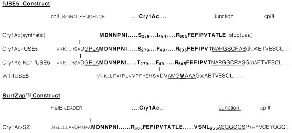
The entire cry1Ac sequence in the fUSE5 vector and the fusion junctions in both vector systems were verified by DNA sequencing. Figure 1 shows the sequences for the WT-fUSE5, 1Ac-fUSE5, and 1Ac-Kpn-fUSE5 phages and the Cry1Ac-expressing phage from the SurfZAP vector (1Ac-SZ). The fUSE5 Cry1Ac fusion sequences contained five additional amino acids at the N terminus due to the addition of the SfiI cloning site in the vector. In addition, a single nucleotide change from the reported cry1Ac sequence, resulting in the single amino acid change I491→F491, was detected in both the 1Ac-fUSE5 and the 1Ac-Kpn-fUSE5 phage. This change was apparently present in the source plasmid pAGM19, since these two phage constructs were made from cry1Ac fragments amplified in independent PCR mixtures containing pAGM19. Finally, the fusion junction with cpIII in the fUSE5 constructs contained some interesting unplanned changes. Originally designed so that there would be a Gly-Ala-Gly-Ala spacer between the Cry1Ac and cpIII peptide sequences, a deletion inducing a frameshift occurred in the fifth-to-the-last codon of the cry1Ac gene of each construct, possibly by an error in the primer LK02. Selection for viable infective phage particles required that the shift be corrected by matching insertions or deletions downstream to have a functional cpIII section of the protein. The result was the 11-amino-acid spacer underlined in the figure, which is neither Cry1Ac nor cpIII sequence, followed by cpIII starting at amino acid 30. This particular corrective rearrangement was selected three times independently during construction of different recombinant phages, emphasizing the power of the selection for phage viability built into this vector. Possible reasons for selection of this particular rearrangement are discussed below. Phage titers for this construct in JM109 cells were between 109 and 1010 TU per ml of overnight culture.
FIG. 1.
Amino acid sequences of Cry1Ac-pIII fusion proteins as derived from their DNA sequences. W indicates a tryptophan substituted in place of a stop codon by the amber suppressor present in the host JM109 cells. Underlining indicates protein sequence which is present in neither the native Cry1Ac nor the cpIII protein. | indicates the predicted signal sequence protease cleavage site.
The cry1Ac and fusion junction sequences for the 1Ac-SZ construct were found to be as planned (Table 1), with the fusion protein containing 43 additional C-terminal amino acids of Cry1Ac (a protoxin fragment) not in the fUSE5 constructs, an alanine/glycine spacer, and only the last 209 amino acids (198 to 406) of cpIII. The PelB leader peptide is presumed to be cleaved exactly at the N terminus of the Cry1Ac portion of the protein, such that the natural end of the protein is exposed, unlike in our fUSE5 construct. Phage titers of this construct were usually over 1011 phage per ml of culture.
CryIAc-expressing phages, but not wild-type phage, are toxic to M. sexta and H. virescens.
DNA sequencing verified that both the fUSE5 and SurfZAP constructs contained in-frame cry1Ac-cpIII gene fusions. The ability of the resulting fusion proteins to fold into biologically active conformations was shown by the ability of our Cry1Ac-expressing phage preparations, but not control phage preparations, to kill insects susceptible to native B. thuringiensis Cry1Ac. The toxicities of Cry1Ac-expressing phages compared to that of purified HD-73 phage were determined by insect feeding assays. Phages precipitated from E. coli supernatants were titrated to determine their concentrations and then diluted in TBS and applied to the surface of insect diet in doses ranging from 109 to 107 TU per well, with approximately 1.8 cm2 of feeding surface area per well. Twenty larvae (H. virescens or M. sexta) were fed individually for each dose and control group for fUSE5 phage, and mortality was recorded after 7 days. LC50s were determined by probit analysis of the mortality data and are presented for H. virescens in Table 1. Given that there are on average five molecules of the Cry1Ac-cpIII fusion protein per phage particle and that it has been shown that typical filamentous phage preparations contain 20 phage particles for every transducing unit (35), it can be calculated that 109 TU of 1Ac-fUSE5 phage contains approximately 10.4 ng of Cry1Ac protein (17.1 ng of the fusion protein). Therefore, the LC50 of the phage-expressed protein can also be expressed as 7.3 ng/well, in remarkably close agreement with the LC50 for the purified Bacillus protein (7.6 ng/well). Likewise, conversion of the 1Ac-Kpn-fUSE5 LC50 from transducing units to nanograms gives a dose of 7.5 ng per well. If these doses are expressed in terms of surface area, they average 4.2 ng per cm2, in agreement with published toxicity for this Cry protein against H. virescens (10). Single-dose feeding experiments demonstrated that 1Ac-fUSE5 phage is extremely toxic to M. sexta larvae as well (not shown). LC50s were not determined for SurfZAP phage, but 1Ac-SZ was shown to be toxic to H. virescens at adequate dosages.
Fusion proteins are expressed on the phage particles and are recognized by Cry1Ac-specific antibodies.
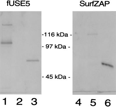
To confirm that the toxin was being expressed as a fusion protein incorporated into phage particles, purified phage was analyzed by immunoblotting. Phage particles produced in both the fUSE5 and SurfZAP systems were concentrated by precipitation in salt and acetic acid, pelleted, boiled in denaturing sample buffer, and subjected to SDS-polyacrylamide gel electrophoresis (PAGE). Proteins electrophoretically transferred to nitrocellulose were detected with rabbit anti-Cry1Ac antibodies (Fig. 2). HD-73 Cry1Ac purified from B. thuringiensis crystals was run in neighboring lanes for comparison of sizes and quantities (lanes 3 and 6). Fusion proteins were detected in the Cry1Ac-expressing phages of both vectors (lanes 1 and 5) but not in control phage (lanes 2 and 4). The mobility of the fUSE5 fusion protein indicated a molecular size of approximately 104 kDa, very close in size to the 107-kDa protein expected (65-kDa Cry1Ac plus 42-kDa cpIII). The difference may indicate some proteolysis of the phage; however, the difference is also within the limits of accuracy for this type of determination and the sharpness of the band does not indicate proteolysis. A second, weaker band with an apparent molecular size of 130 kDa was also recognized by anti-Cry1Ac antibodies whenever immunoblotting was performed on 1Ac-fUSE5 phage. Its components were not positively identified, but the facts that it is twice the molecular mass of the 65-kDa toxin and is recognized by anti-Cry1Ac antiserum suggest that it may be an insoluble toxin dimer, resulting from proteolysis of fusion proteins by endogenous E. coli proteases. It has been previously observed in our lab that toxin associated with membranes can form aggregates that persist through SDS-cracking buffer treatment and appear as a >100-kDa band by SDS-PAGE (24a). Such aggregates are most likely inactive. Immunoblotting of 1Ac-Kpn-fUSE5 phage produced a banding pattern identical to that of 1Ac-fUSE5 phage (not shown). The SurfZAP fusion protein ran with an apparent molecular size of 117 kDa, somewhat larger than the expected molecular mass, since although this construct expresses 43 more amino acids of Cry1Ac than 1Ac-fUSE5 does, it includes only a 25-kDa fragment of cpIII. However, in neither the 1Ac-SZ nor the 1Ac-fUSE5 lane was there any evidence that normal Cry1Ac protein not part of a fusion protein was being expressed. In addition, the relative quantities of Cry1Ac in the phage were as predicted, insofar as the 1Ac-SZ fusion protein was expected to be present at 2% of the level of the fUSE5 fusion protein, and 10 times as many phage particles and twice as much development time were required for 1Ac-SZ to be detected.
FIG. 2.
Immunoblot detection of phage-displayed Cry1Ac fusion proteins. Phages (109 TU for fUSE5 constructs and 1010 TU for SurfZAP constructs) were boiled for 4 min in 30 μl of Laemmli denaturing sample buffer and size separated by SDS–8% PAGE. The proteins were transferred to nitrocellulose and detected by a rabbit polyclonal anti-Cry1Ac antibody and then by an alkaline phosphatase-conjugated goat anti-rabbit antibody. Lane 1, 1Ac-fUSE5 phage; lane 2, WT-fUSE5 phage; lane 3, 10 ng of purified trypsin-activated HD-73 Cry1Ac; lane 4, SurfZAP-FAb phage; lane 5, SurfZAP-1Ac phage; lane 6, 10 ng of purified trypsin-activated HD-73 Cry1Ac. The blot was divided through the molecular mass markers between lanes 3 and 4, and the two sides were developed separately. The SurfZAP side of the blot was allowed to develop for twice as long as the fUSE5 side, since SurfZAP-1Ac phage produced a lower-intensity signal. The effect of the longer development time can be observed by comparing the 10-ng toxin bands on each side of the blot (lanes 3 and 6).
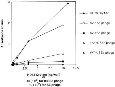
An ELISA was also performed on the Cry1Ac-expressing phages, since unlike in the immunoblot analysis, the proteins would not be denatured before antibody binding and quantitation could be more precise. Phages were serially diluted in Tris buffer, and purified HD-73 Cry1Ac protein served as a standard. Anti-Cry1Ac antibody was again used as the primary antibody. Figure 3 reflects averages from two experiments for fUSE5 phage, both of which determined all points in duplicate. The SZ phage was included in one of these experiments. In this assay, both 1Ac-SZ and 1Ac-fUSE5 phages behaved exactly as expected for equivalent amounts of Cry1Ac protein not attached to phage, except at the highest phage concentrations at which the amount of other phage proteins in the well began to interfere with antibody binding. At those concentrations, however, the HD-73 Cry protein signal was also beyond the most linear part of its response curve. In the linear portion, the above-described calculations accurately determined the amount of Cry1Ac toxin expected to be present on a given amount of phage (i.e., approximately 10 ng of Cry1Ac per 109 TU of 1Ac-fUSE5 and 0.2 ng per 109 TU of 1Ac-SZ). Observe in Fig. 3 that 3 × 10⁸ TU of 1Ac-fUSE5 produced the same signal as 3 ng of Cry1Ac toxin. Likewise, 3 × 10⁹ TU of 1Ac-SZ produced a signal 20% as strong as that of 3 × 10⁸ TU of 1Ac-fUSE5, or 2% as strong when the value was corrected for the number of phage. Therefore, since the amount of Cry1Ac present in our PEG-purified phage preparations was in each case directly related to the phage titer, the Cry1Ac expressed from both the phagemid and phage vectors must be present in these preparations exclusively as fusion protein incorporated into phage particles.
FIG. 3.
ELISA detection of phage-displayed Cry1Ac fusion proteins. Cry1Ac-expressing phage, 1Ac-fUSE5 and SZ-1Ac, and their respective control phages, WT-fUSE5 (no insert) and SZ-FAb (antibody insert), were compared with purified, trypsin-activated HD-73 Cry1Ac in an ELISA. The numbers on the horizontal axis refer to different units per well for the fUSE5 phage, SurfZAP phage, and purified Cry1Ac toxin as indicated on the figure. Ten times more SurfZAP (SZ) phage than fUSE5 phage was applied per well per dilution in order to obtain absorbance readings in the same range. For SurfZAP phage, the results from one trial are shown (n = 2). For fUSE5 phage and HD-73 Cry1Ac, the means of results from two different trials are plotted (n = 4). Mean A450 (n = 2) values for the individual trials for purified Cry1Ac were (trial 1) 0.021, 0.112, 0.366, 1.137, and 2.422 and (trial 2) 0.029, 0.109, 0.333, 1.170, and 3.426, for 0.1, 0.3, 1.1, 3.3 and 11.0 ng of toxin, respectively. Mean A450 (n = 2) values for the individual trials for 1Ac-fUSE5 phage were (trial 1) 0.173, 0.506, 1.243, and 1.521 and (trial 2) 0.213, 0.308, 1.04, and 2.255 for 3.3 × 10⁷, 1.1 × 10⁸, 3.3 × 10⁸, and 1 × 10⁹ TU, respectively. All A450 values for WT-fUSE5 phage in both trials were below 0.050.
In addition, both immunoassay results demonstrated that B. thuringiensis toxin displayed on phage is suitable for these assays without any need to separate it from the phage particle and therefore that the toxin-expressing phage can go from overnight cultures into such assays with less than 30 min of preparation time.
Micropanning experiments.
The possibility of affinity selection of B. thuringiensis toxins by biopanning was tested under several conditions in micropanning experiments, since a library of phage-displayed toxins had not been completed at this time. Micropanning is a comparison of the numbers of Cry1Ac-phage and control phage bound by a toxin-specific target, such as anti-Cry1Ac antibody. It is a useful first step in biopanning, since it helps establish the binding and elution conditions best suited to the affinity of the protein interaction under selection. Micropanning is also used to compare multiple candidate phages selected by library biopanning, where addition of a competing ligand allows determination of the degree of specific binding of each phage (3).
Micropanning against an anti-Cry1Ac antibody was performed in duplicate in microtiter wells (1 μg of antibody per well) with bovine serum albumin as a blocking agent and control ligand. Results are summarized in Table 2. The 1Ac-fUSE5 phage was preferentially retained by the antibody compared to control phage, and this binding was competed quite well (68%) with free Cry1Ac protein, indicating specific binding (Table 2).
TABLE 2.
Micropanning with Cry1Ac-expressing phage against anti-Cry1Ac antibody or BBMV
| Targeta | Relative yieldb of:
|
|
|---|---|---|
| WT-fUSE5 phage | 1Ac-fUSE5 phage | |
| Bovine serum albumin | 1.0 | 1.0 |
| Anti-Cry1Ac Ab R118 | 0.5 | 7.1 |
| Anti-Cry1Ac Ab + 1 μg of Cry1Ac | 0.2 | 2.3 |
| 100 μg of BBMV | 1.0 | 1.0 |
| 100 μg of BBMV + 1 μg of Cry1Ac | 0.24 | 1.7 |
Ab, antibody.
Relative yields of control samples are set to 1.0.
Micropanning with various preparations of toxin receptors proved more complex. When purified M. sexta aminopeptidase N, a known protein receptor for Cry1Ac in this species (19, 30), was micropanned in microtiter plates, no enrichment of 1Ac phage (fUSE5 or SurfZAP) over control phage was observed (data not shown). However, binding of the receptor protein directly to microtiter plates was probably not the ideal method to present this target for two reasons. First, it provides no control over the orientation of the protein on the plate. Second, binding of Cry1Ac toxin to soluble aminopeptidase N has been shown to be only partly reversible (45%) (25), which probably interferes with recovery of the affinity-selected phage since our elution conditions are not expected to release the receptor from the plates. Use of a biotin-streptavidin biopanning method (27) should overcome these problems.
BBMV have been used as a source of toxin receptor in situ and therefore were also tried as a biopanning target. In the absence of a suitable negative control, an excess of free Cry1Ac toxin was added to half the samples as a competitor. As with the purified receptor, there was no significant difference between the number of Cry1Ac-expressing phage and that of control phage eluted after unbound phage was washed away (Table 2). Two experiments reported below, a binding assay with iodinated phage and an immunoblot analysis of phage incubated with BBMV, uncovered possible reasons for this lack of affinity selectivity for BBMV.
Competition binding of 125I-radiolabeled phage to BBMV.
HD-73 Cry1Ac purified from B. thuringiensis, 1Ac-fUSE5 phage, and WT-fUSE5 control phage were iodinated by standard methods. Equal amounts of M. sexta BBMV, with or without unlabelled competitor HD-73 Cry1Ac, were incubated with the iodinated phages or toxin. The results are presented in Table 3. 1Ac-fUSE5 phage and HD-73 Cry1Ac showed similar percentages bound, namely, about 12%, which was approximately twice the percentage bound of the control phage, indicating some selective advantage of the Cry1Ac-expressing phage. However, whereas iodinated HD-73 Cry1Ac was competed quite well by unlabelled toxin, the 1Ac-fUSE5 phage was not. One interpretation of this result is that nonspecific binding interactions occurred between the BBMV and the phage capsid, interfering with affinity selection under our binding conditions. It is possible, of course, that other conditions will be found which will eliminate this problem.
TABLE 3.
Competition binding of 125I-radiolabelled phage to BBMV
| Radiolabelled toxin or phage added to 10 μg of BBMV | 100-fold excess of unlabelled Cry1Ac | cpm added | Mean cpm bound (n = 2) | % cpm bound | % Compe- tition |
|---|---|---|---|---|---|
| 125I-Cry1Ac protein | − | 98,242 | 12,690 | 12.9 | 66.5 |
| + | 98,242 | 4,256 | 4.3 | ||
| 125I-1Ac-fUSE5 phage | − | 105,252 | 12,293 | 11.7 | 8.5 |
| + | 105,252 | 11,212 | 10.7 | ||
| 125I-WT-fUSE5 phage | − | 62,319 | 3,784 | 6.1 | None |
| + | 62,319 | 4,743 | 7.6 |
Incubation of 1Ac-fUSE5 phage with BBMV results in proteolysis of the Cry1Ac-cpIII fusion protein.
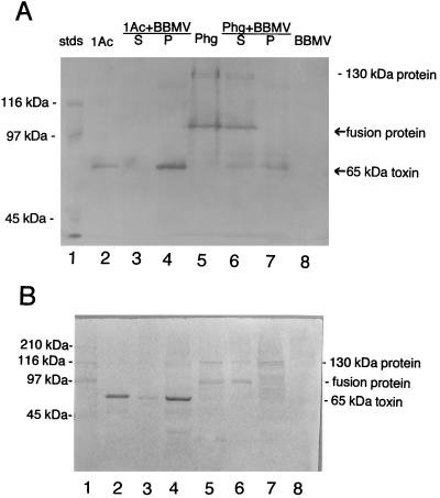
An essential step in the toxic mechanism of B. thuringiensis crystal proteins, such as Cry1Ac, is proteolytic processing in the insect gut. BBMV prepared from insect midgut in the absence of proteinase inhibitors (as they were in the experiments described above) may more closely approximate the conditions the toxin encounters when it is ingested than proteinase-inhibited preparations. However, the proteinases associated with BBMV might interfere with biopanning by degrading phage capsid proteins or the fusion protein. To determine the fate of the Cry1Ac-cpIII fusion protein in the presence of BBMV, 1Ac-fUSE5 phage was incubated at room temperature with BBMV without proteinase inhibitors, as if in micropanning, but then subjected to immunoblot analysis to detect Cry1Ac and fusion protein. Equivalent amounts of Cry1Ac toxin were analyzed in parallel. To separate toxin or phage bound to the BBMV from their unbound counterparts, the BBMV were pelleted and the supernatants were analyzed separately from the vesicle pellets (which were also washed before analysis). The results were surprising (Fig. 4A). Purified toxin (Fig. 4A, lane 2), when it was incubated with BBMV, was found to segregate with the pellet (Fig. 4A, lane 4), apparently bound to the vesicles. In contrast, after incubation of 1Ac-fUSE5 phage (Fig. 4A, lane 5) with BBMV, the 104-kDa fusion protein was detected only in the supernatant (Fig. 4A, lane 6). Also in the supernatant were smaller amounts of a 65-kDa protein recognized by anti-Cry1Ac antibodies, which was most likely the Cry1Ac portion of the fusion protein released by proteolysis. The pellet contained only the 65-kDa protein (Fig. 4A, lane 7). No proteins were recognized by anti-Cry1Ac antibodies in the BBMV control (Fig. 4A, lane 8). The presence of some free toxin in the supernatant (Fig. 4A, lane 6) and the absence of fusion protein in the pellet (Fig. 4A, lane 7) suggested that proteolysis of the fusion protein occurs before binding of the toxin portion to BBMV. To test this hypothesis, purified toxin and 1Ac-fUSE5 binding experiments were repeated in the presence of proteinase inhibitors (Fig. 4B). Proteinase inhibitors did not alter purified toxin binding to BBMV (Fig. 4B, lane 4). However, in contrast to results seen without inhibitors, no 65-kDa protein was detected in the phage samples by anti-Cry1Ac antibodies in either the supernatant (Fig. 4B, lane 6) or the BBMV pellet (Fig. 4B, lane 7). The 104-kDa fusion protein was clearly detected in the supernatant, but no clear fusion protein band was seen in the pellet. The faint 130-kDa band was detected in both the pellet and supernatant fractions. With regard to biopanning, these results indicate that BBMV failed as a source for target ligands because they did not bind whole phage.
FIG. 4.
Immunoblot detection of Cry1Ac and Cry1Ac phage binding to BBMV in the presence and absence of proteinase inhibitors. Cry1Ac-fUSE5 phage (3 × 10⁹ TU) or purified and trypsin-activated HD-73 Cry1Ac toxin (50 ng) was incubated with M. sexta BBMV (100 μg of total protein) in a total volume of 25 μl for 90 min at 22°C. Equal aliquots of phage, toxin, and BBMV were also held separately at 22°C for 90 min, after which all the tubes containing BBMV were microcentrifuged for 5 min to pellet the vesicles. Supernatants were removed to new tubes and mixed with equal volumes of denaturing sample buffer as were the samples containing phage or toxin alone. BBMV pellets were resuspended in 200 μl of TBS and repelleted, and the wash supernatants were discarded. Sample buffer (20 μl) was added to the pellets, and all samples were boiled for 3 min before electrophoresis on an SDS–8% (A) or 10% (B) polyacrylamide gel. The proteins were then electrophoretically transferred to nitrocellulose membrane, and Cry1Ac and Cry1Ac fusion protein were detected by immunoblotting as described for Fig. 2. Experiments shown in panels A and B are identical except that in panel B proteinase inhibitors (500 μM phenylmethylsulfonyl fluoride and 5 mM benzamidine) were added to the BBMV prior to addition of toxin or phage. Lanes 1, prestained standard molecular mass markers (stds; sizes are indicated on the left); lanes 2, HD-73 Cry1Ac toxin (50 ng); lanes 3, HD-73 Cry1Ac toxin plus BBMV supernatant; lanes 4, HD-73 Cry1Ac toxin plus BBMV pellet; lanes 5, 1Ac-fUSE5 phage; lanes 6, 1Ac-fUSE5 phage plus BBMV supernatant; lanes 7, Ac-fUSE5 phage plus BBMV pellet; lanes 8, BBMV alone (100 μg of total protein). Phg, phage; S, supernatant; P, pellet.
DISCUSSION
We have demonstrated here two phage display systems for expression of biologically active Cry1Ac toxin in E. coli hosts. Phage-displayed Cry1Ac produced by either system performed as well as purified natural toxin in ELISA, immunoblotting, and insect toxicity assays. Significant differences in the construction of the SurfZAP and fUSE5 translational fusions (e.g., different leader peptides, fusion junction sequences, and sizes of cpIII fragments) indicate that this method of toxin expression is relatively insensitive to structural details and should be adaptable to many different toxins.
Whether incorporation of the fusion protein into phage particles is essential for successful toxin expression is currently unknown. Our PEG-precipitated phage preparations did not contain detectable amounts of free fusion protein, since dose-response curves in both the ELISA and toxicity assays always correlated closely with phage titers. Yet, it is possible that fusion of the cry1Ac sequence to cpIII alone would allow high-level expression of soluble Cry1Ac toxin in E. coli. While this paper was in preparation, an attempt to express such a fusion protein from a phagemid (not SurfZAP) in the absence of helper phage was reported for another B. thuringiensis toxin, Cry1Aa (26). In those constructs, a nearly complete cry1Aa gene was fused to a cpIII sequence and, in a separate construct, to a glutathione S-transferase gene. Only degraded fragments of fusion protein could be detected in both cases, but addition of helper phage also did not allow expression of intact Cry1Aa-cpIII fusion protein from these constructs. B. thuringiensis toxin fusion proteins which retained insecticidal activity and which were not associated with phage have been reported previously (16, 39). Nevertheless, association of the toxin with phage particles has advantages for rapid purification, quantitation, DNA sequence analysis, and biopanning.
Another point which remains to be clarified is whether the toxin must be released from the phage in order to be toxic. Our results indicate that it does. It has been theorized that B. thuringiensis toxins are required to form multimers in cell membranes to be toxic (20). Separation of the toxin from the phage particle would no doubt enable this process. Incubation of Cry1Ac phage with membrane vesicles from insect midguts in vitro released a protein identical in mobility to B. thuringiensis-derived Cry1Ac toxin and recognized by anti-Cry1Ac antiserum, suggesting that insect proteinases cleave the fusion protein at the Cry1Ac-cpIII junction in vivo. In addition, immunoblotting of BBMV incubated with Cry1Ac-expressing phage did not detect intact fusion protein bound to BBMV in vitro. While the addition of proteinase inhibitors to the Cry1Ac phage plus BBMV mixture eliminated cleavage of the fusion protein, the intact fusion protein still did not detectably bind BBMV. Given the nearly identical LC50s for 1Ac-fUSE5 phage and purified Cry1Ac in H. virescens, proteolytic processing of the fusion junction in the gut seems to be extremely efficient in yielding intact toxin.
The results of our micropanning experiments with phage-displayed Cry1Ac suggest that biopanning for improved or novel B. thuringiensis toxin specificities is possible but may be complex. B. thuringiensis toxins recognize protein receptors in the insect midgut with high affinity (Kd = 1 × 10⁻⁹ to 20 × 10−9 M); some of these receptors have been isolated (15, 19, 30, 40). However, biopanning with purified receptors as targets is complicated by glycolipids which complex with the protein (33) and also sometimes bind the toxin in vitro (11), as well as carbohydrates of unknown function displayed on the receptor proteins, which are difficult to monitor (30). BBMV from insect midgut tissue presumably contain all these molecules and might be used as a biopanning target if several problems can be overcome. A nonspecific-binding problem was indicated in the iodinated-phage binding assay. Also, intact Cry1Ac phage did not detectably bind BBMV, as was revealed in the phage binding and immunoblotting experiment (Fig. 4). BBMV are a technically convenient substrate for biopanning, since they can be pelleted with brief microcentrifugation. Unfortunately, binding affinity of purified toxins for BBMV has not always correlated well with toxicity in the intact insect (10, 22).
One issue about which there is little doubt is the significant advantage of phage display over conventional toxin expression systems for the purposes of producing libraries of genetically modified toxins. This advantage is twofold. First, unlike conventional expression systems which produce protoxins in crystals or inclusion bodies which must be solubilized, phage-displayed toxins are soluble and ready to assay. Based on the apparent absence of free fusion protein in our preparations, it may be that incorporation of the toxin into the relatively rigid structure of the phage capsid is what prevents the accumulation of insoluble toxin precipitates in this type of system. Second, since the phage purification protocol acts on the phage particle rather than on the particular toxin which makes up only a very small portion of it, individual requirements for solubility and activation are not needed and therefore thousands of samples can theoretically be processed in parallel. Other, less consequential advantages include the rapid and inexpensive purification procedure, the ability to quantitate toxin by phage titrating, the ease of preparing single-stranded DNA for sequencing, and the assurance that most frameshift mutations will be eliminated automatically from toxin libraries since they fail to produce viable phage.
In conclusion, there are now many reports in which domain swapping and site-directed protein engineering have shown that B. thuringiensis toxins with novel activities can be produced by genetic manipulation. Expression of these modified toxin genes has often proved problematic, with insolubility and subsequent inactivity routinely affecting more than 80% of modified toxin genes expressed in traditional E. coli vectors (2, 41). It is not possible to prove that all toxin genes will be able to be expressed successfully in phages, but the 65-kDa active core of Cry1Ac was not soluble in conventional systems and it was readily expressed on phage, as was a Cry1Ac mutant. With this advantage of increased solubility, coupled with the capacity to produce large numbers of samples in parallel, toxin expression by phage display may therefore eliminate this obstacle to understanding and improving B. thuringiensis toxins by genetic engineering.
ACKNOWLEDGMENTS
We thank Howard Benjamin at Praecis Pharmaceutical, Inc., for many helpful comments on phage display and Ke Luo at the University of Georgia for gifts of antibody R118 and purified aminopeptidase N.
L.M.K. was supported by a Biotechnology Research Grant from the University of Georgia.
REFERENCES
- 1.Adang M J. Bacillus thuringiensis insecticidal crystal proteins: gene structure, action, and utilization. In: Maramorosch K, editor. Biotechnology for biological control of pests and vectors. Boca Raton, Fla: CRC Press; 1991. pp. 3–24. [Google Scholar]
- 1a.Benjamin, H. Unpublished data.
- 2.Bosch D, Schipper B, van der Kleij H, de Maagd R A, Steikema W J. Recombinant Bacillus thuringiensis crystal proteins with new properties: possibilities for resistance management. Biotechnology. 1994;12:915–918. doi: 10.1038/nbt0994-915. [DOI] [PubMed] [Google Scholar]
- 3.Cabilly S, editor. Methods in molecular biology: combinatorial peptide library protocols. Clifton, N.J: Humana Press; 1997. p. 320. [Google Scholar]
- 4.Chen X J, Lee M K, Dean D H. Site-directed mutations in a highly conserved region of Bacillus thuringiensis δ-endotoxin affect inhibitions of short circuit current across Bombyx mori midguts. Proc Natl Acad Sci USA. 1993;90:9041–9045. doi: 10.1073/pnas.90.19.9041. [DOI] [PMC free article] [PubMed] [Google Scholar]
- 5.Dean D H, Rajamohan F, Lee M K, Wu S J, Chen X J, Alcantara E, Hussain S R. Probing the mechanism of action of Bacillus thuringiensis insecticidal proteins by site-directed mutagenesis—a minireview. Gene. 1996;179:111–117. doi: 10.1016/s0378-1119(96)00442-8. [DOI] [PubMed] [Google Scholar]
- 6.de Maagd R A, van der Kleij H, Bakker P, Stiekema W J, Bosch D. Different domains of Bacillus thuringiensis δ-endotoxins can bind to insect midgut membrane proteins on ligand blots. Appl Environ Microbiol. 1996;62:2753–2757. doi: 10.1128/aem.62.8.2753-2757.1996. [DOI] [PMC free article] [PubMed] [Google Scholar]
- 7.English L, Readdy T L. Delta endotoxin inhibits a phosphatase in midgut epithelial membranes of Heliothis virescens. Insect Biochem. 1989;19:145–152. [Google Scholar]
- 8.Estruch J J, Carozzi N B, Desai N, Duck N B, Warren G W, Koziel M G. Transgenic plants: an emerging approach to pest control. Nat Biotechnol. 1997;15:137–141. doi: 10.1038/nbt0297-137. [DOI] [PubMed] [Google Scholar]
- 9.Feitelson J S, Payne J, Kim L. Bacillus thuringiensis: insects and beyond. Biotechnology. 1992;10:271–275. [Google Scholar]
- 10.Garczynski S F, Crim J W, Adang M J. Identification of putative insect brush border membrane-binding molecules specific to Bacillus thuringiensis δ-endotoxin by protein blot analysis. Appl Environ Microbiol. 1991;57:2816–2820. doi: 10.1128/aem.57.10.2816-2820.1991. [DOI] [PMC free article] [PubMed] [Google Scholar]
- 11.Garczynski S F, Adang M J. Society for Invertebrate Pathology 29th Annual Meeting and IIIrd International Colloquium on Bacillus thuringiensis. 1996. Interactions of Bacillus thuringiensis toxins with lipids isolated from the midgut of Manduca sexta larvae; p. 28. [Google Scholar]
- 12.Ge A Z, et al. Location of the Bombyx mori specificity domain on a Bacillus thuringiensis delta-endotoxin protein. Proc Natl Acad Sci USA. 1989;86:4037–4041. doi: 10.1073/pnas.86.11.4037. [DOI] [PMC free article] [PubMed] [Google Scholar]
- 13.Grochulski P, Masson L, Borisova S, Pusztai-Carey M, Schwartz J-L, Brousseau R, Cygler M. Bacillus thuringiensis Cry1A(a) insecticidal toxin crystal structure and channel formation. J Mol Biol. 1995;254:447–464. doi: 10.1006/jmbi.1995.0630. [DOI] [PubMed] [Google Scholar]
- 14.Harlow E, Lane D. Antibodies: a laboratory manual. Cold Spring Harbor, N.Y: Cold Spring Harbor Laboratory; 1988. [Google Scholar]
- 15.Hofmann C, Vanderbruggen H, Hofte H, Van Rie J, Jansens S, Van Mellaert H. Specificity of Bacillus thuringiensis δ-endotoxins is correlated with the presence of high affinity binding sites in the brush border membrane of target insect midguts. Proc Natl Acad Sci USA. 1988;85:7844–7848. doi: 10.1073/pnas.85.21.7844. [DOI] [PMC free article] [PubMed] [Google Scholar]
- 16.Hofte H, Buyssens S, Vaeck M, Leemans J. Fusion proteins with both insecticidal and neomycin phosphotransferase II activity. FEBS Lett. 1988;226:364–370. [Google Scholar]
- 17.Hogrefe H H, Amberg J R, Hay B N, Sorge J A, Shopes B. Cloning in a bacteriophage lambda vector for the display of binding proteins on filamentous phage. Gene. 1993;137:85–91. doi: 10.1016/0378-1119(93)90255-2. [DOI] [PubMed] [Google Scholar]
- 18.Hunter W, Greenwood F. Preparation of iodine-131 labelled human growth hormone of high specific activity. Nature. 1962;194:495–496. doi: 10.1038/194495a0. [DOI] [PubMed] [Google Scholar]
- 18a.Kasman, L. M. Unpublished data.
- 19.Knight P J, Crickmore N, Ellar D J. The receptor for Bacillus thuringiensis Cry1A(c) delta-endotoxin in the brush border membrane of the lepidopteran Manduca sexta is aminopeptidase N. Mol Microbiol. 1994;11:429–436. doi: 10.1111/j.1365-2958.1994.tb00324.x. [DOI] [PMC free article] [PubMed] [Google Scholar]
- 20.Knowles B H, Dow J A T. The crystal delta-endotoxins of Bacillus thuringiensis—models for their mechanism of action on the insect gut. Bioessays. 1993;15:469–476. [Google Scholar]
- 21.Lambert B, Peferoen M. Insecticidal promise of Bacillus thuringiensis. Bioscience. 1992;42:112–122. [Google Scholar]
- 22.Lee M K, Rajamohan F, Gould F, Dean D H. Resistance to Bacillus thuringiensis Cry1A δ-endotoxins in a laboratory-selected Heliothis virescens strain is related to receptor alteration. Appl Environ Microbiol. 1995;61:3836–3842. doi: 10.1128/aem.61.11.3836-3842.1995. [DOI] [PMC free article] [PubMed] [Google Scholar]
- 23.Lee M K, Young B A, Dean D H. Domain III exchanges of Bacillus thuringiensis Cry1A toxins affect binding to different gypsy moth midgut receptors. Biochem Biophys Res Commun. 1995;216:306–312. doi: 10.1006/bbrc.1995.2625. [DOI] [PubMed] [Google Scholar]
- 24.Li J, Carroll J, Ellar D J. Crystal structure of insecticidal δ-endotoxin from Bacillus thuringiensis at 2.5 Å resolution. Nature. 1991;353:815–821. doi: 10.1038/353815a0. [DOI] [PubMed] [Google Scholar]
- 24a.Lu, Y.-J., and M. Adang. Unpublished data.
- 24b.Luo, K. (University of Georgia, Athens). Unpublished data.
- 25.Masson L, Lu Y-J, Mazza A, Brosseau R, Adang M J. The Cry1A(c) receptor purified from Manduca sexta displays multiple specificities. J Biol Chem. 1995;270:20309–20315. doi: 10.1074/jbc.270.35.20309. [DOI] [PubMed] [Google Scholar]
- 26.Marzari R, Edomi P, Bhatjagar R K, Ahmad S, Selvapandiyan A, Bradbury A. Phage display of Bacillus thuringiensis Cry1A(a) toxin. FEBS Lett. 1997;411:27–31. doi: 10.1016/s0014-5793(97)00647-9. [DOI] [PubMed] [Google Scholar]
- 27.Parmley S F, Smith G P. Antibody-selectable filamentous phage vectors: affinity purification of target genes. Gene. 1988;73:305–318. doi: 10.1016/0378-1119(88)90495-7. [DOI] [PubMed] [Google Scholar]
- 28.Rajamohan F, Cotrill J A, Gould F, Dean D H. Role of domain II, loop 2 residues of Bacillus thuringiensis Cry1Ab δ-endotoxin in reversible and irreversible binding to Manduca sexta and Heliothis virescens. J Biol Chem. 1996;271:2390–2396. doi: 10.1074/jbc.271.5.2390. [DOI] [PubMed] [Google Scholar]
- 29.Raymond M. Presentation d’un programme d’analyse log-probit pour micro-ordinateur. Cah O R S T O M Ser Entomol Med Parasitol. 1985;22:117–121. [Google Scholar]
- 30.Sangadala S, Walters F S, English L H, Adang M J. A mixture of Manduca sexta aminopeptidase and phosphatase enhances Bacillus thuringiensis insecticidal Cry1A(c) toxin binding and 86RbŒ+ efflux in vitro. J Biol Chem. 1994;269:10088–10092. [PubMed] [Google Scholar]
- 31.Schmitz J, Preiser H, Maestracci D, Ghosh B K, Cerda J J, Crane R K. Purification of the human intestinal brush border membrane. Biochim Biophys Acta. 1973;323:98–112. doi: 10.1016/0005-2736(73)90434-3. [DOI] [PubMed] [Google Scholar]
- 32.Schnepf H E. Bacillus thuringiensis toxins: regulation, activities and structural diversity. Curr Opin Biotechnol. 1995;6:305–312. [Google Scholar]
- 33.Schwartz J-L, Lu Y J, Soehnlein P, Brousseau R, Masson L, Laprade R, Adang M J. Ion channels formed in planar lipid bilayers by Bacillus thuringiensis toxins in the presence of Manduca sexta midgut receptors. FEBS Lett. 1997;412:270–276. doi: 10.1016/s0014-5793(97)00801-6. [DOI] [PubMed] [Google Scholar]
- 34.Scott J K, Smith G P. Searching for peptide ligands with an epitope library. Science. 1990;249:386–390. doi: 10.1126/science.1696028. [DOI] [PubMed] [Google Scholar]
- 35.Smith G P. Filamentous phage assembly: morphogenetically defective mutants that do not kill the host. Virology. 1988;167:156–165. doi: 10.1016/0042-6822(88)90065-7. [DOI] [PubMed] [Google Scholar]
- 36.Smith G P, Ellar D J. Mutagenesis of two surface exposed loops of the Bacillus thuringiensis Cry1C δ-endotoxin affects insecticidal specificity. Biochem J. 1994;302:611–616. doi: 10.1042/bj3020611. [DOI] [PMC free article] [PubMed] [Google Scholar]
- 37.Stewart C N, Adang M J, All J N, Boerma H R, Cardineau G, Tucker D, Parrott W A. Genetic transformation, recovery, and characterization of fertile soybean transgenic for a synthetic Bacillus thuringiensis cry1Ac gene. Plant Physiol. 1996;112:121–129. doi: 10.1104/pp.112.1.121. [DOI] [PMC free article] [PubMed] [Google Scholar]
- 38.Tabashnik B E, Malvar T, Liu Y-B, Finson N, Boethakur D, Shin B S, Park S-H, Masson L, DeMaagd R, Bosch D. Cross-resistance of diamondback moth implies altered interactions with domain II of Bacillus thuringiensis toxins. Appl Environ Microbiol. 1996;62:2839–2844. doi: 10.1128/aem.62.8.2839-2844.1996. [DOI] [PMC free article] [PubMed] [Google Scholar]
- 39.Thompson M A, Schnepf H E, Feitelson J S. Structure, function, and engineering of Bacillus thuringiensis toxins. Genet Eng. 1995;17:99–117. [PubMed] [Google Scholar]
- 40.Van Rie J, Jansens S, Hofte H, Degheele D, Van Melleart H. Receptors on the brush border membrane of the insect midgut as determinants of the specificity of B. thuringiensis delta-endotoxins. Appl Environ Microbiol. 1990;56:1378–1385. doi: 10.1128/aem.56.5.1378-1385.1990. [DOI] [PMC free article] [PubMed] [Google Scholar]
- 41.Wabiko H, Yasuda E. Bacillus thuringiensis protoxin location of toxic border and requirement of non-toxic domain for high level in vivo production of active toxin. Microbiology. 1995;141:629–639. doi: 10.1099/13500872-141-3-629. [DOI] [PubMed] [Google Scholar]
- 42.Wolfersberger M G, Chen X J, Dean D H. Site-directed mutations on the third domain of Bacillus thuringiensis δ-endotoxin Cry1Aa affect its ability to increase the permeability of Bombyx mori midgut brush border membrane vesicles. Appl Environ Microbiol. 1996;62:279–282. doi: 10.1128/aem.62.1.279-282.1996. [DOI] [PMC free article] [PubMed] [Google Scholar]
- 43.Wolfersberger M G, Luthy P, Maurer A, Parenti P, Sacchi V F, Giordana B, Hanozet G M. Preparation and partial characterization of amino acid transporting brush border membrane vesicles from the larval midgut of the cabbage butterfly (Pieris brassicae) Comp Biochem Physiol A. 1987;86:301–308. [Google Scholar]


