Abstract
Introduction
Knee joint distraction (KJD) is a potential technique for cartilage regeneration in young patients with osteoarthritis of the knee. Static distraction has been utilised typically; however, a significant proportion of patients complain of knee stiffness post-distractor removal. The use of a hinged distractor may reduce the duration and severity of post-treatment knee stiffness by maintaining the range of motion during distraction. Furthermore, improved cartilage regeneration has been demonstrated in hinged ankle joint distraction as compared to static, and this may also be demonstrated at the knee. An evidence review was undertaken to inform further research and a potential change in practice.
Aim
A systematic review of all primary research on hinged knee joint distraction for cartilage regeneration.
Methods
An online systematic search of citation databases was conducted. Quality assessment and data extraction were undertaken by two separate researchers.
Results
The literature search returned a small number of relevant studies, of which 7 were included. Three of these were animal studies, two cadaveric and two case series. The study quality was low or very low. There was significant methodological heterogeneity with difficulties encountered in the transfer of constructs from animal and cadaveric studies to humans. Issues faced included difficulties with hinge placement and pin site pain in motion.
Conclusion
The feasibility of hinged knee joint distraction has yet to be proven. Any further research attempting to establish the benefits of hinged-over static knee distraction will have to take construct design considerations into account.
How to cite this article
Lineham B, van Duren B, Harwood P, et al. The Feasibility of Hinged Knee Arthrodiastasis for Cartilage Regeneration: A Systematic Review of the Literature. Strategies Trauma Limb Reconstr 2023;18(1):37–43.
Keywords: Arthrodiastasis, Arthrodistraction, Cartilage, Knee joint distraction, Range of motion
Introduction
Knee joint distraction (KJD) is an emerging joint-sparing technique for the management of knee osteoarthritis (OA). Temporary unloading of the joint has been shown to enhance endogenous cartilage repair mechanisms and promote regeneration.1,2 Promising outcomes have been demonstrated, following KJD with clinical improvements lasting up to at least 9 years.3,4 Initial deterioration in function following treatment has been described particularly with regard to knee stiffness but this tends to improve at 1 year.5 Constructs used for arthrodiastasis at the knee usually provide static distraction. In contrast, fixators at the ankle typically employ dynamic hinged distraction.6,7 For ankle arthritis, constructs that facilitate motion during distraction have been associated with superior clinical outcomes when compared with static distraction.8 Despite this, hinged distraction at the knee has not been widely employed. There is less experience employing this technique at the knee as the joint kinematics are more complex and thus contributory.
Various theories of knee movement have been described. There is no fixed axis for knee movement, with the axis of rotation changing orientation throughout flexion and extension.9 Around 2 mm of femoral rollback occur between 15 and 90 degrees of flexion.10 This produces relative translation of the tibia and femur and, as such, the motion of the knee cannot be rationalised to a single axis. Modern concepts describe rotation around two fixed axes, the flexion–extension axis (FEA) and longitudinal rotation axis (LRA). Flexion and extension occur at the FEA, which is fixed in the femur, with simultaneous rotation occurring at the LRA, which passes through the medial compartment of the joint. The FEA is difficult to approximate, and therefore the transepicondylar axis (TEA) has been used as a substitute.11 Functional analysis indicates TEA does not demonstrate vertical displacement during motion, suggesting it is not interchangeable with the FEA.12 Also the TEA is difficult to locate with high variability demonstrated.13 A cadaveric study demonstrated anteroposterior and balanced tension axes were more reliable to determine flexion–extension axis to set rotational alignment in TKA.13 An alternative theory is the use of a cylinder axis. The co-axis of two cylinders fitted to the medial and lateral posterior condyles of the femur has been demonstrated to be more closely associated with FEA.14 The existence of multiple theories of knee kinematics indicates the complexity of the joint. This makes construction of appropriate devices to facilitate hinged distraction more difficult. Given the potential to improve cartilage regeneration in OA and reduce joint stiffness following fixator removal, it is important to consider the feasibility of hinged KJD in a human population.
The aim of this review was to systematically review all studies on hinged knee joint distraction for cartilage regeneration to evaluate the feasibility of the constructs used and clinical relevance.
Fig. 1.
Study quality assessment
Materials and Methods
Protocol and Registration
This systematic review was performed in accordance with the Preferred Reporting Items for Systematic Reviews and Meta-analysis (PRISMA) statement.15 The protocol was registered on the International Prospective Register of Systematic Reviews database (CRD346374).
Search Strategy
Citation databases PubMed, Institute of Science Index, Scopus, Cochrane Central Register of Controlled Trials and EMBASE were searched electronically using the terms: (distraction OR arthrodiastasis OR arthrodiastasis) AND (knee) AND (hinge OR mobile OR motion). A manual search was then completed using references and citations of previous reviews and included trials. All studies reporting hinged knee distraction constructs for cartilage regeneration were included. There were no restrictions regarding age, sex or race, or the date or country of publication. Non-English language studies were excluded.
Quality Assessment
The quality of studies was evaluated using the Grading of Recommendations, Assessment, Development and Evaluations (GRADE) tool16 with risk of bias evaluated using the Systematic Review Centre for Laboratory Animal Experimentation (SCYRLE) risk of bias assessment for animal studies,17 the Quality Appraisal for Cadaveric Studies (QUACS) score for cadaveric studies18 and the Joanna Briggs Institute (JBI) critical appraisal tool for the case series,19 by two researchers with a discussion of specific issues and uncertainties. The level of evidence was determined independently by two researchers with discrepancies discussed.
Data Extraction
All extracted data were entered into an electronic database (GraphPad Prism). The following were compared across different studies: (i) type of construct, (ii) amount of distraction, (iii) time in construct, (iv) range of motion (ROM) enabled in construct and (v) complications. Meta-analysis was not undertaken due to heterogeneity of participants, interventions and outcomes.
Results
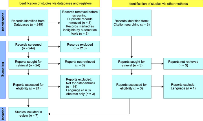
In total, 252 studies were identified following initial searches. Following screening, 7 studies were included in the final review as outlined in the PRISMA flowchart.
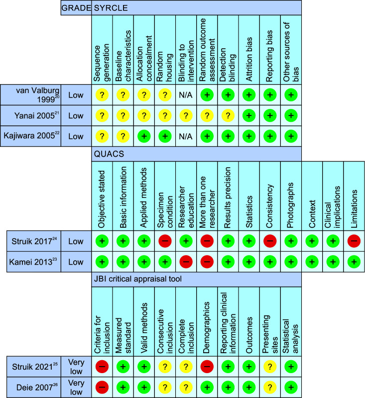
The study quality was generally low. The animal studies were randomised control trials (RCTs), but uncertainties regarding the randomisation and blinding processes and lack of applicability to human populations reduced quality. Of the observational studies, none were upgraded to a higher quality using the GRADE tool; therefore, overall study quality was low or very low. Quality assessment using the specific study type tools SYRCLE, QUACS and JBI is outlined in Flowchart 1. The authors of these tools do not recommend giving an overall score.
Flowchart 1.
PRISMA flowchart
The study characteristics of the included studies are outlined in Table 1.
Table 1.
Study demographics
| Study | Study type | Sample | Sample | Age | Gender | Control |
|---|---|---|---|---|---|---|
| van Valburg 200020 |
RCT | Canine ACL transection |
13 | N/A | N/A |
N = 5 articulating N = 3 blocked hinges N = 5 control no treatment |
| Yanai 200521 | RCT | Rabbit Cartilage defect |
33 | N/A | N/A |
N = 15 articulating N = 6 no distraction N = 6 articulating and ACBMT N = 6 atelocollagen gel |
| Kajiwara 200522 | RCT | Rabbit Osteochondral defect |
36 | N/A | N/A |
N = 36 articulating N = 36 control (contralateral knee) |
| Kamei 201323 | Observational | Cadaveric | 10 | N/A | N/A | No control |
| Struik 201724 | Observational | Cadaveric | 3 | N/A | N/A | No control |
| Deie 200726 | Case series | Human | 7 | 49 (42–63) | 2M 4F | No control |
| Struik 202125 | Case series | Human | 3 | ND | ND | No control |
ND, not documented
Hinged Distraction for OA
Animal Studies
Three animal RCTs met the inclusion criteria. A canine model of OA reported by van Valburg et al.20 used an articulating modified Ilizarov device for distraction. This study compared three groups, articulating distraction, non-articulating distraction and no treatment, following anterior cruciate ligament transection. Treatment was continued for 8 weeks. Intra-articular pressures were measured, which demonstrated changes in intra-articular pressures throughout movement of the hinges.20 Yanai et al.21 examined 33 rabbits with artificially created full-thickness cartilage defects and compared groups with and without a collagen gel and weight-bearing.21 The construct used was a modified Ilizarov construct using half rings. Treatment was continued for 12 weeks. A further animal study evaluated hinged knee joint distraction in rabbits combined with microfracture. The device was limited to 20 degrees of extension but full flexion.22 In the three animal studies, the devices used were similar to those in current clinical practice, although modified for use in animals. All constructs demonstrated feasibility with consistency in the maintenance of the frame and range of motion throughout the study period. No significant complications were recorded; however, this was not specifically documented in two studies.
Human Studies
Two cadaveric and two clinical studies were included. All appraised novel constructs were designed specifically for use in hinged knee joint distraction for cartilage regeneration. Kamei et al.23 utilised a magnetic distraction arthroplasty system centred on a temporary wire inserted from the apex of the lateral epicondyle to the apex of the medial epicondyle in 10 cadaveric knees. On loading with 15 or 30 kg, distraction was maintained through a range of motion. The construct used by Struik et al.24 negated the need for an intra-articular positioning wire by using a personalised joint-specific approach. Joint kinematics were measured prior to application of the frame and personalised frame cams were produced according to these specifications. Maintenance of 5 mm joint distraction was demonstrated within 30 degrees of joint flexion and upon axial loading in a cadaveric sample.24 A further study used this approach in a clinical setting; after initial distraction in a static frame for 2–4 weeks, three patients had a personalised articulating frame assembled based on joint kinematics. However, only 15 degrees of joint flexion was obtained due to painful motion in soft tissues around pin sites, particularly in the thigh. This did not allow for placement of the frame, and thus the authors concluded that the risks of hinged knee joint distraction outweigh the potential benefits.25
Deie et al.26 utilised an articulating construct based on a device previously described in an animal study.22 All patients had microfracture treatment of OA lesions then hinged joint distraction for 2–3 months. None of the 6 participants had significant complications except for one superficial pin site infection. The follow-up period ranged from 1 year 2 months to 4 years 3 months. There was no static control.
The study methodology and outcomes are outlined in Table 2.
Table 2.
Distraction methods and outcomes
| Study | Construct | Hinge identification | Distraction | ROM in frame | Time in frame | Complications |
|---|---|---|---|---|---|---|
| van Valburg 200020 | Ilizarov external fixator | ND | ND | Unlimited | 8 weeks | No significant complications |
| Yanai 200521 | Study specific – 2 half ring, 4 half pins: 2 in femur 2 in tibia | Wire passed through distal femur at origin of lateral collateral ligament | ND | Unlimited | 12 weeks | ND |
| Kajiwara 200522 | Modified mini external fixator (Meira, Nagoya, Japan) | Wire passed through insertion point of medial collateral ligament to centre of curvature of lateral femoral condyle | 1.5 mm | Unlimited | 4, 8, and 12 weeks | ND |
| Kamei 201323 | Magnetic distraction arthroplasty system (Hitachi Metals, Tokyo, Japan) | Wire inserted apex of lateral epicondyle to apex of medial epicondyle | 5 mm | Almost full range | N/A | N/A |
| Struik 201724 | Joint-specific articulating distraction device attached with bone pins and bone pin clamps (Triax Monotube, Stryker, Switzerland) | Personalised cam fabrication | 5 mm | 0–30 degrees | N/A | N/A |
| Deie 200726 | Articulated arthroplasty device (Meira, Nagoya, Japan | Two guide pins inserted apex of medial and lateral epicondyles | 1.6 mm (0–3 mm) | ~30 degrees | 8–12 weeks | 1 superficial skin infection |
| Struik 202125 | Joint-specific articulating distraction device | Personalised joint-specific hinges | 5 mm | 0–15 degrees | 6 weeks | Significant pain leading abandonment of device in all patients |
ND, not documented
Discussion
Multiple animal, cadaveric and clinical studies were identified investigating the use of hinged knee joint constructs for the treatment of OA. However, study quality was generally low. There is significant variability in methodology, including the timing of hinged distraction, the range of motion possible and the constructs used. Neither of the human studies had controls, and both had small sample sizes.
The range of motion aimed for and achieved varied significantly between studies. All animal studies achieved unlimited range in the frames, with animals able to fully weight-bear whilst mobilising. Of the studies in humans, only Kamei et al.23 attempted to achieve unlimited range of motion in their cadaveric study and achieved near full flexion, although this was measured on video only and not formally reported. The other cadaveric and clinical studies limited flexion to 30 degrees to reduce soft-tissue complications related to pin-site irritation on motion. Most studies were evaluating novel devices for hinged joint distraction with none of the devices utilised in clinical use currently. There was significant heterogeneity in construct designs attributable to the complex kinematics of the knee. One approach demonstrated feasibility in cadaveric testing,24 but this could not be translated to clinical practice due to the significant pain experienced by the participants.25 A critical issue appears to be hinge placement. If this is incorrect, joint compression or distraction will occur during range of motion. This may result in discomfort and, particularly if compression occurs, be detrimental to cartilage regeneration by negating the offloading effect. Different methods for placement of hinges have been demonstrated in these studies, including use of temporary intra-articular wires for positioning. This raises concerns of increased potential for infection, although the small samples included in these studies do not reflect this. It is also important to consider the effect of wire placement on other soft tissues, which may result in increased pain through a range of motion.
Another consideration is fixator construct stability. The use of hinges may reduce stability which leads to increased motion at the knee when compared with static distraction constructs. The effect of this on cartilage regeneration is unclear as the underlying mechanisms are not fully understood. In general, however, unstable constructs will tend to result in increased pain which will decrease weight-bearing. This may have an indirect effect if repeated axial loading with resultant pulsatile changes in articular pressure is important.27 Furthermore, instability will result in increased soft tissue irritation, which will increase pain and the risk of infection and pin loosening. This consideration is particularly relevant for the elective management of OA where pin site osteomyelitis may jeopardise future arthroplasty.
Hinged External Fixation for Other Indications
This review is limited by the low number and heterogeneity of studies investigating the use of hinged constructs for knee distraction in cartilage regeneration. It is potentially useful to discuss the use of hinged knee constructs for alternative clinical indications, whilst maintaining that these findings may not be generalisable to knee joint distraction.
Several studies have examined hinged external fixation across the knee without distraction for trauma. These focus predominantly on fixators to control sagittal plane translation in ligament deficient knees.28–36 Single axis hinge fixators have been widely utilised. In a cadaveric study, only a limited range of motion was possible with on-axis hinges permitting only 19–79 degrees of flexion without increased forces within the knee. A 5 mm posteriorly translated hinge gave movement to 86 degrees.29 A further cadaveric study28 utilising an EBI-Orthofix (EBI Corp, Persipanny, NJ) unilateral frame in PCL deficient knees demonstrated difficulties in siting the hinges.28 Stannard et al.33 described the use of the COMPASS knee hinge (Smith and Nephew, UK), which uses an isometric centring wire for hinge placement.33 A further clinical study that compared the COMPASS hinge to a hinged knee brace demonstrated that where ROM beyond 60 degrees was permitted, pin-site complications increased.35
A number of studies have also investigated knee joint distraction in select cases for flexion contractures.37–41 These are small series or case reports which demonstrated gradual correction of the flexed position of the knee with additional joint distraction to avoid cartilage compression during correction. Varying amounts of distraction were placed across the knee joint with fixed motors posteriorly being distracted to achieve correction gradually. All constructs utilised external hinges placed with radiography without intra-articular wires.37–42 Due to the nature of the conditions treated, with active correction through hinges, these fixator constructs are less relevant to a fixator which permits passive range during distraction for OA.
Conclusions
These studies provide some insight into issues for the further development of hinged distractors for osteoarthritis. Study heterogeneity and methodology limit the conclusions that can be drawn from this review. A hinged knee joint fixator does appear to be feasible, but difficulties in positioning of the constructs, achieving a significant range of motion and avoiding complications have not been overcome. Multiple unanswered questions remain, including construct stability, hinge placement, hinge design, range of motion achievable and the potential for significant complications. Furthermore, any potential benefits in the treatment of OA by arthrodistraction would need to be demonstrated. Further research is required to address all these issues before hinged distraction for this purpose were to be considered in clinical practice.
Footnotes
Source of support: Nil
Conflict of interest: Dr Paul Harwood is associated as Editor-in-Chief of this journal and this manuscript was subjected to this journal's standard review procedures, with this peer review handled independently of the Editor-in-Chief and his research group.
References
- 1.Besselink NJ, Vincken KL, Bartels LW, et al. Cartilage quality (dGEMRIC Index) following knee joint distraction or high tibial osteotomy. Cartilage. 2020;11(1):19–31. doi: 10.1177/1947603518777578. [DOI] [PMC free article] [PubMed] [Google Scholar]
- 2.Baboolal TG, Mastbergen SC, Jones E, et al. Synovial fluid hyaluronan mediates MSC attachment to cartilage, a potential novel mechanism contributing to cartilage repair in osteoarthritis using knee joint distraction. Ann Rheum Dis. 2016;75(5):908–915. doi: 10.1136/annrheumdis-2014-206847. [DOI] [PMC free article] [PubMed] [Google Scholar]
- 3.Goh EL, Lou WCN, Chidambaram S, et al. The role of joint distraction in the treatment of knee osteoarthritis: A systematic review and quantitative analysis. Orthop Res Rev. 2019;11:79–92. doi: 10.2147/ORR.S211060. [DOI] [PMC free article] [PubMed] [Google Scholar]
- 4.Jansen MP, Boymans TAEJ, Custers RJH, et al. Knee joint distraction as treatment for osteoarthritis results in clinical and structural benefit: A systematic review and meta-analysis of the limited number of studies and patients available. Cartilage. 2021;13(1 Suppl):1113S–1123S. doi: 10.1177/1947603520942945. [DOI] [PMC free article] [PubMed] [Google Scholar]
- 5.Jansen MP, Mastbergen SC, van Heerwaarden RJ, et al. Knee joint distraction in regular care for treatment of knee osteoarthritis: A comparison with clinical trial data. PLoS One. 2020;15(1):e0227975. doi: 10.1371/journal.pone.0227975. [DOI] [PMC free article] [PubMed] [Google Scholar]
- 6.Herrera-Perez M, Alrashidi Y, Galhoum AE, et al. Debridement and hinged motion distraction is superior to debridement alone in patients with ankle osteoarthritis: A prospective randomized controlled trial. Knee Surg Sports Traumatol Arthrosc. 2019;27(9):2802–2812. doi: 10.1007/s00167-018-5156-3. [DOI] [PubMed] [Google Scholar]
- 7.Bernstein M, Reidler J, Fragomen A, et al. Ankle distraction arthroplasty: Indications, technique, and outcomes. J Am Acad Orthop Surg. 2017;25(2):89–99. doi: 10.5435/JAAOS-D-14-00077. [DOI] [PubMed] [Google Scholar]
- 8.Saltzman CL, Hillis SL, Stolley MP, et al. Motion versus fixed distraction of the joint in the treatment of ankle osteoarthritis: A prospective randomized controlled trial. J Bone Joint Surg Am. 2012;94(11):961–970. doi: 10.2106/JBJS.K.00018. [DOI] [PMC free article] [PubMed] [Google Scholar]
- 9.Sheehan FT. The finite helical axis of the knee joint (a non-invasive in vivo study using fast-PC MRI). J Biomech. 2007;40(5):1038–1047. doi: 10.1016/j.jbiomech.2006.04.006. [DOI] [PubMed] [Google Scholar]
- 10.Todo S, Kadoya Y, Moilanen T, et al. Anteroposterior and rotational movement of femur during knee flexion. Clin Orthop Relat Res. 1999;(362):162–170. 10335295 [PubMed] [Google Scholar]
- 11.Churchill DL, Incavo SJ, Johnson CC, et al. The transepicondylar axis approximates the optimal flexion axis of the knee. Clin Orthop Relat Res. 1998;(356):111–118. doi: 10.1097/00003086-199811000-00016. [DOI] [PubMed] [Google Scholar]
- 12.Mochizuki T, Sato T, Blaha JD, et al. The clinical epicondylar axis is not the functional flexion axis of the human knee. J Orthop Sci. 2014;19(3):451–456. doi: 10.1007/s00776-014-0536-0. [DOI] [PubMed] [Google Scholar]
- 13.Katz MA, Beck TD, Silber JS, et al. Determining femoral rotational alignment in total knee arthroplasty: reliability of techniques. J Arthroplasty. 2001;16(3):301–305. doi: 10.1054/arth.2001.21456. [DOI] [PubMed] [Google Scholar]
- 14.Yin L, Chen K, Guo L, et al. Identifying the functional flexion-extension axis of the knee: An in-vivo kinematics study. PLoS One. 2015;10(6):e0128877. doi: 10.1371/journal.pone.0128877. [DOI] [PMC free article] [PubMed] [Google Scholar]
- 15.Liberati A, Altman DG, Tetzlaff J, et al. The PRISMA statement for reporting systematic reviews and meta-analyses of studies that evaluate health care interventions: Explanation and elaboration. J Clin Epidemiol. 2009;62(10):e1–e34. doi: 10.1016/j.jclinepi.2009.06.006. [DOI] [PubMed] [Google Scholar]
- 16.Schünemann H, Brożek J, Guyatt G, et al. The GRADE Handbook; 2013. [Google Scholar]
- 17.Hooijmans CR, Rovers MM, de Vries RB, et al. SYRCLE's risk of bias tool for animal studies. BMC Med Res Methodol. 2014;14:43. doi: 10.1186/1471-2288-14-43. [DOI] [PMC free article] [PubMed] [Google Scholar]
- 18.Wilke J, Krause F, Niederer D, et al. Appraising the methodological quality of cadaveric studies: validation of the QUACS scale. J Anat. 2015;226(5):440–446. doi: 10.1111/joa.12292. [DOI] [PMC free article] [PubMed] [Google Scholar]
- 19.Munn Z, Barker TH, Moola S, et al. Methodological quality of case series studies: An introduction to the JBI critical appraisal tool. JBI Evid Synth. 2020;18(10):2127–2133. doi: 10.11124/JBISRIR-D-19-00099. [DOI] [PubMed] [Google Scholar]
- 20.van Valburg AA, van Roermund PM, Marijnissen AC, et al. Joint distraction in treatment of osteoarthritis (II): Effects on cartilage in a canine model. Osteoarthritis Cartilage. 2000;8(1):1–8. doi: 10.1053/joca.1999.0263. [DOI] [PubMed] [Google Scholar]
- 21.Yanai T, Ishii T, Chang F, et al. Repair of large full-thickness articular cartilage defects in the rabbit: The effects of joint distraction and autologous bone-marrow-derived mesenchymal cell transplantation. J Bone Joint Surg Br. 2005;87(5):721–729. doi: 10.1302/0301-620X.87B5.15542. [DOI] [PubMed] [Google Scholar]
- 22.Kajiwara R, Ishida O, Kawasaki K, et al. Effective repair of a fresh osteochondral defect in the rabbit knee joint by articulated joint distraction following subchondral drilling. J Orthop Res. 2005;23(4):909–915. doi: 10.1016/j.orthres.2004.12.003. [DOI] [PubMed] [Google Scholar]
- 23.Kamei G, Ochi M, Okuhara A, et al. A new distraction arthroplasty device using magnetic force: A cadaveric study. Clin Biomech (Bristol, Avon) 2013;28(4):423–428. doi: 10.1016/j.clinbiomech.2013.02.003. [DOI] [PubMed] [Google Scholar]
- 24.Struik T, Jaspers JEN, Besselink NJ, et al. Technical feasibility of personalized articulating knee joint distraction for treatment of tibiofemoral osteoarthritis. Clin Biomech (Bristol, Avon) 2017;49:40–47. doi: 10.1016/j.clinbiomech.2017.08.002. [DOI] [PubMed] [Google Scholar]
- 25.Striuk T, Custers RJH, Besselink NJ, et al. Clinical feasibility of personalized articulating knee joint distraction. Austin J Orthop Rheumatol. 2021;8(2):1104. [Google Scholar]
- 26.Deie M, Ochi M, Adachi N, et al. A new articulated distraction arthroplasty device for treatment of the osteoarthritic knee joint: A preliminary report. Arthroscopy. 2007;23(8):833–838. doi: 10.1016/j.arthro.2007.02.014. [DOI] [PubMed] [Google Scholar]
- 27.van Valburg AA, van Roy HL, Lafeber FP, et al. Beneficial effects of intermittent fluid pressure of low physiological magnitude on cartilage and inflammation in osteoarthritis. An in vitro study. J Rheumatol. 1998;25(3):515–520. 9517773 [PubMed] [Google Scholar]
- 28.Wroble RR, Grood ES, Cummings JS. Changes in knee kinematics after application of an articulated external fixator in normal and posterior cruciate ligament-deficient knees. Arthroscopy. 1997;13(1):73–77. doi: 10.1016/s0749-8063(97)90212-7. [DOI] [PubMed] [Google Scholar]
- 29.Sommers MB, Fitzpatrick DC, Kahn KM, et al. Hinged external fixation of the knee: Intrinsic factors influencing passive joint motion. J Orthop Trauma. 2004;18(3):163–169. doi: 10.1097/00005131-200403000-00007. [DOI] [PubMed] [Google Scholar]
- 30.Fitzpatrick DC, Sommers MB, Kam BC, et al. Knee stability after articulated external fixation. Am J Sports Med. 2005;33(11):1735–1741. doi: 10.1177/0363546505275132. [DOI] [PubMed] [Google Scholar]
- 31.Gatti G. Conceptual design and implantation of an external fixator with improved mobility for knee rehabilitation. Comput Methods Biomech Biomed Engin. 2017;20(8):884–892. doi: 10.1080/10255842.2017.1307342. [DOI] [PubMed] [Google Scholar]
- 32.Richter M, Lobenhoffer P. Chronic posterior knee dislocation: Treatment with arthrolysis, posterior cruciate ligament reconstruction and hinged external fixation device. Injury. 1998;29(7):546–549. doi: 10.1016/s0020-1383(98)00095-3. [DOI] [PubMed] [Google Scholar]
- 33.Stannard JP, Sheils TM, McGwin G, et al. Use of a hinged external knee fixator after surgery for knee dislocation. Arthroscopy. 2003;19(6):626–631. doi: 10.1016/s0749-8063(03)00125-7. [DOI] [PubMed] [Google Scholar]
- 34.Zaffagnini S, Iacono F, Lo Presti M, et al. A new hinged dynamic distractor, for immediate mobilization after knee dislocations: Technical note. Arch Orthop Trauma Surg. 2008;128(11):1233–1237. doi: 10.1007/s00402-007-0515-4. [DOI] [PubMed] [Google Scholar]
- 35.Stannard JP, Nuelle CW, McGwin G, et al. Hinged external fixation in the treatment of knee dislocations: A prospective randomized study. J Bone Joint Surg Am. 2014;96(3):184–191. doi: 10.2106/JBJS.L.01603. [DOI] [PubMed] [Google Scholar]
- 36.Angelini FJ, Helito CP, Bonadio MB, et al. External fixator for treatment of the sub-acute and chronic multi-ligament-injured knee. Knee Surg Sports Traumatol Arthrosc. 2015;23(10):3012–3018. doi: 10.1007/s00167-015-3719-0. [DOI] [PubMed] [Google Scholar]
- 37.Damsin JP, Ghanem I. Treatment of severe flexion deformity of the knee in children and adolescents using the Ilizarov technique. J Bone Joint Surg Br. 1996;78(1):140–144. 8898146 [PubMed] [Google Scholar]
- 38.Kiely PD, McMahon C, Smith OP, et al. The treatment of flexion contracture of the knee using the Ilizarov technique in a child with haemophilia B. Haemophilia. 2003;9(3):336–339. doi: 10.1046/j.1365-2516.2003.00753.x. [DOI] [PubMed] [Google Scholar]
- 39.Kumar A, Logani V, Neogi DS, et al. IIlizarov external fixator for bilateral severe flexion deformity of the knee in haemophilia: Case report. Arch Orthop Trauma Surg. 2010;130(5):621–625. doi: 10.1007/s00402-009-0968-8. [DOI] [PubMed] [Google Scholar]
- 40.Balci HI, Kocaoglu M, Eralp L, et al. Knee flexion contracture in haemophilia: Treatment with circular external fixator. Haemophilia. 2014;20(6):879–883. doi: 10.1111/hae.12478. [DOI] [PubMed] [Google Scholar]
- 41.Zhai J, Weng X, Zhang B, et al. Management of knee flexion contracture in haemophilia with the Ilizarov technique. Knee. 2019;26(1):201–206. doi: 10.1016/j.knee.2018.08.006. [DOI] [PubMed] [Google Scholar]
- 42.Qin SH, Chen JW, Zheng XJ, et al. Ilizarov technique for correcting flexion deformity of the knee of arthrogryposis multiplex congenita. Zhonghua Wai Ke Za Zhi. 2004;42(16):993–996. 153632327 [PubMed] [Google Scholar]


