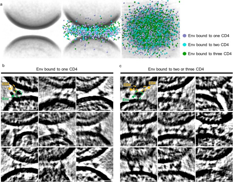
Extended Data Fig. 5. HIV-1 Env trimers bound to one, two and three CD4 receptors on biological membranes.
a Individual subtomograms of all the membrane-membrane interfaces were aligned and averaged (left panel). In the middle panel, the coordinates of Env bound to one (purple), two (cyan) and three (green) CD4 molecules are overlaid onto the averaged volumes. The top-down view is shown to the right. b, c Gallery of HIV-1 Env trimers bound to CD4 receptors on biological membranes. Representative images from cryo-tomograms of the HIV-1 Env trimers bound to one CD4 (b) or two and three CD4 (c) molecules at the membrane-membrane interfaces between HIV-1 and MLV-CD4 particles. Scale bar = 20 nm.

