Abstract
Cannabis sativa has gained popularity as a “natural substance”, leading many to falsely assume that it is not harmful. This assumption has been documented amongst pregnant mothers, many of whom consider Cannabis use during pregnancy as benign. The purpose of this study was to validate a Cannabis smoke exposure model in pregnant rats by determining the plasma levels of cannabinoids and associated metabolites in the dams after exposure to either Cannabis smoke or injected cannabinoids. Maternal and fetal cytokine and chemokine profiles were also assessed after exposure. Pregnant Sprague–Dawley rats were treated daily from gestational day 6–20 with either room air, i.p. vehicle, inhaled high-Δ9-tetrahydrocannabinol (THC) (18% THC, 0.1% cannabidiol [CBD]) smoke, inhaled high-CBD (0.7% THC, 13% CBD) smoke, 3 mg/kg i.p. THC, or 10 mg/kg i.p. CBD. Our data reveal that THC and CBD, but not their metabolites, accumulate in maternal plasma after repeated exposures. Injection of THC or CBD was associated with fewer offspring and increased uterine reabsorption events. For cytokines and chemokines, injection of THC or CBD up-regulated several pro-inflammatory cytokines compared to control or high-THC smoke or high-CBD smoke in placental and fetal brain tissue, whereas smoke exposure was generally associated with reduced cytokine and chemokine concentrations in placental and fetal brain tissue compared to controls. These results support existing, but limited, knowledge on how different routes of administration contribute to inconsistent manifestations of cannabinoid-mediated effects on pregnancy. Smoked Cannabis is still the most common means of human consumption, and more preclinical investigation is needed to determine the effects of smoke inhalation on developmental and behavioural trajectories.
Subject terms: Development of the nervous system, Pharmacology, Toxicology
Introduction
General trends of Cannabis legalization/decriminalization have changed social narratives surrounding use and risk perception in Canada and around the world. As social perception has changed, rates of use have increased for all ages by 7.3% since legalization, where smoking still accounts for 79% of Cannabis consumption in Canada1. Importantly, Cannabis has gained popularity as a “natural substance,” with the false assumption that it is therefore safe. Emerging human data indicate that children exposed to Cannabis in utero are at a higher risk of being born pre-term, underweight, and developing persistent behavioural psychopathologies and cognitive deficits across their lifetime2–11. Although long-term outcomes are still not understood, these data indicate the need for more preclinical research to establish the risks and investigate the molecular and physiological consequences of in utero Cannabis smoke exposure.
Over 120 unique phytocannabinoids are produced by the Cannabis sativa plant, with the acid forms of Δ9-tetrahydrocannabinol (THC) and cannabidiol (CBD) present at the highest relative concentrations12. These compounds, along with the lesser-known phytocannabinoids, terpenes, flavonoids, and alkaloids have a wide array of pharmacological activities via our endogenous cannabinoid system (eCS) and other biological systems13. The eCS is comprised of the G protein-coupled receptors (GPCR) cannabinoid 1 receptor (CB1R) and the cannabinoid 2 receptor (CB2R), their endogenous ligands [2-arachidonoylglycerol (2-AG) and anandamide (AEA)], and the enzymes that synthesize [diacylglycerolipase (DAGL) and N-arachidonoyl phosphatidyl ethanolamine-specific phospholipase D (NAPE-PLD)] and degrade [monoacylglycerol lipase (MAGL) and fatty acid amide hydrolase (FAAH)] those endogenous ligands, respectively14.
THC, the main intoxicating constituent of Cannabis, accounts for the distinct “high” associated with Cannabis use via CB1R partial agonism15. CBD, by comparison, acts in vitro as a possible negative allosteric modulator at CB1R, partial agonist at CB2R, positive allosteric modulator of µ-opioid receptor, partial agonist of serotonin 1a receptors, and a modulator of the signaling of several other GPCRs and non-GPCR targets16–18. Evidence suggests that most of CBD’s in vivo activity occurs at non-cannabinoid receptor targets, such as the serotonin 1a receptor and the transient receptor potential vanilloid 1 (TRPV1)19, further contributing to the pharmacological complexity of Cannabis.
Pregnancy is in part orchestrated by the intricate communication and coordination of maternal and fetal immune cells by cytokines and chemokines to establish and maintain the maternal–fetal interface20. Cytokines and chemokines play an important role in embryogenesis through the mediation of cell survival, proliferation, differentiation, and promotion of cell adhesion to specific sites to coordinate invasion of fetal trophoblasts into the maternal decidua21–24. Dysregulation in the establishment of a healthy placenta can result in maternal and fetal pathologies such as pre-eclampsia, spontaneous pre-term birth, intra-uterine growth restrictions, and resorption25–29.
The eCS plays a formative role in the coordination of pregnancy and fetal neurodevelopment. Decreasing levels of intra-uterine AEA and concomitant signaling via CB1R and CB2R may dictate successful implantation30, and changes in receptor expression and AEA throughout reproductive tissue and plasma has been associated with ectopic pregnancy, miscarriage, and pre-eclampsia31. CB1R also coordinate directional migration and elongation of axons32,33, and the maturation of astrocytes and oligodendrocytes34–36. Cannabis constituents cross the blood-placenta barrier to accumulate in the developing fetus37–40. THC administration in utero interferes with endogenous cannabinoid orchestration of neuronal growth cones, cytoskeletal architecture, and the development of novel synapses41, as well as in increased incidents of fetal growth restriction42. Preclinical studies have focused on the downstream effects of gestational cannabinoid exposure by using intravenous (i.v.), oral, intraperitoneal (i.p.), and subcutaneous (s.c.) administration methods of THC (0.5–30 mg/kg)42–69 and high-potency synthetic CB1R agonists (e.g. WIN-55,212-2)47,63, 70–76, with some recent studies examining inhalation of THC vapour (100 ng/mL)40,77–80, high-THC Cannabis smoke81, and injected CBD82–86.
Plasma cannabinoid pharmacokinetics have been studied in healthy adult humans for multiple forms of consumption87–90; however, these metrics are poorly characterized in pregnancy39. Reported peak serum concentrations of THC after smoking a Cannabis joint achieve tmax approximately 12 min after inhalation and vary widely (Cmax 60–200 ng/mL), due to inter-individual variability, variation in THC concentrations and frequency of use, among other factors87–90. In studies with humans, Cannabis consumption by expectant mothers is often measured by the number of joints smoked per day39. This method of measuring Cannabis consumption in humans makes comparing cannabinoid doses and exposures between clinical assessments and pre-clinical smoke exposure models challenging. Whereas prior preclinical studies have explored injection, inhalation, and oral administration of isolated THC or CBD, administering THC and/or CBD alone fails to take into consideration the potential effects of other phytocannabinoids, terpenes, and flavonoids of the Cannabis plant as well as the smoke of a combusted product13,91, 92. Our group has previously worked to establish rat models of Cannabis smoke exposure in the contexts of absence epilepsy93, cognition94,95, and prenatal exposure during pregnancy on offspring health and behaviour81. In the present study, we build on this previous work to directly compare the effects of repeated inhalation of smoke from Cannabis products high in THC or CBD to repeated injections of THC or CBD in pregnant rats. Specifically, the pharmacokinetics of THC, CBD, and their metabolites; as well as the effects of Cannabis exposure on an array of 27 cytokines and chemokines measured in the placenta and fetuses pregnant Sprague–Dawley rats treated with Cannabis or cannabinoids daily from gestational day (GD) 6–20. The aim of our studies was a validation of a whole plant combustion method in gestation to enhance translation and address unanswered questions regarding the effects of Cannabis exposure during gestation.
Results
Pharmacokinetic evaluation and comparison of maternal plasma phytocannabinoid levels during gestation: accumulation of cannabinoids over time
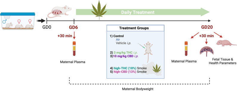
Pregnant dams were treated daily between GD6 – 20 with either vehicle (i.p.), room air, 3 mg/kg i.p. THC, 10 mg/kg i.p. CBD, Skywalker Kush Cannabis flower smoke (high-THC smoke, 18% THC:0.1% CBD) 300 mg inhaled, or Treasure Island Cannabis flower smoke (high-CBD smoke, 0.7% THC:13% CBD) 300 mg inhaled (Fig. 1). Maternal plasma samples were collected from separate groups of rats 30 min after the first (GD6) and last (GD20) treatment to evaluate maternal phytocannabinoid levels (Figs. 1, 2, 3). For specific details on Cannabis combustion protocol refer to “Smoke exposure” in the Methods section below.
Figure 1.
Experimental timeline for gestational Cannabis exposure. Rats were bred and treated once daily between gestational day (GD) 6 and 20 for 15 consecutive days of treatment. Maternal and fetal tissues were harvested 30 min following the first (GD6) and last (GD20) treatment. THC, CBD, and their metabolites were quantified in maternal plasma using high performance liquid chromatography tandem mass spectroscopy (HPLC–MS/MS). Protein samples from placenta and whole fetal brain were prepared for cytokine and chemokine quantification. Created with Biorender.com.
Figure 2.
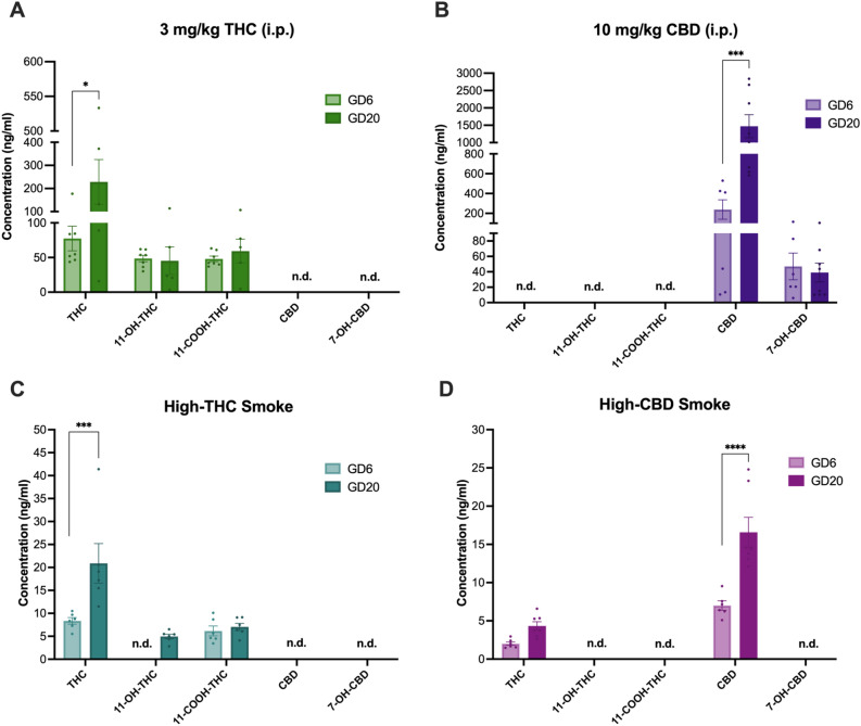
Comparison of THC, CBD, and metabolite levels in maternal plasma according to treatment between first exposure on GD6 and last exposure on GD20. Plasma levels of phytocannabinoids were quantified on GD6 and GD20 30 min after cannabinoid exposure in the following treatment groups: (A) 3 mg/kg/day THC i.p., (B) 10 mg/kg/day CBD i.p., (C) High-THC smoke, and (D) High-CBD smoke. Note the different scaling of the y-axes of the panels. Data are mean ± S.E.M. n = 5–8 dams/per treatment. Values falling below LLOQ were deemed not detectable (n.d.).
Figure 3.
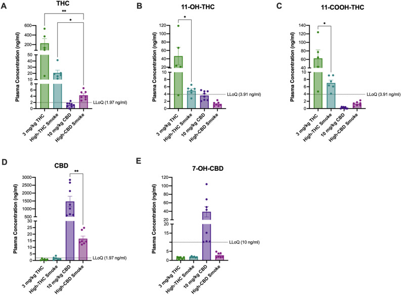
Comparison of THC, CBD, and metabolite levels among treatments in maternal plasma on GD20 after repeated exposure to either 3 mg/kg/day THC i.p., high THC smoke, 10 mg/kg/day CBD i.p., or high CBD smoke. Mean maternal plasma concentrations of (A) THC, (B) 11-OH-THC, (C) 11-COOH-THC, (D) CBD, and (E) 7-OH-CBD. All samples were collected on GD20, 30 min following final treatment. Note the different scaling of the y-axes of the panels. Data are mean ± S.E.M. n = 5–8 dams/treatment; *p < 0.05, ** p < 0.01 as determined by Kruskal–Wallis (KW) one-way ANOVA or Kolmogorov–Smirnov (KS) test.
THC, CBD, and their metabolite levels were compared between GD6 and GD20 within treatments to assess whether levels changed across the gestational period (Fig. 2). For 3 mg/kg i.p. THC exposed dams, mean plasma levels of THC, 11-OH-THC, and 11-COOH-THC were above the lower limit of quantification (LLOQ) (THC: 1.97 ng/mL; 11-OH-THC and 11-COOH-THC: 3.91 ng/mL) on GD6 and GD20. A 3 × 2 ANOVA (Analyte by Day) demonstrated a significant main effect of Analyte (F(2,30) = 5.70, p = 0.008), but not Day (F(1,30) = 3.41, p = 0.08), and no interaction (F(2, 30) = 2.93, p = 0.07). Holm-Šídák’s multiple comparisons test revealed a significant difference between plasma levels of THC on GD6 vs. GD20 in i.p. THC exposed dams, with higher levels on GD20 (p = 0.015) (Fig. 2A).
For 10 mg/kg i.p. CBD exposed dams, mean plasma levels of CBD and 7-OH-CBD were above the LLOQ (CBD: 1.97 ng/mL; 7-OH-CBD: 10 ng/mL). A 2 × 2 ANOVA (Analyte by Day), demonstrated significant main effects of Analyte (F(1,24) = 16.83, p = 0.0004) and Day (F(1,24) = 9.57, p = 0.005), as well as a significant interaction (F(1,24) = 9.82, p = 0.0045). Holm-Sidak’s multiple comparisons test revealed a significant difference between plasma levels of CBD on GD6 vs. GD20 in i.p. CBD exposed dams, with higher levels on GD20 (p = 0.0004) (Fig. 2B).
For high-THC smoke exposed dams, mean plasma levels of THC and 11-COOH-THC were above the LLOQ on GD6 and GDG20; however, 11-OH-THC was only above the LLOQ on GD20. A 2 × 2 ANOVA (Analyte by Day) demonstrated significant main effects of Analyte (F(1,20) = 12.36, p < 0.0022) and Day (F(1,20) = 8.64, p = 0.0081), as well as a significant interaction (F(1,20) = 6.52, p = 0.019). Holm-Šídák’s multiple comparisons test revealed a significant difference between plasma levels of THC on GD6 vs. GD20 in high-THC smoke exposed dams, with higher levels on GD20 (p = 0.0018) (Fig. 2C).
For high-CBD smoke-exposed dams, mean plasma levels of THC and CBD were above the LLOQ. The 2 × 2 ANOVA (Analyte by Day) demonstrated significant main effects of Analyte (F(1,22) = 56.59, p < 0.0001) and Day (F(1,22) = 26.91, p < 0.0001), as well as a significant interaction (F(1,22) = 10.07, p = 0.004). Holm-Šídák’s multiple comparisons test revealed a significant difference between plasma levels of CBD only on GD6 vs. GD20 in high-CBD exposed dams, with higher levels on GD20 (p < 0.0001) (Fig. 2D).
From these data, we conclude that circulating levels of THC or CBD, but not their respective metabolites, increased by approximately threefold between GD6 to GD20 in all treatments, regardless of administration method.
Pharmacokinetic evaluation and comparison of maternal plasma phytocannabinoid levels during gestation: route of exposure influences observed concentrations
Mean GD6 and GD20 plasma levels of THC, CBD, and their metabolites were compared among treatments. As these analyses revealed similar differences among the treatments at each timepoint, we depicted the data from GD20 in Fig. 3 and the data from GD6 in Supp. Fig S1. For GD20 samples, THC levels were above the LLOQ for i.p THC, high-THC smoke, and high-CBD smoke. The Kruskal–Wallis (KW) test revealed a significant difference among treatments (H(3) = 13.70, p < 0.0001), where the post-hoc showed that levels of THC were significantly higher following i.p. THC and high-THC smoke than high-CBD smoke (Fig. 3A). It is noteworthy that mean levels of THC in the i.p. THC-exposed dams are approximately tenfold higher than those of the high-THC smoke group; however, due to unequal variances between treatments, this difference did not emerge as significant in the analysis. Mean plasma levels of 11-OH-THC were above the LLOQ for the i.p. THC and high-THC smoke groups only (Fig. 3B). Levels of 11-OH-THC in the i.p. THC treatment were significantly higher than levels in the high-THC smoke group (KS: p = 0.047). Mean plasma levels of 11-COOH-THC were greater than the LLOQ in the i.p. THC and high-THC smoke treatments only (Fig. 3C). Plasma levels of 11-COOH-THC were higher in the i.p. THC group compared to high-THC smoke (KS: p = 0.047) (Fig. 3C).
Plasma levels of CBD were greater than the LLOQ for the i.p. CBD and high-CBD smoke groups only and were significantly higher with i.p. CBD compared to the high-CBD smoke-exposed group (KS: p = 0.0003) (Fig. 3D). 7-OH-CBD was only detectable above the LLOQ in rats of the i.p. CBD group (Fig. 3E).
From these data, we conclude that exposure of pregnant dams to high-THC smoke was associated with quantifiable levels of THC, 11-OH-THC, and 11-COOH-THC that were approximately 10% of the respective levels of THC, 11-OH-THC, and 11-COOH-THC in pregnant dams treated with 3 mg/kg/day THC i.p. (Fig. 3A–C). Similarly, treatment with high-CBD smoke was associated with quantifiable levels of CBD on GD20. Levels of CBD and 7-OH-CBD in high-CBD smoke-exposed dams were 1% and 10% of the respective plasma levels observed in pregnant dams treated with 10 mg/kg/day CBD i.p. (Fig. 3D,E). Therefore, the smoke exposure paradigm used in these studies yields quantifiable levels of plasma cannabinoids in pregnant dams at levels lower than those observed with conventionally used rodent injection models when collected 30 min after the final treatment.
Fetal resorptions and litter size—but not other maternal health outcomes—were affected by phytocannabinoid injection
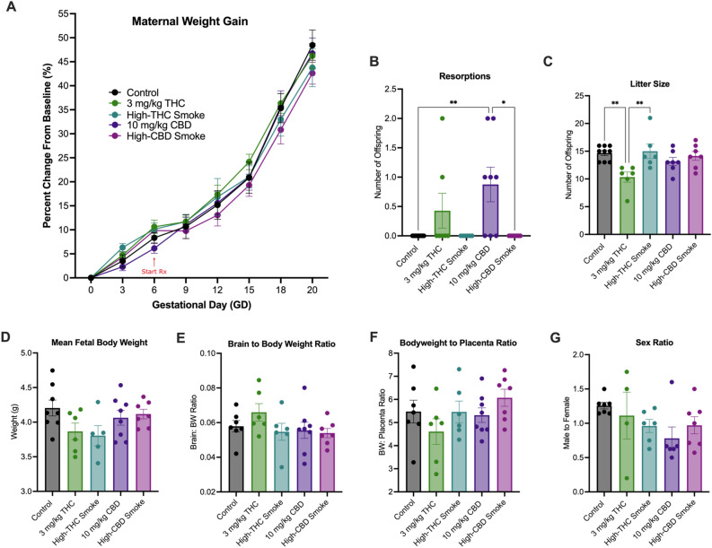
Maternal weight gain was measured every other day beginning on GD0. Uterine resorptions, litter sizes, fetal weight, fetal brain to body weight (BW), and fetal BW to placenta ratios were collected on GD20. For maternal weight gain (Fig. 4A), there was a significant main effect of Time (F(2.11, 57.57) = 527.7, p < 0.0001) but no main effect of Treatment (F(4,28) = 0.34, p = 0.85) or Time by Treatment interaction (F(28, 191) = 0.93, p = 0.57). There was a significant difference in fetal-placenta resorptions between treatments (one-way Kruskal–Wallis test: H(5) = 15.32, p < 0.0041), where the post-hoc demonstrated that i.p. CBD treatment caused significantly more resorptions than control and high-CBD smoke exposure, but not i.p. THC (Fig. 4B). In total, five of eight litters had resorptions in the CBD group, and two of six had resorptions in the THC group, where no litters exposed to i.p. VEH had resorptions. Litter sizes were smaller for dams in the i.p. THC group compared to control animals (one-way ANOVA followed by Holm-Šídák post-hoc analysis; F(4,28) = 4.50, p = 0.0062) (Fig. 4C). There was no difference in fetal BW between treatments (F(4, 29) = 2.14), p = 0.10) (Fig. 4D). No differences were observed between treatment groups in mean fetal BW (Fig. 4D), brain to BW ratio (F(4, 27) = 1.07, p = 0.39) (Fig. 4E), or BW to placenta ratio (F(4, 29) = 1.37, p = 0.27) (Fig. 4E). No differences in the ratios of male to female offspring were observed among the treatments (F(4, 25) = 1.56, p = 0.22) (Fig. 4G).
Figure 4.
Markers of maternal and litter health during gestation. (A) Rates of maternal weight gain, (B) uterine resorption discovered, (C) litter size (D), mean fetal body weight per litter, (E) mean fetal brain to body weight ratio per litter, and (F) mean fetal bodyweight to placenta ratio per litter, and (G) Ratio of male to female offspring per litter. Data are mean ± S.E.M, n = 4–8 dams/per treatment. Measurements in panels B-G were taken on GD20. *p < 0.05, as determined by one-way ANOVA followed by Holm-Šídák post-hoc analysis.
THC injection was unique among treatment groups increasing or maintaining cytokine and chemokine levels relative to controls whereas other treatments were associated with reduced cytokine and chemokine levels
To survey the inflammatory state of placental and fetal tissue after phytocannabinoid treatment, 27 cytokines and chemokines were quantified in placental and fetal brain tissue collected on GD20, 30 min after final treatment.
Placenta
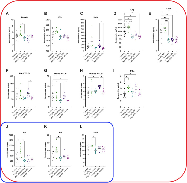
Significant main effect differences in cytokine or chemokine concentrations were observed for 15 of 27 cytokines and chemokines {epidermal growth factor (EGF); eotaxin; granulocyte colony-stimulating factor (G-CSF); interferon γ (IFNγ); IFNγ-induced protein 10 (IP-10, CXCL10); interleukins (IL) IL-1α, IL-1β, IL-4, IL-6, IL-10, and IL-17A; lipopolysaccharide-Induced CXC chemokine (LIX) or (CXCL5); macrophage inflammatory protein [MIP-1α; C–C motif chemokine (CCL3)], regulated up activation, normal T cell expressed and presumably secreted (RANTES, CCL5); and tumor necrosis factor α (TNFα)} (Suppl. Table S1). Post-hoc testing detected significant differences in 12 of 27 cytokines and chemokines (Fig. 5; Suppl. Table S1). Differences were not detected between groups in placental tissue for the remaining 15 cytokines and chemokines (Suppl. Table S1). The following is a summary of statistically significant post-hoc differences observed for cytokine and chemokine concentrations for placental tissue (Fig. 5). Data have been organized as being broadly defined as pro-inflammatory (red shape), anti-inflammatory (blue shape), or both depending on context (encapsulated by red and blue) (Fig. 5)96, 97.
Figure 5.
Placental cytokine and chemokine levels 30 min after final treatment on GD20. Cytokines and chemokines are organized by type: species within the red squircle are broadly considered pro-inflammatory, species within the blue squircle are broadly considered anti-inflammatory. Because the role of IL-4, IL-6 and IL-10 can be pro- or anti-inflammatory depending on context, these species are within both squircles. Data are displayed as mean ± S.E.M, n = 6–8 dams/group. An average of two replicates was used when possible. Note the y-axis varies in each panel depending on the cytokine measured. *p < 0.05, **p < 0.01, ***p < 0.001, ****p < 0.0001 as indicated between groups.
Compared to control, the pro-inflammatory cytokines IL-1β (p < 0.05) and TNFα (p < 0.05) were elevated in placenta of dams receiving injected THC (Fig. 5D,I). Likewise, RANTES (p < 0.05) and TNFα (p < 0.05) were elevated in the placenta of dams receiving injected CBD (Fig. 5H,I). In contrast—and compared to control—the pro-inflammatory cytokines and chemokines IL-1β (p < 0.01 for high-THC smoke and high-CBD smoke, p < 0.05 for CBD injection) and IL-17A (p < 0.01 for high-THC smoke and high-CBD smoke, p < 0.001 for CBD injection), were decreased in the placenta of dams receiving high-THC smoke, high-CBD smoke, and injected CBD (Fig. 5D,E). Levels of the pro-inflammatory chemokine MIP-1α were also decreased in the placenta of dams receiving high-THC smoke or high-CBD smoke compared to control (p < 0.01, Fig. 5G). Four differences were observed in levels of pro-inflammatory cytokines and chemokines when placenta from dams receiving injected THC were compared to other groups. Eotaxin (p < 0.01), IFNγ (p < 0.001), IL-1β (p < 0.0001), and IL-17A (p < 0.0001) levels were higher in the placenta of dams receiving THC injections compared to the placenta of dams receiving high-THC smoke (Fig. 5A,B,D,E). Similarly, IL-1α (p < 0.01), LIX (p < 0.05), MIP-1α (p < 0.05), and RANTES (p < 0.05) were all higher in the placenta of dams receiving CBD injections compared to the placenta of dams receiving high-CBD smoke (Fig. 5C,F–H).
IL-4, IL-6 and IL-10 are pro- and anti-inflammatory cytokines depending on how they interact with various cell types in pregnancy and other conditions29. In this study, IL-6 levels were elevated in the placenta of dams receiving THC injections compared to control (p < 0.01) or compared to dams receiving high-THC smoke (p < 0.001) (Fig. 5J). IL-4 (p < 0.05) and IL-10 (p < 0.05) levels were elevated in the placenta of dams receiving THC injections compared to dams receiving high-THC smoke (Fig. 5K,L).
From these data, three general trends emerge: (i) the majority of changes observed were for pro-inflammatory and not anti-inflammatory cytokines; (ii) when comparing to controls, injection of either THC or CBD was associated with the upregulation of four species (IL-1β, RANTES, TNFα, and IL-6), three of which are pro-inflammatory, whereas smoke exposure was generally associated with decreases in pro-inflammatory species; and (iii) upregulation of a subset of cytokines (eotaxin, IFNγ, IL-1β, IL-4, IL-6, IL-10, IL-17A, and TNFα) was observed in placenta tissue of rats receiving THC injection relative to high-THC smoke and three species (LIX, MIP-1α, and RANTES) were similarly up-regulated in rats receiving CBD injection relative to high-CBD smoke. Together, these data highlight the widespread changes in inflammatory state that occurred in both injection and inhalation rodent models of prenatal cannabinoid exposure and illustrate that those injection and smoke exposure produced disparate outcomes in placental tissue.
Fetal brain
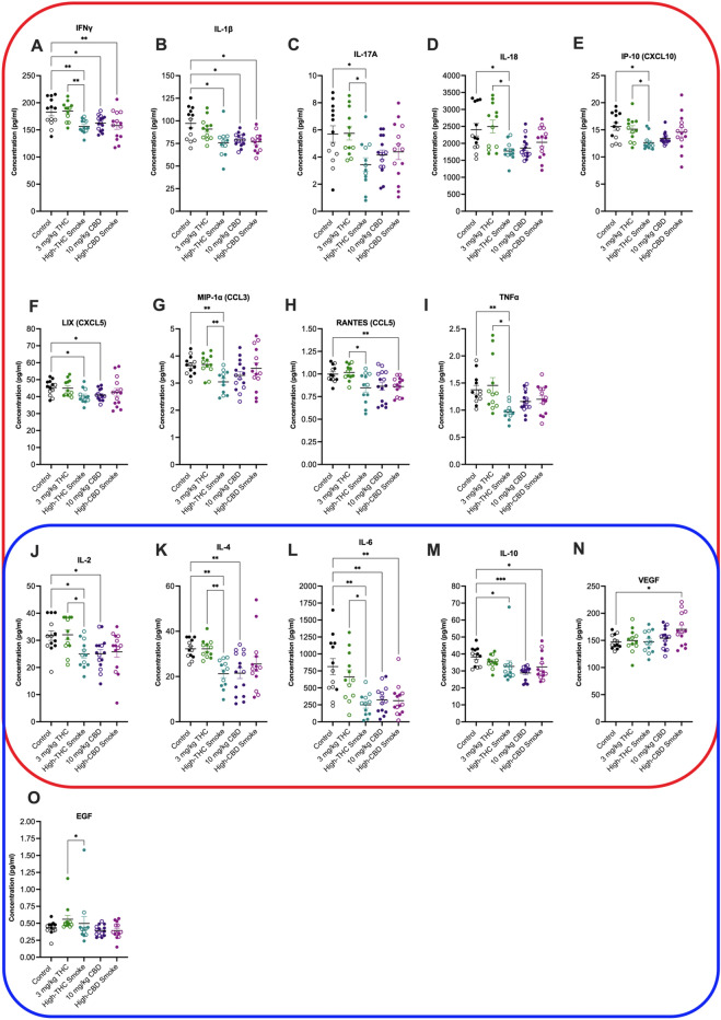
Significant main effect differences in cytokine or chemokine concentrations were observed for 16 of 27 cytokines and chemokines [EGF; IFNγ; IP-10 (CXCL10); ILs IL-1α, IL-1β, IL-2, IL-4, IL-6, IL-10, IL-17A, and IL-18; LIX (CXCL5); MIP-1α (CCL3); RANTES (CCL5); TNFα; and vascular endothelial growth factor (VEGF)] (Suppl. Table S2). Post-hoc testing detected significant differences in 15 of 27 cytokines and chemokines (Fig. 6; Suppl. Table S2). Differences were not detected between groups in fetal brain tissue for the remaining 12 cytokines and chemokines (Suppl. Fig. S2; Suppl. Table S2). The following is a summary of statistically significant post-hoc differences observed for cytokine and chemokine concentrations for fetal brain tissue (Fig. 6). Data have been organized as being broadly defined as pro-inflammatory (red squircle), anti-inflammatory (blue squircle), or both depending on context (encapsulated by red and blue) (Fig. 6)70, 71.
Figure 6.
Fetal brain cytokine and chemokine concentrations 30 min after final treatment on GD20. Cytokines and chemokines are organized by type: species within the red squircle are broadly considered pro-inflammatory, species within the blue squircle are broadly considered anti-inflammatory. Because the role of IL-2, IL-4, IL-6, IL-10 and VEGF can be pro- or anti-inflammatory depending on context, these species are within both squircles. Data are displayed as mean ± S.E.M, n = 12–15 samples/group. Closed circles are data points from males. Open circles are data points from females. An average of two replicates was used when possible. Note the y-axis varies in each panel depending on the cytokine measured. *p < 0.05, **p < 0.01 as indicated between groups.
Compared to control, the pro-inflammatory cytokines IFNγ (p < 0.01 compared to high-THC or high-CBD smoke, p < 0.05 compared to CBD injection) and IL-1β (p < 0.01) were lower in fetal brains of dams receiving high-THC smoke, high-CBD smoke, and CBD injections (Fig. 6A,B). Levels of the pro-inflammatory cytokines IL-17A (p < 0.05), IL-18 (p < 0.05), IP-10 (p < 0.05), LIX (p < 0.05), and MIP-1α (p < 0.01), TNFα (p < 0.01) were similarly lower in fetal brains of dams receiving high-THC smoke compared to control (Fig. 6C–E,G,I). LIX levels were also reduced in fetal brains from dams that received CBD injections (Fig. 6F); and RANTES levels were lower in fetal brains from dams that received high-CBD smoke compared to controls (p < 0.01, Fig. 6H). Between treatment groups IFNγ (p < 0.01), IL-1β (p < 0.05), IL-17A (p < 0.05), IL-18 (p < 0.05), IP-10 (p < 0.05), MIP-1α (p < 0.01), RANTES (p < 0.05) and TNFα (p < 0.05) were all elevated in fetal brains from THC injected grouped compared to high-THC smoke exposure groups (Fig. 6A–E,G–I).
IL-2, IL-4, IL-6, IL-10, and VEGF behave as pro- and anti-inflammatory cytokines depending on cellular context97. In this study, IL-2 (p < 0.05) and IL-4 (p < 0.01) levels were lower in fetal brain tissue of dams receiving high-THC smoke or CBD injections compared to control (Fig. 6J,K). Likewise, IL-6 (p < 0.01) and IL-10 (p < 0.05 for high-THC and high-CBD smoke, p < 0.001 for CBD injections) were lower in fetal brain tissue of dams treated with high-THC smoke, high-CBD smoke, or CBD injections (Fig. 6L,M). VEGF levels were elevated in fetal brain tissues exposed to high-CBD smoke compared to controls (p < 0.05, Fig. 6N). IL-2 (p < 0.05), IL-4 (p < 0.01), and IL-6 (p < 0.05) levels were higher in the fetal brain tissues of dams treated with THC injections compared to high-THC exposure (Fig. 6J–N).
EGF is considered an anti-inflammatory chemokine98. EGF levels were elevated in the fetal brain tissues of dams that received THC injections compared to dams that received CBD injections (p < 0.05, Fig. 6O).
From the fetal brain samples two patterns arise: (i) exposure to Cannabis smoke (high-THC or high-CBD) or injected CBD was associated with widespread downregulation of several cytokines and chemokines in fetal brains; and (ii) several analytes were notably elevated in fetal brain tissue from THC-injected groups relative to high-THC Cannabis smoke, but no post-hoc elevation was observed when THC-injected sample means were compared to control means. These data further demonstrate the widespread changes in inflammatory state that occur in both injection and inhalation rodent models of prenatal cannabinoid exposure and present several instances where changes observed in the placenta are similarly observed in the fetal brain.
Discussion
In the present study, pregnant female rats were treated daily from GD6 until GD20 with either a control (i.p. vehicle injection or room air), 3 mg/kg/day i.p. THC, 300 mg/day high-THC Cannabis smoke (SW), 10 mg/kg/day i.p. CBD, or 300 mg/day high-CBD Cannabis smoke (TI). Results indicate that the duration of exposure (Fig. 2) and route of administration (Fig. 3) significantly affect the levels of cannabinoids and their metabolites in maternal plasma 30 min following administration. Analyses of maternal and fetal bodyweight, resorptions, litter size, fetal brain to BW and BW to placenta and sex ratios, over the course of pregnancy suggest relatively subtle effects of these treatment regimes, except for some evidence of reduced fetal viability following repeated injections of either THC or CBD (Fig. 4). Following repeated treatment of the dams, levels of cytokines and chemokines in the placenta (Fig. 5) and fetal brain (Fig. 6) were also altered in a cannabinoid- and route-dependent manner. Taken together, our results suggest that the route of administration of cannabinoids in developmental studies is an important factor in determining the specific effects obtained. Thus, care should be taken to use methods of administration to closely resemble those of humans to improve the construct validity of preclinical models.
Comparing plasma levels of THC and CBD following repeated injections or smoke administrations to pregnant rats
Analyses of plasma levels of cannabinoids 30 min after exposure revealed that plasma levels of THC and CBD, but not their metabolites, were significantly higher when sampled on GD20, after 15 administrations, compared to GD6, after one administration (Fig. 2). Selection of the 30 min timepoint enabled comparisons to existing plasma concentrations measured in adult male rats from our lab93,94. To the best of our knowledge, this is the first-time plasma cannabinoid levels have been directly compared between acute and repeated administrations during pregnancy in rats. While the potential for accumulation during pregnancy is interesting, these results may be explained by several factors including—but not limited to—homeostatic changes to the metabolism of cannabinoids during repeated exposures or binding and depositing of these cannabinoids on plasma proteins and other tissue during repeated dosing. It is noteworthy that levels of THC and CBD following acute exposure on GD6 were lower than typically observed in male, but not female, rats. Barnard and colleagues94 reported plasma levels of THC of 125 ng/ml (vs. approximately 77 ng/ml for GD6 here) and 15 ng/ml (vs. approximately 8 ng/ml for GD6 here) 30 min following acute treatment of male rats with the same THC injection and high-THC smoke protocols used in the present study. Plasma levels of CBD following high-CBD smoke exposure were approximately 15 ng/ml in the same study94, which is also considerably higher than the roughly 5 ng/ml reported following the first administration of high-CBD smoke to the pregnant rats in this study. However, Baglot and colleagues99 reported sex differences in the plasma concentrations of injected THC (2.5 mg/kg, i.p.) with females showing levels similar to those reported herein, and males showing higher levels similar to the Barnard study94. In a comparison of THC (dronabinol) Volcano® inhalation treatment to i.p. injection in adult male rats, Manwell and colleagues100 reported comparable plasma levels of THC at 20 min and at 40 min time points between 1 mg/pad THC and 2.5 mg/kg i.p. THC to males injected in Barnard94. Using a different protocol of vaporized THC exposure, Baglot and colleagues found higher plasma levels of THC in male and female rats 30 min after inhalation than reported in the present study and showed no difference in circulating concentrations between their inhalation and injected THC groups at this time point99. Notably, Sandini and colleagues detected behavioural changes in adult offspring after repeated prenatal exposures to high-THC smoke like those used in the present study81. In another inhalation model, Jenkins and colleagues101 observed suppressed oscillatory power and coherence with acute THC flower vapour exposure in male rats; however, neither Jenkins et al.101 nor Sandini et al.81 provide pharmacokinetic support for their models.
To the best of our knowledge, there are no previous reports of CBD accumulation during pregnancy and the direct comparison of plasma levels of CBD following injected CBD or inhalation of high-CBD smoke in this study is novel. We found maternal plasma CBD levels at 30 min following 10 mg/kg/day i.p. were 239 ng/mL on GD6, increasing to 1,473 ng/mL after 15 daily injections on GD20 (Fig. 3). In the high-CBD smoke group, levels of CBD were dramatically lower than in injected CBD dams, where on GD6 plasma levels were 7 ng/ml which increased to 17 ng/mL on GD20 at 30 min after administration. Our injected CBD GD6 plasma values more closely align with acute vapor administration in female rats at 100 ng/mL and 10 mg/kg i.p102. whereas our GD20 injected CBD plasma values resemble those from Ochiai and colleagues (2021), where a single dose of 10 mg/kg i.v. of CBD resulted in a Cmax in maternal plasma of ~ 1200 ng/mL and 900 ng/mL in whole fetal tissue82. Of note, Ochiai and colleagues (2021) reported the half-life of injected CBD (10 mg/kg; i.v.) as 5 h in pregnant mice82, which casts doubt on significant accumulation from repeated once daily injections. Thus, comparing plasma cannabinoid levels over a time course by sex and reproductive status will be an important consideration for future research. In addition, assessing plasma levels of cannabinoids without treatment on GD20 would confirm whether accumulation of the cannabinoids was occurring over repeated exposures.
Our experimental design also allowed for a direct comparison of plasma levels of cannabinoids following either smoke exposure or injections. Levels of THC and CBD, as well as some of their metabolites, were significantly higher 30 min following repeated injections than repeated smoke exposure on GD20. In general, we chose to compare injected doses of THC and CBD commonly used in rodent models41,42, 45, 49, 51, 103–106, with the smoke inhalation protocol developed in our laboratory81,93–95. The inhalation protocol resembles other ‘hotbox’ methods100,101, 107, and has been shown to result in acute behavioural and oscillatory effects93–95, as well as long-term changes in the offspring following exposure during pregnancy81. Despite previous acute and chronic behavioural disturbances associated with inhalation methods, this is the first pharmacokinetic evaluation comparing acute (GD6) and chronic (GD20) exposure in a prenatal model of whole cannabis flower smoke exposure. As reported previously for injected THC and smoke exposure94, levels of THC were lower following inhalation than injections. Determining the peak and total exposure is difficult with one sampling point and the passive exposure methods we used with smoke administration; however, it is likely that our smoke exposure protocol results in lower levels of exposure when compared to some other inhalation methods40. In addition, different pharmacokinetics following these administration methods may dramatically affect the peak and total exposure of the pregnant woman and fetus to cannabinoids. Our selection of timepoint and quantification of metabolites indicate that the doses of major phytocannabinoids received from smoked whole flower differs from injected phytocannabinoids and may contribute to different long-term outcomes in the offspring.
Relatively subtle effects of repeated cannabinoid administration on maternal and fetal parameters
Injected cannabinoids and inhaled Cannabis did not significantly impact maternal weight gain, fetal weight, fetal brain to BW or BW to placenta ratios, or sex ratios in this study. Previously, we observed no effect of repeated high-THC smoke exposures on maternal weight gain, litter size, or litter weight following high-THC smoke exposure81, although in that study an increase in the ratio of male:female offspring was significant. A limitation of this study is the relatively small sample sizes for the treatment groups that were carried to GD20, which may have limited the emergence of more nuanced differences in markers of maternal and fetal health. However, injected CBD did increase fetal resorptions and injected THC decreased litter size. Natale and colleagues (2020) reported increased rates of uterine resorption in mice treated with 3 mg/kg/day i.p. THC, alongside indicators of intra-uterine growth restriction (IUGR) and placenta abnormalities42. Although CBD has not been previously investigated in these models in vivo, in vitro assays of trophoblasts show that CBD has a dose-dependent negative impact on cell cycle progression, viability, and migration; all crucial mechanisms in fetal trophoblast development108. Analysis of clinical maternal and childhood health outcomes has yielded different results due to confounding variables such as polysubstance use and inaccurate reporting of Cannabis use frequency. However, clinical retrospectives and pre-clinical investigation have been associated with adverse neonatal outcomes such as decreased birthweight and APGAR scores, preterm births, miscarriages, growth restrictions, and placenta abnormalities (reviewed in36), and new epidemiological data show geospatial associations in the US between congenital abnormalities and Cannabis availability, specifically CBD products109. It is therefore possible that injected THC and CBD produce similar restrictions to healthy litter growth resulting in the loss of pups. Because neither smoke treatment used in the current study produced any differences in measures of maternal and fetal health, we suggest that injected and inhaled Cannabis models differ in their effects on maternal and fetal health.
Repeated cannabinoid treatments differentially alter cytokine levels in placenta and fetal brain tissue
In GD20 placental tissues from rats repeatedly exposed to either THC or CBD, we observed significant differences in levels of 13 of 27 cytokines and chemokines compared to control (Fig. 5). Injected THC treatment upregulated 4 inflammatory cytokines and chemokines, whereas injected CBD only elevated TNFα (also observed with injected THC) and RANTES (not observed for injected THC). Smoke exposure, regardless of the cannabinoid content, generally decreased cytokine and chemokine levels. Analyses of fetal brain tissue harvested from the same pregnancies as the placental tissue revealed significant alterations in levels of 14 of 27 cytokines and chemokines tested among the groups (Fig. 6). Although the effects of maternal Cannabis exposure on immune mechanisms in the mother and offspring are of current interest110, limited in vivo data exist regarding the maternal and fetal immune response to repeated cannabinoid treatments, and potential sex dependant differences. Our results show intriguing changes 30 min following the final treatment on G20. Determining the time course and significance of these changes for placental and fetal health will require additional experimentation. IL-1β, IL-6, IP-10 (CXCL10), and TNFα are all pro-inflammatory cytokines and chemokines important for macrophage and natural killer (NK) cell activation, where the presence of NK cells correlate with fetal resorption, due to their damaging effects on trophoblast proliferation26. Elevations in IL-6, TNFα, and IP-10 (CXCL10), specifically amongst placenta and fetal tissue, have been associated with preterm births, intrauterine fetal death, and resorptions in addition to other downstream pathophysiological and behaviour changes111–113.
Conclusion
Our results show that injection of THC or CBD during gestation produces markedly different physiological effects when compared to smoke exposure with high-THC or high-CBD Cannabis. Smoke exposure did not cause any large-scale changes in markers of maternal or fetal health but did change the levels of immunomodulatory cytokines and chemokines in both placenta and fetal brain samples. Further in-depth behavioural analyses of offspring following exposure to Cannabis smoke compared to injected cannabinoids is required. An important limitation of our current model is that cannabinoids were administered to rats during the equivalent of the late first and second trimester of humans, and therefore our exposure model does not reflect potential use patterns and effects during critical neuro-developmental periods before conception, early in gestation, or the third trimester. Additionally, our model of smoke inhalation uses passive exposure, which standardizes the exposure that dams received. Active self-administration models (e.g.,78), may better represent voluntary usage patterns and reduce exposure stress. In addition, analysis of rodent breathing patterns during hot box exposure methods may also help to determine whether rodents make attempts to titrate exposure by limiting inhalation following repeated treatments. Overall, these data highlight the fundamental differences in outcomes that route, dose, and formulation of Cannabis can have. These results should be considered in our interpretations of rodent model data for gestation and other applications of smoke inhalation and cannabinoid injection.
Methods
Animals
Virgin female (n = 76) and male Sprague–Dawley rats (n = 24; Charles River, Senneville, QC) arrived at our animal housing facility and were allowed to habituate for at least 1 week in ventilated cages in a climate-controlled vivarium under a 12 h:12 h light–dark cycle (lights on at 07:00). Ad libitum food (Purina rat chow) and water was provided throughout all experiments. Animals were then handled for 1 week before males and females (8–10 weeks of age) were paired at 17:00 and left overnight to breed. Evidence of sperm following vaginal swab at 09:00 the following morning was considered GD0. Dams were left undisturbed until GD6, when drug treatments began (Fig. 1). Dams were randomized and assigned treatment with either high-THC Cannabis smoke (Skywalker Kush [SW]: 18% THC, 0.1% CBD, Aphria Inc., Lot# 5142072190); high-CBD Cannabis smoke (Treasure Island Kush [TI]: 13% CBD, 0.7% THC, Aphria Inc., Lot# 5142071556); or i.p. injections of 3 mg/kg/day THC (Cayman Chemical, Ann Arbor, MI Cat. No. 9003740), 10 mg/kg/day CBD (Cayman Chemical, Cat. No. 9003741), vehicle (i.p.) (1:1:18 ratio ethanol:kolliphor:phosphate buffered saline) or air exposure once daily between GD6-20 (Fig. 1). Once pregnant, dams were singly housed in standard cages with adequate cage enrichment. Dams and offspring were euthanized via overdose with 5% isoflurane, followed by cardiac puncture for plasma collection for dams and decapitation for offspring. All experiments were conducted according to standards set by Canadian Council on Animal Care and the University of Saskatchewan Research Ethics Board (AREB), and in accordance with the ARRIVE guidelines114. All experimental methods and protocols were approved by the University of Saskatchewan AREB (Protocol numbers 20210009, 20190067).
Smoke exposure
Pregnant rats (n = 5–8/group) were treated using a well-validated inhalation system (La Jolla Research Inc.) and method established in81,93–95. Cannabis was shredded and ground using a coffee grinder (10 s). Each bowl (ceramic coil) was packed with 300 mg of the ground Cannabis flower. Airtight chambers (33 cm × 30.5 cm × 51 cm) equivalent to approximately 50 L each, could house a maximum of 2 rats, separated into plastic cages with metal grate roof used exclusively in the smoke chambers. Rats were habituated to the smoke chamber and pumps for 2 days for 20 min prior to smoke exposure. Before combustion, rats were placed in the chambers with pumps turned on for 5 min. Air was pumped through the chambers at 10 L/min and exhausted into a fume hood. Cannabis was combusted over a period of 1 min and the resulting smoke continuously pumped through the system. Pumps were turned off for 1 min following the combustion protocol before the smoke was vented gradually over 13 min.
Tissue collection
Maternal blood collection was performed via cardiac puncture. Rats were anesthetized with 5% isoflurane, and maternal blood, organs and fetal tissues were collected 30 min following the start of their last drug treatment. Fetal and maternal tissues were placed in 5 mL LoBind Eppendorf tubes (Cat. No. 0030122356), frozen in liquid nitrogen, and stored at – 80 °C. Cardiac blood was collected by syringe and immediately transferred to 4 mL BD Vacutainer tube (Cat. No. CABD367884). All blood samples were centrifuged for 10 min at 4 °C at 2000×g under 1 h from time of collection. Plasma was collected, aliquoted at 300 µL into 1.5 mL Lobind Eppendor tubes (Cat. No.0030108442), frozen and stored at − 80 °C until needed.
HPLC–MS/MS
The pharmacokinetic investigation of maternal plasma concentrations of THC, CBD and their associated metabolites ± 11-OH- 9- THC (11-OH-THC), ± 11-nor-9-carboxy-9− THC (11-COOH-THC), 6α-OH-CBD and 7-OH-CBD was performed using high performance liquid chromatography tandem mass spectrometry (HPLC–MS/MS)93,94 (Fig. 2). Phytocannabinoids were quantified using Agilent 1260 binary pump LC system (Agilent Technologies Canada, Mississauga, Ontario, Canada) coupled to an ABSciex 4000QTRAP (ABSciex, Concord, ON) mass spectrometer using an Eclipse Plus phenyl hexyl column (4.6 Å 100 mm, 5 μm column, Agilent) and 1290 Infinity II inline filter (0.3 µm). HPLC–MS/MS was run and peaks analyzed using Analyst (version 1.7).
CBD, CBD-D3, THC, THC-D3, 11-OH-THC, 11-COOH-THC working and internal standards were purchased from Cerilliant, where 6α-OH-CBD and 7-OH-CBD and 7-OH-CBD-D9 were purchased from Toronto Research Chemicals and diluted to 1 mg/mL and stored at − 20 °C. Working standards and quality control dilutions were prepared from a stock solution of 5000 ng/mL and diluted to final working concentrations between 39.06 and 5000 ng/mL.
Representative standard curve and quality control samples were prepared by spiking 10 µL of working standards to 190 µL of blank plasma resulting in a 20 × dilution from the working standards. This gave rise to a final LLOQ sample concentration for THC and CBD of 1.97 ng/mL. For 11-OH-THC, 11-COOH-THC, and 6α-OH-CBD the LLOQ was 3.91 ng/mL and for 7-OH-CBD the LLOQ was 10 ng/mL. LLOQ’s were determined as 5–10 × signal to noise for each compound. The low quality control (LQC) for THC and CBD was 5 ng/mL. For 11-OH-THC, 11-COOH-THC, and 6α-OH-CBD the LQC was 10 ng/mL. For 7-OH-CBD it was 20 ng/mL. All phytocannabinoids and their metabolites shared a medium quality control (MQC) of 100 ng/mL, a high-quality control of 175 ng/mL and upper limit of quantification of 250 ng/mL. All 6α-OH-CBD was below our LLOQ.
Six hundred µL of prepared internal standard solution at 12 ng/mL was added to all standards and QCs, vortexed and centrifuged at 14,000×g at 4 °C to precipitate proteins. Supernatant was collected, vacuum filtered through an Agilent Captiva EMR-Lipid 96-well plate (Cat. No. 5190–1001) and transferred to an amber autosampler vial for testing. To prepare maternal plasma, samples were thawed at room temperature, 600 μL of internal standard solution was added, samples were vortexed, centrifuged and filtered as above. The mobile phase was a solution of 0.01% ammonium formate and 0.1% formic acid prepared fresh daily in 100% methanol for the organic phase or prepared in water for the aqueous phase.
Cytokine and chemokine array
Whole fetal brains were dissected, and a transverse dissection of the placenta was collected and homogenized (Rotor–Stator, OMNI International) over ice using RIPA buffer (Cat. No.89901 ThermoFisher) and 1:100 HALT Protease and Phosphatase Inhibitors (Cat#: 78440 ThermoFisher, Burlington, ON) at a ratio of 750 µL RIPA to 150 mg of tissue. Samples were centrifuged at 12,000×g for 10 min at 4 °C and the supernatant was transferred to a new Lobind Ependorf tube (Cat. No. 0030108442), this step was repeated twice. Protein concentrations were quantified using the Pierce™ BCA protein assay kit (Thermofisher). Four thousand µg/mL of sample was aliquoted, and samples were analyzed using the Rat Cytokine 27-Plex (Millipore Milliplex) by Eve Technologies (Calgary, AB). A maximum of one placenta per litter and two pup brains per litter (one male/ one female) were used in this analysis.
Statistical analyses
All data analyses for this study were conducted and graphed with Prism (GraphPad, v. 9.0). The pharmacokinetic data (Figs. 2 and 3) were analyzed two separate ways. One injected THC dam was removed from analysis on GD20 due to low plasma THC levels. Plasma cannabinoid concentrations were expressed as a mean ± S.E.M. n = 5–8 litters per group. Only samples that were above the LLOQs were analyzed. Data in Figs. 2 and 3, as well as Supp Fig. S1, were analyzed with ANOVA or nonparametric tests as appropriate (see Results for specific details). Percentage maternal weight gain across gestation was calculated as: ((Final-Baseline)/Baseline*100)). Data were analyzed with a 2 level (7 × 5: Day x Treatment) mixed effects model (REML), where data are expressed as a mean ± S.E.M., n = 6–7 dams per treatment. Other maternal and pup health data were assessed for normality (KS-test) and homoscedasticity (Brown-Forsyth test) and analyzed with one-way ANOVA with Greenhouse–Geisser correction or Kruskal–Wallis (KW) test (non-parametric) with maternal Treatment as a between-subjects factor. Planned comparisons between control to injected-THC, control to injected-CBD, injected-THC to high-THC smoke, injected-CBD to high-CBD smoke, and high-THC to high-CBD smoke were performed using Holm-Šídák's (parametric) or Dunn’s (non-parametric) multiple comparisons tests. The multiplex data were assessed for normality (KS-test) and homoscedasticity (Brown-Forsyth test). When data met these assumptions, they were analyzed according to an ordinary one-way ANOVA with Greenhouse–Geisser correction. If data violated the KS-test, outliers were identified using Grubbs’, and data were further analyzed using ordinary one-way ANOVA with Greenhouse–Geisser correction and KW-test if not normally distributed. If data were heteroscedastic, Brown-Forsythe ANOVA was used. Planned comparisons (as listed above) were performed using Holm-Šídák's (parametric), Dunn’s (non-parametric), or Dunnett’s T3 tests (heteroscedastic) to compare mean concentrations of each treatment group.
Supplementary Information
Acknowledgements
The authors acknowledge Maddie Stewart for input and assistance regarding interpretation of the multiplex experiment.
Author contributions
T.B. planned and executed the experiment, analyzed all data, wrote, and edited the manuscript with the technical and practical assistance of S.L.B., I.L.B., E.F., D.L.M., F.V.L.A.-S., and Q.G. for animal work. D.M. and A.Z. provided assistance and oversight for the pharmacokinetic experiments and data analysis. J.G.H. and R.B.L. designed and planned the experiments, supervised all highly qualified personnel, assisted with data analysis, and contributed to the writing and editing of the manuscript.
Funding
TB is funded by a Doctoral Award from the Canadian Institutes of Health Research. SLB is supported by a Mitacs award and the College of Pharmacy and Nutrition. ILB and FVLA-S were supported by scholarships from the Natural Sciences and Engineering Research Council of Canada. DLM was supported by a scholarship from the College of Medicine, University of Saskatchewan. DM and AZ are supported by funds from the College of Pharmacy and Nutrition. This project was funded by a Brain Canada Future Leaders fund to RBL and a grant to JGH and RBL from the College of Medicine, University of Saskatchewan.
Data availability
The datasets used and/or analysed during the current study available from the corresponding author on reasonable request.
Competing interests
RBL has worked as a consultant on recent medico-legal cases involving Cannabis in Canada and currently serves as a consultant on the scientific advisory board for Shackleford Pharma Inc. All Cannabis used in this study was purchased from Aphria-Tilray Inc. and their corporation was not involved in the research conducted. All other authors declare no competing interests.
Footnotes
Publisher's note
Springer Nature remains neutral with regard to jurisdictional claims in published maps and institutional affiliations.
Contributor Information
John G. Howland, Email: john.howland@usask.ca
Robert B. Laprairie, Email: robert.laprairie@usask.ca
Supplementary Information
The online version contains supplementary material available at 10.1038/s41598-023-47861-8.
References
- 1.Rotermann, M. Looking back from 2020, how cannabis use and related behaviours changed in Canada. Health Rep.32, (2020). [DOI] [PubMed]
- 2.Marchand G, et al. Birth outcomes of neonates exposed to marijuana in utero: A systematic review and meta-analysis. JAMA Netw. Open. 2022;5:e2145653. doi: 10.1001/jamanetworkopen.2021.45653. [DOI] [PMC free article] [PubMed] [Google Scholar]
- 3.Gunn JKL, et al. The effects of prenatal cannabis exposure on fetal development and pregnancy outcomes: A protocol. BMJ Open. 2015;5:e007227. doi: 10.1136/bmjopen-2014-007227. [DOI] [PMC free article] [PubMed] [Google Scholar]
- 4.Gunn JKL, et al. Prenatal exposure to cannabis and maternal and child health outcomes: A systematic review and meta-analysis. BMJ Open. 2016;6:e009986. doi: 10.1136/bmjopen-2015-009986. [DOI] [PMC free article] [PubMed] [Google Scholar]
- 5.Corsi DJ, et al. Maternal cannabis use in pregnancy and child neurodevelopmental outcomes. Nat. Med. 2020;26:1536–1540. doi: 10.1038/s41591-020-1002-5. [DOI] [PubMed] [Google Scholar]
- 6.Paul SE, et al. Associations between prenatal cannabis exposure and childhood outcomes: Results from the ABCD study. JAMA Psychiatry. 2021;78:64. doi: 10.1001/jamapsychiatry.2020.2902. [DOI] [PMC free article] [PubMed] [Google Scholar]
- 7.Davis E, Lee T, Weber JT, Bugden S. Cannabis use in pregnancy and breastfeeding: The pharmacist’s role. Can. Pharm. J. 2020;153:95–100. doi: 10.1177/1715163519893395. [DOI] [PMC free article] [PubMed] [Google Scholar]
- 8.Rompala, G., Nomura, Y. & Hurd, Y. L. Maternal cannabis use is associated with suppression of immune gene networks in placenta and increased anxiety phenotypes in offspring. Proc. Natl. Acad. Sci. USA.118, e2106115118 (2021). [DOI] [PMC free article] [PubMed]
- 9.Luke S, et al. Cannabis use in pregnancy and maternal and infant outcomes: A Canadian cross-jurisdictional population-based cohort study. PLOS ONE. 2022;17:e0276824. doi: 10.1371/journal.pone.0276824. [DOI] [PMC free article] [PubMed] [Google Scholar]
- 10.Shi Y, Zhu B, Liang D. The associations between prenatal cannabis use disorder and neonatal outcomes. Addiction. 2021;116:3069–3079. doi: 10.1111/add.15467. [DOI] [PMC free article] [PubMed] [Google Scholar]
- 11.Koto, P., Allen, V. M., Fahey, J. & Kuhle, S. Maternal cannabis use during pregnancy and maternal and neonatal outcomes: A retrospective cohort study. BJOG Int. J. Obstet. Gynaecol.129, 1687–1694 (2022). [DOI] [PubMed]
- 12.Morales P, Hurst DP, Reggio PH. Molecular targets of the phytocannabinoids: A complex picture. Prog. Chem. Organic Nat. Prod. 2017;103:103–131. doi: 10.1007/978-3-319-45541-9_4. [DOI] [PMC free article] [PubMed] [Google Scholar]
- 13.Russo EB. Taming THC: potential cannabis synergy and phytocannabinoid-terpenoid entourage effects. Br. J. Pharmacol. 2011;163:1344–1364. doi: 10.1111/j.1476-5381.2011.01238.x. [DOI] [PMC free article] [PubMed] [Google Scholar]
- 14.Howlett AC, Abood ME. CB1 and CB2 receptor pharmacology. Adv. Pharmacol. 2017;80:169–206. doi: 10.1016/bs.apha.2017.03.007. [DOI] [PMC free article] [PubMed] [Google Scholar]
- 15.Howlett AC. The cannabinoid receptors. Prostaglandins Other Lipid Mediat. 2002;68–69:619–631. doi: 10.1016/S0090-6980(02)00060-6. [DOI] [PubMed] [Google Scholar]
- 16.Thomas A, et al. Cannabidiol displays unexpectedly high potency as an antagonist of CB1 and CB2 receptor agonists in vitro. Br. J. Pharmacol. 2007;150:613–623. doi: 10.1038/sj.bjp.0707133. [DOI] [PMC free article] [PubMed] [Google Scholar]
- 17.Pertwee RG. The diverse CB1 and CB2 receptor pharmacology of three plant cannabinoids: delta9-tetrahydrocannabinol, cannabidiol and delta9-tetrahydrocannabivarin. Br. J. Pharmacol. 2008;153:199–215. doi: 10.1038/sj.bjp.0707442. [DOI] [PMC free article] [PubMed] [Google Scholar]
- 18.Laprairie RB, Bagher AM, Kelly MEM, Denovan-Wright EM. Cannabidiol is a negative allosteric modulator of the cannabinoid CB1 receptor. Br. J. Pharmacol. 2015;172:4790–4805. doi: 10.1111/bph.13250. [DOI] [PMC free article] [PubMed] [Google Scholar]
- 19.de Almeida DL, Devi LA. Diversity of molecular targets and signaling pathways for CBD. Pharmacol. Res. Perspect. 2020;8:e00682. doi: 10.1002/prp2.682. [DOI] [PMC free article] [PubMed] [Google Scholar]
- 20.Red-Horse K, Drake PM, Gunn MD, Fisher SJ. Chemokine ligand and receptor expression in the pregnant uterus. Am. J. Pathol. 2001;159:2199–2213. doi: 10.1016/S0002-9440(10)63071-4. [DOI] [PMC free article] [PubMed] [Google Scholar]
- 21.Du M-R, Wang S-C, Li D-J. The integrative roles of chemokines at the maternal–fetal interface in early pregnancy. Cell Mol. Immunol. 2014;11:438–448. doi: 10.1038/cmi.2014.68. [DOI] [PMC free article] [PubMed] [Google Scholar]
- 22.Vinketova K, Mourdjeva M, Oreshkova T. Human decidual stromal cells as a component of the implantation niche and a modulator of maternal immunity. J. Pregn. 2016;2016:1–17. doi: 10.1155/2016/8689436. [DOI] [PMC free article] [PubMed] [Google Scholar]
- 23.Salamonsen LA, Hannan NJ, Dimitriadis E. Cytokines and chemokines during human embryo implantation: Roles in implantation and early placentation. Semin. Reprod. Med. 2007;25:437–444. doi: 10.1055/s-2007-991041. [DOI] [PubMed] [Google Scholar]
- 24.Shukla V, Soares MJ. Modeling trophoblast cell-guided uterine spiral artery transformation in the rat. Int. J. Mol. Sci. 2022;23:2947. doi: 10.3390/ijms23062947. [DOI] [PMC free article] [PubMed] [Google Scholar]
- 25.Yockey LJ, Iwasaki A. Interferons and proinflammatory cytokines in pregnancy and fetal development. Immunity. 2018;49:397–412. doi: 10.1016/j.immuni.2018.07.017. [DOI] [PMC free article] [PubMed] [Google Scholar]
- 26.Gendron RL, Nestel FP, Lapp WS, Baines MG. Lipopolysaccharide-induced fetal resorption in mice is associated with the intrauterine production of tumour necrosis factor-alpha. Reproduction. 1990;90:395–402. doi: 10.1530/jrf.0.0900395. [DOI] [PubMed] [Google Scholar]
- 27.Reister F, et al. Macrophage-induced apoptosis limits endovascular trophoblast invasion in the uterine wall of preeclamptic women. Lab. Invesig. 2001;81:1143–1152. doi: 10.1038/labinvest.3780326. [DOI] [PubMed] [Google Scholar]
- 28.Gomez-Lopez N, Laresgoiti-Servitje E, Olson DM, Estrada-Gutiérrez G, Vadillo-Ortega F. The role of chemokines in term and premature rupture of the fetal membranes: A Review. Biol. Reprod. 2010;82:809–814. doi: 10.1095/biolreprod.109.080432. [DOI] [PubMed] [Google Scholar]
- 29.Zhang S, et al. Regulation and function of chemokines at the maternal–fetal interface. Front. Cell Dev. Biol. 2022;10:826053. doi: 10.3389/fcell.2022.826053. [DOI] [PMC free article] [PubMed] [Google Scholar]
- 30.Paria BC, et al. Dysregulated cannabinoid signaling disrupts uterine receptivity for embryo implantation. J. Biol. Chem. 2001;276:20523–20528. doi: 10.1074/jbc.M100679200. [DOI] [PubMed] [Google Scholar]
- 31.Maia, J., Fonseca, B. M., Teixeira, N. & Correia-da-Silva, G. The fundamental role of the endocannabinoid system in endometrium and placenta: implications in pathophysiological aspects of uterine and pregnancy disorders. Hum. Rep. Update26, 586–602 (2020). [DOI] [PMC free article] [PubMed]
- 32.Berghuis P, et al. Endocannabinoids regulate interneuron migration and morphogenesis by transactivating the TrkB receptor. Proc. Natl. Acad. Sci. U.S.A. 2005;102:19115–19120. doi: 10.1073/pnas.0509494102. [DOI] [PMC free article] [PubMed] [Google Scholar]
- 33.Berghuis P, et al. Hardwiring the brain: Endocannabinoids shape neuronal connectivity. Science. 2007;316:1212–1216. doi: 10.1126/science.1137406. [DOI] [PubMed] [Google Scholar]
- 34.Aguado T, et al. The endocannabinoid system promotes astroglial differentiation by acting on neural progenitor cells. J. Neurosci. 2006;26:1551–1561. doi: 10.1523/JNEUROSCI.3101-05.2006. [DOI] [PMC free article] [PubMed] [Google Scholar]
- 35.Harkany T, et al. The emerging functions of endocannabinoid signaling during CNS development. Trends Pharmacol. Sci. 2007;28:83–92. doi: 10.1016/j.tips.2006.12.004. [DOI] [PubMed] [Google Scholar]
- 36.Bara A, Ferland J-MN, Rompala G, Szutorisz H, Hurd YL. Cannabis and synaptic reprogramming of the developing brain. Nat. Rev. Neurosci. 2021;22:423–438. doi: 10.1038/s41583-021-00465-5. [DOI] [PMC free article] [PubMed] [Google Scholar]
- 37.Bailey JR, Cunny HC, Paule MG, Slikker W. Fetal disposition of delta 9-tetrahydrocannabinol (THC) during late pregnancy in the rhesus monkey. Toxicol. Appl. Pharmacol. 1987;90:315–321. doi: 10.1016/0041-008X(87)90338-3. [DOI] [PubMed] [Google Scholar]
- 38.Hutchings DE, Martin BR, Gamagaris Z, Miller N, Fico T. Plasma concentrations of delta-9-tetrahydrocannabinol in dams and fetuses following acute or multiple prenatal dosing in rats. Life Sci. 1989;44:697–701. doi: 10.1016/0024-3205(89)90380-9. [DOI] [PubMed] [Google Scholar]
- 39.Grant KS, Petroff R, Isoherranen N, Stella N, Burbacher TM. Cannabis use during pregnancy: Pharmacokinetics and effects on child development. Pharmacol. Ther. 2018;182:133–151. doi: 10.1016/j.pharmthera.2017.08.014. [DOI] [PMC free article] [PubMed] [Google Scholar]
- 40.Baglot SL, et al. Maternal-fetal transmission of delta-9-tetrahydrocannabinol (THC) and its metabolites following inhalation and injection exposure during pregnancy in rats. J. Neurosci. Res. 2022;100:713–730. doi: 10.1002/jnr.24992. [DOI] [PubMed] [Google Scholar]
- 41.Tortoriello G, et al. Miswiring the brain: 9-tetrahydrocannabinol disrupts cortical development by inducing an SCG10/stathmin-2 degradation pathway. EMBO J. 2014;33:668–685. doi: 10.1002/embj.201386035. [DOI] [PMC free article] [PubMed] [Google Scholar]
- 42.Natale BV, et al. Δ9-tetrahydrocannabinol exposure during rat pregnancy leads to symmetrical fetal growth restriction and labyrinth-specific vascular defects in the placenta. Sci. Rep. 2020;10:544. doi: 10.1038/s41598-019-57318-6. [DOI] [PMC free article] [PubMed] [Google Scholar]
- 43.Silva L, Zhao N, Popp S, Dow-Edwards D. Prenatal Tetrahydrocannabinol (THC) alters cognitive function and amphetamine response from weaning to adulthood in the rat. Neurotoxicol. Teratol. 2012;34:63–71. doi: 10.1016/j.ntt.2011.10.006. [DOI] [PMC free article] [PubMed] [Google Scholar]
- 44.Spano MS, Ellgren M, Wang X, Hurd YL. Prenatal cannabis exposure increases heroin seeking with allostatic changes in limbic enkephalin systems in adulthood. Biol. Psychiatry. 2007;61:554–563. doi: 10.1016/j.biopsych.2006.03.073. [DOI] [PubMed] [Google Scholar]
- 45.de Salas-Quiroga A, et al. Prenatal exposure to cannabinoids evokes long-lasting functional alterations by targeting CB1 receptors on developing cortical neurons. Proc. Natl. Acad. Sci. 2015;112:13693–13698. doi: 10.1073/pnas.1514962112. [DOI] [PMC free article] [PubMed] [Google Scholar]
- 46.de Salas-Quiroga A, et al. Long-term hippocampal interneuronopathy drives sex-dimorphic spatial memory impairment induced by prenatal THC exposure. Neuropsychopharmacology. 2020;45:877–886. doi: 10.1038/s41386-020-0621-3. [DOI] [PMC free article] [PubMed] [Google Scholar]
- 47.Vargish GA, et al. Persistent inhibitory circuit defects and disrupted social behaviour following in utero exogenous cannabinoid exposure. Mol. Psychiatry. 2017;22:56–67. doi: 10.1038/mp.2016.17. [DOI] [PMC free article] [PubMed] [Google Scholar]
- 48.Singh ME, McGregor IS, Mallet PE. Perinatal exposure to Δ9-tetrahydrocannabinol alters heroin-induced place conditioning and Fos-immunoreactivity. Neuropsychopharmacology. 2006;31:58–69. doi: 10.1038/sj.npp.1300770. [DOI] [PubMed] [Google Scholar]
- 49.Sarikahya, M. H. et al. Prenatal THC exposure induces sex-dependent neuropsychiatric endophenotypes in offspring and long-term disruptions in fatty-acid signaling pathways directly in the mesolimbic circuitry. eNeuro9, ENEURO.0253–22.2022 (2022). [DOI] [PMC free article] [PubMed]
- 50.Sarikahya, M. H. et al. Prenatal THC exposure induces long-term, sex-dependent cognitive dysfunction associated with lipidomic and neuronal pathology in the prefrontal cortex-hippocampal network. Mol. Psychiatr. 1–17 (2023). 10.1038/s41380-023-02190-0 [DOI] [PubMed]
- 51.Gillies R, et al. Maternal exposure to Δ9-tetrahydrocannabinol impairs female offspring glucose homeostasis and endocrine pancreatic development in the rat. Reprod. Toxicol. 2020;94:84–91. doi: 10.1016/j.reprotox.2020.04.070. [DOI] [PubMed] [Google Scholar]
- 52.Di Bartolomeo M, et al. Crosstalk between the transcriptional regulation of dopamine D2 and cannabinoid CB1 receptors in schizophrenia: Analyses in patients and in perinatal Δ9-tetrahydrocannabinol-exposed rats. Pharmacol. Res. 2021;164:105357. doi: 10.1016/j.phrs.2020.105357. [DOI] [PubMed] [Google Scholar]
- 53.Trezza V, et al. Effects of perinatal exposure to delta-9-tetrahydrocannabinol on the emotional reactivity of the offspring: a longitudinal behavioral study in Wistar rats. Psychopharmacology. 2008;198:529–537. doi: 10.1007/s00213-008-1162-3. [DOI] [PubMed] [Google Scholar]
- 54.Campolongo P, et al. PRECLINICAL STUDY: Perinatal exposure to delta-9-tetrahydrocannabinol causes enduring cognitive deficits associated with alteration of cortical gene expression and neurotransmission in rats. Addict. Biol. 2007;12:485–495. doi: 10.1111/j.1369-1600.2007.00074.x. [DOI] [PubMed] [Google Scholar]
- 55.González B, et al. Effects of perinatal exposure to Δ9-tetrahydrocannabinol on operant morphine-reinforced behavior. Pharmacol. Biochem. Behav. 2003;75:577–584. doi: 10.1016/S0091-3057(03)00115-1. [DOI] [PubMed] [Google Scholar]
- 56.Rubio P, et al. Maternal exposure to low doses of Δ9-tetrahydrocannabinol facilitates morphine-induced place conditioning in adult male offspring. Pharmacol. Biochem. Behav. 1998;61:229–238. doi: 10.1016/S0091-3057(98)00099-9. [DOI] [PubMed] [Google Scholar]
- 57.Rubio P, et al. Long-term behavioral effects of perinatal exposure to Δ9-tetrahydrocannabinol in rats: Possible role of pituitary adrenal axis. Life Sci. 1995;56:2169–2176. doi: 10.1016/0024-3205(95)00204-J. [DOI] [PubMed] [Google Scholar]
- 58.Vela G, et al. Maternal exposure to Δ9-tetrahydrocannabinol facilitates morphine self-administration behavior and changes regional binding to central μ opioid receptors in adult offspring female rats. Brain Res. 1998;807:101–109. doi: 10.1016/S0006-8993(98)00766-5. [DOI] [PubMed] [Google Scholar]
- 59.Vela, G., Fuentes, J., Bonnin, A., Ferna´ndez-Ruiz, J. & Ruiz-Gayo, M. Perinatal exposure to Δ9-tetrahydrocannabinol (Δ9-THC) leads to changes in opioid-related behavioral patterns in rats. Brain Res.680, 142–147 (1995). [DOI] [PubMed]
- 60.Frau R, Melis M. Sex-specific susceptibility to psychotic-like states provoked by prenatal THC exposure: Reversal by pregnenolone. J. Neuroendocrinol. 2023;35:e13240. doi: 10.1111/jne.13240. [DOI] [PubMed] [Google Scholar]
- 61.Frau R, et al. Prenatal THC exposure produces a hyperdopaminergic phenotype rescued by pregnenolone. Nat. Neurosci. 2019;22:1975–1985. doi: 10.1038/s41593-019-0512-2. [DOI] [PMC free article] [PubMed] [Google Scholar]
- 62.Sagheddu C, et al. Mesolimbic dopamine dysregulation as a signature of information processing deficits imposed by prenatal THC exposure. Prog. Neuro-Psychopharmacol. Biol. Psychiatry. 2021;105:110128. doi: 10.1016/j.pnpbp.2020.110128. [DOI] [PubMed] [Google Scholar]
- 63.Bara, A. et al. Sex-dependent effects of in utero cannabinoid exposure on cortical function. eLife7, e36234 (2018). [DOI] [PMC free article] [PubMed]
- 64.Traccis F, et al. Prenatal THC does not affect female mesolimbic dopaminergic system in Preadolescent Rats. Int. J. Mol. Sci. 2021;22:1666. doi: 10.3390/ijms22041666. [DOI] [PMC free article] [PubMed] [Google Scholar]
- 65.Brancato A, Castelli V, Lavanco G, Marino RAM, Cannizzaro C. In utero Δ9-tetrahydrocannabinol exposure confers vulnerability towards cognitive impairments and alcohol drinking in the adolescent offspring: Is there a role for neuropeptide Y? J. Psychopharmacol. 2020;34:663–679. doi: 10.1177/0269881120916135. [DOI] [PubMed] [Google Scholar]
- 66.Newsom RJ, Kelly SJ. Perinatal delta-9-tetrahydrocannabinol exposure disrupts social and open field behavior in adult male rats. Neurotoxicol. Teratol. 2008;30:213–219. doi: 10.1016/j.ntt.2007.12.007. [DOI] [PMC free article] [PubMed] [Google Scholar]
- 67.Abel EL, Subramanian MG. Effects of low doses of alcohol on delta-9-tetrahydrocannabinol’s effects in pregnant rats. Life Sci. 1990;47:1677–1682. doi: 10.1016/0024-3205(90)90374-Z. [DOI] [PubMed] [Google Scholar]
- 68.DiNieri, J. A. & Hurd, Y. L. Rat models of prenatal and adolescent cannabis exposure in Psychiatric Disorders: Methods and Protocols (ed. Kobeissy, F.H.) 829, 231–242 (Humana Press, 2012). [DOI] [PubMed]
- 69.Molina-Holgado F, Alvarez FJ, Gonzalez I, Antonio MT, Leret ML. Maternal Exposure to Δ9-Tetrahydrocannabinol (Δ9-THC) Alters Indolamine Levels and Turnover in Adult Male and Female Rat Brain Regions. Brain Res. Bull. 1997;43:173–178. doi: 10.1016/S0361-9230(96)00434-0. [DOI] [PubMed] [Google Scholar]
- 70.Manduca A, et al. Sex-specific behavioural deficits induced at early life by prenatal exposure to the cannabinoid receptor agonist WIN55, 212–2 depend on mGlu5 receptor signalling. Br. J. Pharmacol. 2020;177:449–463. doi: 10.1111/bph.14879. [DOI] [PMC free article] [PubMed] [Google Scholar]
- 71.Scheyer, A. F., Borsoi, M., Pelissier-Alicot, A.-L. & Manzoni, O. J. J. Maternal exposure to the cannabinoid agonist WIN 55,12,2 during lactation induces lasting behavioral and synaptic alterations in the rat adult offspring of both sexes. eNeuro7, ENEURO.0144–20.2020 (2020). [DOI] [PMC free article] [PubMed]
- 72.Shabani M, Hosseinmardi N, Haghani M, Shaibani V, Janahmadi M. Maternal exposure to the CB1 cannabinoid agonist WIN 55212–2 produces robust changes in motor function and intrinsic electrophysiological properties of cerebellar Purkinje neurons in rat offspring. Neuroscience. 2011;172:139–152. doi: 10.1016/j.neuroscience.2010.10.031. [DOI] [PubMed] [Google Scholar]
- 73.Antonelli T, et al. Prenatal exposure to the CB1 receptor agonist WIN 55,212–2 causes learning disruption associated with impaired cortical NMDA receptor function and emotional reactivity changes in rat offspring. Cerebral Cortex. 2005;15:2013–2020. doi: 10.1093/cercor/bhi076. [DOI] [PubMed] [Google Scholar]
- 74.Saez TMM, Aronne MP, Caltana L, Brusco AH. Prenatal exposure to the CB1 and CB2 cannabinoid receptor agonist WIN 55,212–2 alters migration of early-born glutamatergic neurons and GABAergic interneurons in the rat cerebral cortex. J. Neurochem. 2014;129:637–648. doi: 10.1111/jnc.12636. [DOI] [PubMed] [Google Scholar]
- 75.Borsoi, M. et al. Sex Differences in the behavioral and synaptic consequences of a single in vivo exposure to the synthetic cannabimimetic WIN55,212–2 at puberty and adulthood. Front. Behav. Neurosci.13, (2019). [DOI] [PMC free article] [PubMed]
- 76.Ferraro L, et al. Short- and long-term consequences of prenatal exposure to the cannabinoid agonist WIN55,212–2 on rat glutamate transmission and cognitive functions. J. Neural Transm. 2009;116:1017–1027. doi: 10.1007/s00702-009-0230-0. [DOI] [PubMed] [Google Scholar]
- 77.Weimar HV, et al. Long-term effects of maternal cannabis vapor exposure on emotional reactivity, social behavior, and behavioral flexibility in offspring. Neuropharmacology. 2020;179:108288. doi: 10.1016/j.neuropharm.2020.108288. [DOI] [PubMed] [Google Scholar]
- 78.Weimar HV, et al. Determining impacts of prenatal cannabis exposure on cannabis vapor self-administration using a novel response-contingent vapor model in pregnant rat dams. Addict. Neurosci. 2023;6:100071. doi: 10.1016/j.addicn.2023.100071. [DOI] [Google Scholar]
- 79.Breit KR, Rodriguez CG, Lei A, Thomas JD. Combined vapor exposure to THC and alcohol in pregnant rats: Maternal outcomes and pharmacokinetic effects. Neurotoxicol. Teratol. 2020;82:106930. doi: 10.1016/j.ntt.2020.106930. [DOI] [PMC free article] [PubMed] [Google Scholar]
- 80.Breit, K. R. et al. A model of combined exposure to nicotine and tetrahydrocannabinol via electronic cigarettes in pregnant rats. Front. Neurosci.16, (2022). [DOI] [PMC free article] [PubMed]
- 81.Sandini, T. M. et al. Repeated exposure to high-THC Cannabis smoke during gestation alters sex ratio, behavior, and amygdala gene expression of Sprague Dawley rat offspring. eNeuro 2023. Nov 10: ENEURO.0100-23.2023. doi: 10.1523/ENEURO.0100-23.2023. 03.23.533930. [DOI] [PMC free article] [PubMed]
- 82.Ochiai W, et al. Maternal and fetal pharmacokinetic analysis of cannabidiol during pregnancy in mice. Drug Metab. Dispos. 2021;49:337–343. doi: 10.1124/dmd.120.000270. [DOI] [PubMed] [Google Scholar]
- 83.Maciel, I. de S. et al. Perinatal CBD or THC exposure results in lasting resistance to fluoxetine in the forced swim test: Reversal by fatty acid amide hydrolase inhibition. Cannabis Cannabinoid Res.7, 318–327 (2022). [DOI] [PMC free article] [PubMed]
- 84.Iezzi D, Caceres-Rodriguez A, Chavis P, Manzoni OJJ. In utero exposure to cannabidiol disrupts select early-life behaviors in a sex-specific manner. Transl. Psychiatry. 2022;12:501. doi: 10.1038/s41398-022-02271-8. [DOI] [PMC free article] [PubMed] [Google Scholar]
- 85.Wanner NM, Colwell M, Drown C, Faulk C. Developmental cannabidiol exposure increases anxiety and modifies genome-wide brain DNA methylation in adult female mice. Clin. Epigenet. 2021;13:4. doi: 10.1186/s13148-020-00993-4. [DOI] [PMC free article] [PubMed] [Google Scholar]
- 86.Swenson, K. S. et al. Fetal cannabidiol (CBD) exposure alters thermal pain sensitivity, cognition, and prefrontal cortex excitability. 2022.12.06.519350. 10.1101/2022.12.06.519350 (2022) [DOI] [PMC free article] [PubMed]
- 87.Huestis MA. Human cannabinoid pharmacokinetics. C&B. 2007;4:1770–1804. doi: 10.1002/cbdv.200790152. [DOI] [PMC free article] [PubMed] [Google Scholar]
- 88.Newmeyer, M. et al. Free and Glucuronide whole blood cannabinoids’ pharmacokinetics after controlled smoked, vaporized, and oral cannabis administration in frequent and occasional cannabis users: identification of recent cannabis intake. Clin. Chem.62, (2016). [DOI] [PubMed]
- 89.Bidwell, L. C. et al. A naturalistic study of orally administered vs. inhaled legal market cannabis: cannabinoids exposure, intoxication, and impairment. Psychopharmacology239, 385–397 (2022). [DOI] [PMC free article] [PubMed]
- 90.Bidwell LC, et al. Association of naturalistic administration of cannabis flower and concentrates with intoxication and impairment. JAMA Psychiatry. 2020;77:787–796. doi: 10.1001/jamapsychiatry.2020.0927. [DOI] [PMC free article] [PubMed] [Google Scholar]
- 91.Russo, E. B. The case for the entourage effect and conventional breeding of clinical cannabis: No “strain,” no gain. Front. Plant Sci.9 (2019). [DOI] [PMC free article] [PubMed]
- 92.Ferber SG, et al. The “Entourage Effect”: Terpenes coupled with cannabinoids for the treatment of mood disorders and anxiety disorders. Curr. Neuropharmacol. 2020;18:87–96. doi: 10.2174/1570159X17666190903103923. [DOI] [PMC free article] [PubMed] [Google Scholar]
- 93.Roebuck AJ, et al. Dissociable changes in spike and wave discharges following exposure to injected cannabinoids and smoked cannabis in Genetic Absence Epilepsy Rats from Strasbourg. Eur. J. Neurosci. 2022;55:1063–1078. doi: 10.1111/ejn.15096. [DOI] [PubMed] [Google Scholar]
- 94.Barnard IL, et al. The effects of acute Cannabis smoke or Δ9-THC injections on the trial-unique, nonmatching-to-location and five-choice serial reaction time tasks in male Long-Evans rats. Neurobiol. Learn. Memory. 2022;192:107624. doi: 10.1016/j.nlm.2022.107624. [DOI] [PubMed] [Google Scholar]
- 95.Barnard, I. L. et al. High-THC Cannabis smoke impairs incidental memory capacity in spontaneous tests of novelty preference for objects and odors in male rats. eNeuro. 2023 Nov 15:ENEURO.0115-23.2023. doi: 10.1523/ENEURO.0115-23.2023. [DOI] [PMC free article] [PubMed]
- 96.Hughes CE, Nibbs RJB. A guide to chemokines and their receptors. FEBS J. 2018;285:2944–2971. doi: 10.1111/febs.14466. [DOI] [PMC free article] [PubMed] [Google Scholar]
- 97.Zhang J-M, An J. Cytokines, inflammation and pain. Int. Anesthesiol. Clin. 2007;45:27–37. doi: 10.1097/AIA.0b013e318034194e. [DOI] [PMC free article] [PubMed] [Google Scholar]
- 98.Logan RM, et al. The role of pro-inflammatory cytokines in cancer treatment-induced alimentary tract mucositis: Pathobiology, animal models and cytotoxic drugs. Cancer Treatm. Rev. 2007;33:448–460. doi: 10.1016/j.ctrv.2007.03.001. [DOI] [PubMed] [Google Scholar]
- 99.Baglot SL, et al. Pharmacokinetics and central accumulation of delta-9-tetrahydrocannabinol (THC) and its bioactive metabolites are influenced by route of administration and sex in rats. Sci. Rep. 2021;11:23990. doi: 10.1038/s41598-021-03242-7. [DOI] [PMC free article] [PubMed] [Google Scholar]
- 100.Manwell LA, et al. A vapourized Δ9-tetrahydrocannabinol (Δ9-THC) delivery system part I: Development and validation of a pulmonary cannabinoid route of exposure for experimental pharmacology studies in rodents. J. Pharmacol. Toxicol. Methods. 2014;70:120–127. doi: 10.1016/j.vascn.2014.06.006. [DOI] [PubMed] [Google Scholar]
- 101.Jenkins, B. W., Buckhalter, S., Perreault, M. L. & Khokhar, J. Y. Cannabis vapor exposure alters neural circuit oscillatory activity in a neurodevelopmental model of schizophrenia: exploring the differential impact of cannabis constituents. Schizophrenia Bulletin Open3, sgab052 (2022). [DOI] [PMC free article] [PubMed]
- 102.Javadi-Paydar M, Creehan KM, Kerr TM, Taffe MA. Vapor inhalation of cannabidiol (CBD) in rats. Pharmacol. Biochem. Behav. 2019;184:172741. doi: 10.1016/j.pbb.2019.172741. [DOI] [PMC free article] [PubMed] [Google Scholar]
- 103.Oke SL, Lee K, Papp R, Laviolette SR, Hardy DB. In utero exposure to Δ9-tetrahydrocannabinol leads to postnatal catch-up growth and dysmetabolism in the adult rat liver. Int. J. Mol. Sci. 2021;22:7502. doi: 10.3390/ijms22147502. [DOI] [PMC free article] [PubMed] [Google Scholar]
- 104.Lee K, Laviolette SR, Hardy DB. Exposure to Δ9-tetrahydrocannabinol during rat pregnancy leads to impaired cardiac dysfunction in postnatal life. Pediatr. Res. 2021;90:532–539. doi: 10.1038/s41390-021-01511-9. [DOI] [PMC free article] [PubMed] [Google Scholar]
- 105.Long LE, et al. A behavioural comparison of acute and chronic Δ9-tetrahydrocannabinol and cannabidiol in C57BL/6JArc mice. Int. J. Neuropsychopharmacol. 2010;13:861–876. doi: 10.1017/S1461145709990605. [DOI] [PubMed] [Google Scholar]
- 106.Moore CF, Weerts EM. Cannabinoid tetrad effects of oral Δ9-tetrahydrocannabinol (THC) and cannabidiol (CBD) in male and female rats: Sex, dose-effects and time course evaluations. Psychopharmacology. 2022;239:1397–1408. doi: 10.1007/s00213-021-05995-5. [DOI] [PMC free article] [PubMed] [Google Scholar]
- 107.Moore CF, Stiltner JW, Davis CM, Weerts EM. Translational models of cannabinoid vapor exposure in laboratory animals. Behav. Pharmacol. 2022;33:63–89. doi: 10.1097/FBP.0000000000000592. [DOI] [PMC free article] [PubMed] [Google Scholar]
- 108.Alves P, Amaral C, Teixeira N, Correia-da-Silva G. Cannabidiol disrupts apoptosis, autophagy and invasion processes of placental trophoblasts. Arch. Toxicol. 2021;95:3393–3406. doi: 10.1007/s00204-021-03122-z. [DOI] [PubMed] [Google Scholar]
- 109.Reece AS, Hulse GK. Geotemporospatial and causal inference epidemiological analysis of US survey and overview of cannabis, cannabidiol and cannabinoid genotoxicity in relation to congenital anomalies 2001–2015. BMC Pediatr. 2022;22:47. doi: 10.1186/s12887-021-02996-3. [DOI] [PMC free article] [PubMed] [Google Scholar]
- 110.Dong C, et al. Cannabinoid exposure during pregnancy and its impact on immune function. Cell. Mol. Life Sci. 2019;76:729–743. doi: 10.1007/s00018-018-2955-0. [DOI] [PMC free article] [PubMed] [Google Scholar]
- 111.Hudalla H, et al. LPS-induced maternal inflammation promotes fetal leukocyte recruitment and prenatal organ infiltration in mice. Pediatr. Res. 2018;84:757–764. doi: 10.1038/s41390-018-0030-z. [DOI] [PubMed] [Google Scholar]
- 112.Lins BR, et al. Prospective analysis of the effects of maternal immune activation on rat cytokines during pregnancy and behavior of the male offspring relevant to schizophrenia. eNeuro. 2018;5:1. doi: 10.1523/ENEURO.0249-18.2018. [DOI] [PMC free article] [PubMed] [Google Scholar]
- 113.Ballendine SA, et al. Behavioral alterations in rat offspring following maternal immune activation and ELR-CXC chemokine receptor antagonism during pregnancy: Implications for neurodevelopmental psychiatric disorders. Prog. Neuro-Psychopharmacol. Biol. Psychiatry. 2015;57:155–165. doi: 10.1016/j.pnpbp.2014.11.002. [DOI] [PMC free article] [PubMed] [Google Scholar]
- 114.du Sert NP, et al. The ARRIVE guidelines 2.0: Updated guidelines for reporting animal research. PLOS Biol. 2020;18:e3000410. doi: 10.1371/journal.pbio.3000410. [DOI] [PMC free article] [PubMed] [Google Scholar]
Associated Data
This section collects any data citations, data availability statements, or supplementary materials included in this article.
Supplementary Materials
Data Availability Statement
The datasets used and/or analysed during the current study available from the corresponding author on reasonable request.


