Abstract
Borrelia burgdorferi is a motile spirochete which has been identified as the causative microorganism in Lyme disease. The physiological functions which govern the motility of this organism have not been elucidated. In this study, we found that motility of B. burgdorferi required an environment similar to interstitial fluid (e.g., pH 7.6 and 0.15 M NaCl). Several methods were used to detect and measure chemotaxis of B. burgdorferi. A number of chemical compounds and mixtures were surveyed for the ability to induce positive and negative chemotaxis of B. burgdorferi. Rabbit serum was found to be an attractant for B. burgdorferi, while ethanol and butanol were found to be repellents. Unlike some free-living spirochetes (e.g., Spirochaeta aurantia), B. burgdorferi did not exhibit any observable chemotaxis to common sugars or amino acids. A method was developed to produce spirochete cells with a self-entangled end. These cells enabled us to study the rotation of a single flagellar bundle in response to chemoattractants or repellents. The study shows that the frequency and duration for pausing of flagella are important for chemotaxis of B. burgdorferi.
Borrelia burgdorferi is a spirochete which has been shown to be the causative agent for Lyme disease (22, 25–27). It normally lives in Ixodes ticks which feed on mice or deer. The bacteria are transmitted to humans by tick bites. Infection may spread in the skin, causing the distinctive lesion (erythema migrans). B. burgdorferi then enters the bloodstream and invades a variety of host tissues. Some studies indicate that motility of B. burgdorferi may be important for pathogenesis of this organism (23, 24).
The motility of B. burgdorferi has been well studied (5, 8, 9, 16, 20). Like many spirochetes, B. burgdorferi has multiple internal periplasmic flagella (axial filaments) attached at each cell end that overlap in the cell center (19). Wild-type B. burgdorferi cells swim, reverse, and flex (16). It is believed that the direction of rotation of the two flagellar bundles at two cell ends results in different swim patterns. During smooth swimming, flagellar bundles at both ends may rotate in opposite directions as viewed from the center of the cell. A reversal of direction can occur when both flagellar bundles switch synchronously. A flexing movement can result when the two flagellar bundles rotate in the same direction (9, 16). Some motility and chemotaxis genes of B. burgdorferi have been identified. They exhibit homology with motility and chemotaxis genes of Escherichia coli and Bacillus subtilis (12–14). At present, there is scant understanding of chemotaxis of B. burgdorferi. In this study, we adapted several known chemotaxis assays to analyze the chemotactic behavior of B. burgdorferi.
MATERIALS AND METHODS
Bacterial strain, media, and growth conditions.
The avirulent B. burgdorferi strain ATCC B31 was used in this study. The bacteria were grown in BSK II medium (4) supplemented with 5% young rabbit serum (Pel-Freez Biologicals) at 34°C as described by Foley et al. (10). Log-phase cells (optical density at 605 nm [OD605] = 0.04 to 0.06) were used for the experiments. Chemotaxis buffer consists of 0.15 M NaCl, 10 mM NaH2PO4 (pH 7.6), 10−4 M EDTA, and 10 mg of bovine serum albumin (BSA) per ml. Sometimes methylcellulose was added to a final concentration of 1% to increase the viscosity of the chemotaxis buffer (7).
Spatial chemotaxis assays. (i) Capillary assay.
The capillary assay for measuring chemotaxis is similar to those described by Adler (1) and Greenberg and Canale-Parola (17). To assay chemotaxis, a capillary tube containing a solution of attractant or repellent is inserted into a suspension of motile B. burgdorferi cells (∼108 cells/ml) resuspended in the chemotaxis buffer. After incubation at 25°C for 2 h, the contents of the capillary tube are transferred to 0.2 ml of chemotaxis buffer, and bacteria are counted in a Petroff-Hausser counting chamber. A Petroff-Hausser chamber is used because colony counting for B. burgdorferi is difficult. To reduce the inaccuracy, the results are based on averages of triplicates on each of two separate capillary assays. A capillary tube containing buffer alone is used for a background control.
(ii) Cuvette assay.
The cuvette assay is a modified chemotaxis assay based on the method described by Zhulin et al. (29). A solution of attractant or repellent is mixed with an equal volume of 2% melted agarose solution and placed into the bottom of the cuvette chamber (about 4-mm thickness). After the agarose has solidified, chemotaxis buffer containing bacterial suspension is carefully layered on top of the agar. Black paper is used to direct the light beam through the cuvette 3 mm above the surface of the solidified agarose. The OD605, measured with a Milton Roy Co. model Spectronic 21D spectrophotometer, changes over time due to the positive or negative chemotaxis to chemicals in the bottom agarose. Agarose with buffer alone was used as a control.
(iii) Plug assay.
The plug assay is very similar to the one described by Tso and Adler (28). A plug of hard agarose (1.5%), about 5 mm in diameter, containing the chemical to be tested is placed in the center of a petri dish. Bacteria grown in BSK II medium are harvested by low-speed centrifugation (3,000 rpm) and resuspended with the chemotaxis buffer in a smaller volume that gives visible turbidity (OD605 = 0.2, ∼108 cells/ml). Then the bacterial solution is mixed with an equal volume of 0.4% agarose in chemotaxis buffer (prewarmed at 45°C) and poured into the petri dish. A clear zone around the hard agar plug can be observed in 2 h if the plug contains repellents.
Temporal chemotaxis assay.
Using the methods described by Greenberg and Canale-Parola (17) and Goldstein et al. (16), we studied cellular behavior of B. burgdorferi via videomicroscopy. Motility is observed with a Zeiss phase-contrast microscope (Axiophot) with a 40× objective. The microscopic images are captured by a Zeiss videocamera (ZVS-47DEC) and recorded by a videocassette recorder (VCR; Panasonic AG-6040) to examine bacterial motility.
Analyzing the gyration of cells with a self-entangled end.
Cells with a self-entangled end were produced by resuspending cells in chemotaxis buffer at pH 9.0. These cells only have one active flagellar bundle at the nontangled end. When the free end rotates CCW (counterclockwise) (as viewed from the entangled end), it produces a short-wavelength helical cylinder and pulls the cell forward. When the free end rotates CW (clockwise), it produces a long-wavelength helical wave and pushes the cell backward.
RESULTS
Motility of B. burgdorferi.
Under the microscope, we observed that B. burgdorferi cells grown in BSK II medium (with 5% rabbit serum) between 20 and 37°C exhibited motility. When the growth temperature was >37°C, both the rate of growth and motility were reduced. B. burgdorferi cells grown at 37°C were actively motile from early log phase (OD605 < 0.01, ∼106 cells/ml) to late log phase (OD605 = 0.08 to 0.1). As the cells grew into stationary phase (OD605 > 0.1), a marked reduction in motility was observed.
Under a microscope, B. burgdorferi cells moved at speeds of 2 to 10 μm/s in the growth medium. As reported by Goldstein et al. (16), unstimulated B. burgdorferi cells swam forward or backward, paused, and flexed.
One interesting question is whether B. burgdorferi prefers the use of one particular cell end to swim forward. To answer this question, we used videomicroscopy to follow 20 bacteria cells for a total of 2 h and found that all of them spent about equal amounts of time swimming forward by using either end (Table 1). These data indicate no difference in motility for the two cell ends of B. burgdorferi.
TABLE 1.
Examples of cellular behavior studied by videomicroscopya
| Cell no. | Time (min) of movement with:
|
Time moving forward/time moving reversed | |
|---|---|---|---|
| One end forward | The other end forward | ||
| 1 | 11.3 | 9.8 | 1:0.87 |
| 2 | 7.1 | 8.4 | 1:1.18 |
| 3 | 8.1 | 6.9 | 1:0.85 |
| 4 | 4.5 | 6.4 | 1:1.42 |
| 5 | 10.4 | 7.1 | 1:0.68 |
| 6 | 7.4 | 5.8 | 1:0.78 |
Cellular behavior of strain B31 was observed by videomicroscopy as described in Materials and Methods. Results for only some representative cells are given; more than 20 cells were studied, and similar results were observed.
Chemotaxis assays require a defined buffer which allows bacterial motility. One widely used chemotaxis buffer is phosphate buffer (pH 7.0; 10−2 M) containing potassium EDTA (10−4 M). This buffer has been successfully used for chemotaxis studies of E. coli and Spirochaeta aurantia (1, 17). However, in the present study this buffer did not support motility for B. burgdorferi. We formulated an alternative buffer which contained 0.15 M NaCl, 10 mM NaH2PO4 buffer (pH 7.6), 10−4 M EDTA, and 10 mg of BSA per ml. B. burgdorferi cells were observed to be motile in this buffer for many hours. It is interesting that 0.15 M NaCl and pH 7.6 mimic the conditions for interstitial fluid in the human body (18), a natural environment for B. burgdorferi. A high concentration of NaCl is needed to make B. burgdorferi motile in the chemotaxis buffer. We varied the concentration of NaCl in the buffer and found that 0.1 M NaCl was minimally required for motility whereas 0.15 M NaCl resulted in the greatest motility. When 0.15 M NaCl was replaced with 0.15 M KCl, 0.15 M LiCl, 0.1 M MgCl2, or 0.1 M CaCl2, motility was greatly diminished, suggesting that factors other than the ionic strength or osmolarity of 0.15 M NaCl modulate the motility of B. burgdorferi. The optimal pH for B. burgdorferi is 7.6, although B. burgdorferi cells exhibit some motility in solutions with pHs ranging from 4.0 to 9.0. Heavy metal ions such as Ni2+, Co2+, and Cu2+ were very toxic to B. burgdorferi motility (data not shown); therefore, EDTA (10−4 M) was added to chelate these heavy metal ions. The addition of BSA, a major protein in serum, was found to help B. burgdorferi cells retain motility for a longer time. It has been reported that addition of 1% methylcellulose to medium facilitates spirochetal motility (9, 16). We found that B. burgdorferi cells were very motile in chemotaxis buffer with addition of 1% methylcellulose; however, the removal of 1% methylcellulose had no observable effect on motility. Therefore, most of the experiments described below were done in chemotaxis buffer without methylcellulose.
Motility of bacteria can be driven either by proton motive force or by sodium motive force (3, 15). Since a high concentration of NaCl was required for motility of B. burgdorferi, we tested whether a sodium-driven flagellar motor played a role in motility of B. burgdorferi. Proton motive force inhibitors such as 2,4-dinitrophenol (1 mM) and dicyclohexylcarbodiimide (1 mM) strongly inhibited motility, while 1 mM amiloride, a known inhibitor of sodium motive force, had no effect on motility. These data suggest that the flagellar motor of B. burgdorferi may be driven by the proton motive force.
Chemotaxis of B. burgdorferi.
Selected spatial chemotaxis assays consisting of a capillary assay, a cuvette assay, and a plug assay (see Materials and Methods) were used to survey the chemotactic properties of a number of chemical compounds and mixtures. The capillary assay is a very sensitive assay which enables the detection of chemoattraction. Usually after 2 h, there are 70 to 200 bacteria in a capillary containing chemotaxis buffer alone, while there are more than 1,000 cells in the capillary containing chemoattractants. Chemicals which attract five or more times the bacteria as the control are considered attractants. The cuvette assay is a very simple and effective way to study chemotaxis of B. burgdorferi. Unfortunately, it is not very sensitive; the biggest change in OD605 recorded was about threefold (in response to undiluted serum). Furthermore, cell precipitation and cell lysis may also affect the results. The plug assay works particularly well for negative chemotaxis: repellents in the hard agar plug repel bacteria, creating a clear zone around the plug.
Table 2 lists the chemicals tested. BSK II medium (with 5% rabbit serum) was found to be an attractant for B. burgdorferi. Further analyzing the components of BSK II medium, we found that the major chemoattraction resulted from the rabbit serum (Table 2). Serum diluted 200-fold (0.5%) still showed chemoattraction (Table 2), indicating that the attractant(s) in the serum must be able to act at a very low concentration. Some of the known components of serum, such as Fe2+ and hemin, were tested and found not to be attractants (data not shown). Sugars have been found to be good attractants for many other bacteria such as E. coli and S. aurantia (2, 17). A number of sugars (glucose, lactose, maltose, galactose, and sucrose) were tested and found not to induce chemotaxis of B. burgdorferi (Table 2). Many amino acids such as serine and aspartate are excellent attractants for bacteria such as E. coli (21). In this study, single amino acids as well as complex mixtures like peptone and yeast extract were also tested, and none were found to attract B. burgdorferi (Table 2).
TABLE 2.
Chemicals used for attraction and repulsion of B. burgdorferi B31
| Categorya | Chemical compound, mixture, or condition | Threshold |
|---|---|---|
| Attractant | BSK II medium with serum | |
| Rabbit serum | 0.5% solution | |
| Repellent | High pH | >8.5 |
| Low pH | <6.5 | |
| Ethanol | 10−2 M | |
| Butanol | 5 × 10⁻³ M | |
| H2O2 | 1% | |
| CaCl2 | 7.5 × 10⁻² M | |
| KCl | 10−1 M | |
| No effect | Each single amino acid | 10−2 M |
| Glucose | 10−1 M | |
| Galactose | 10−1 M | |
| Maltose | 10−1 M | |
| Lactose | 10−1 M | |
| BSA | 100 mg/ml | |
| Peptone | 1% | |
| Yeast extract | 1% | |
| BSK II medium without serum |
Chemoattraction was tested by the capillary assay. Chemicals attracting >5-fold more bacteria than the control were considered attractants. Repellents were tested by the plug assay or temporal assay as described in Materials and Methods.
The plug assays showed that B. burgdorferi moved away from certain chemicals (negative chemotaxis). Ethanol and other small alcohols were found to be excellent repellents (Table 2). Plugs with pHs less than 6.5 or higher than 8.5 repelled bacteria (Table 2). H2O2 and high concentrations of KCl were also found to have a repellent effect on B. burgdorferi (Table 2). None of these chemicals lysed B. burgdorferi cells at the concentrations tested (data not shown); therefore, the clear zones around the plugs were most likely due to negative chemotaxis.
Cellular response to chemicals and adaptation.
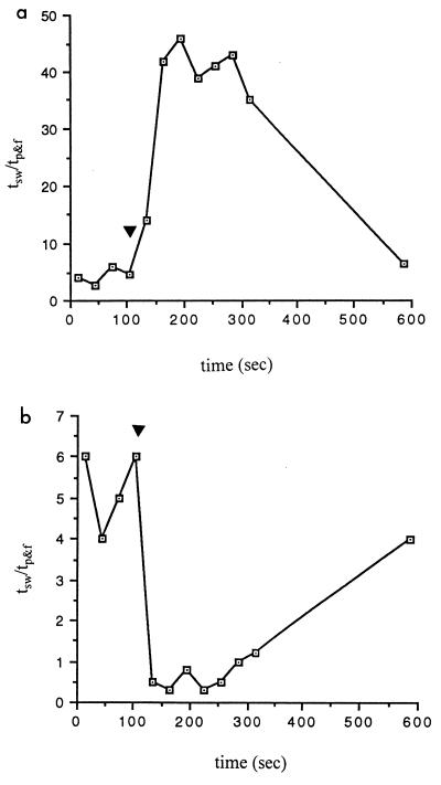
Videomicroscopy was used to study the cellular behavior in response to chemoattractants and repellents. Unstimulated cells swim forward and backward, flex, and pause. Usually a bacterium moving fast (>5 μm/s) swims longer (5 to 9 s) and then pauses and flexes less (1 to 2 s); a bacterium moving slowly (<5 μm/s) swims for a shorter period (3 to 5 s) and spends more time (2 to 4 s) pausing and flexing. Unstimulated bacteria usually spent 80 to 85% of the time for smooth swimming and 15 to 20% of the time for flexing and pausing (Fig. 1). After a chemoattractant (e.g., 1% rabbit serum) was added, the bacteria became almost exclusively smooth swimming, with rare reversing, pausing, or flexing (Fig. 1a). After a repellent (e.g., 100 mM ethanol) was added, the bacteria spent most of time flexing and pausing, with very little time spent smooth swimming (Fig. 1b). In both cases, after a certain period of time (depending on the strength of the stimulus), the bacteria adapted to the stimuli and gradually returned to the regular time ratio for swimming and flexing-pausing, a behavior called adaptation (Fig. 1).
FIG. 1.
Cellular behavioral change in response to a chemoattractant (1% young rabbit serum; a) and a chemorepellent (100 mM ethanol; b). Wild-type B31 cells were used; the testing chemical was added to bacterial suspension at s 120, as indicated by an arrowhead. The behavioral change in response to the chemicals was studied by videomicroscopy, recorded by a VCR, and analyzed frame by frame for the ratio of time spent swimming and pausing-flexing (tsw/tp&f). Each data point represents a time ratio in a 30-s window. Data shown represent the behavioral change of one representative cell; more than 20 cells were studied, and similar results were observed.
Chemotactic behavior of cells with a self-entangled end.
B. burgdorferi has one flagellar bundle at each cell end; the two bundles overlap in the cell center. The observed behavior of swimming, flexing, and pausing is the combined movement of both flagellar bundles. To understand how the cellular behavior is affected by rotation of flagella, it is important to study the behavior of a single flagellar bundle in response to chemoattractants or repellents. To this end, we used a newly developed method to produce spirochetes which contain only one active flagellar bundle.
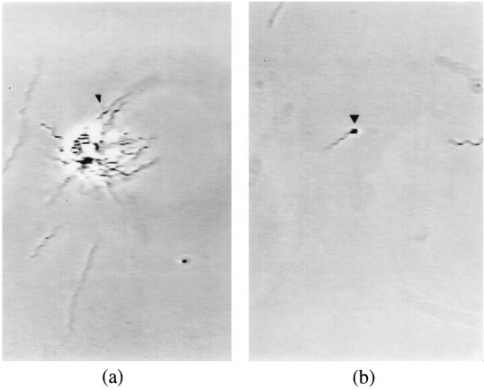
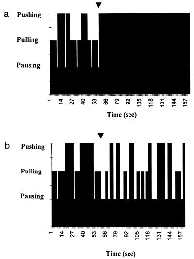
When B. burgdorferi was placed in a solution with a pH of >8.5, the cell surface seemed to become very sticky. When cells were resuspended at high density, many of them adhered to form large cell clumps (Fig. 2a). Cells in the clumps were still alive and motile. When cells were resuspended at low density, the ends of a small portion (∼1 to 5%) of them became self-entangled. Cells with both ends self-entangled were found to be nonmotile (data not shown), indicating that tangling of cell ends with the cell body prevents normal functioning of the flagellar bundle. Cells with one self-entangled end were studied (Fig. 2b). These cells have only one active flagellar bundle located at the nontangled end; the gyration of this free end indicates the direction of rotation of the single flagellar bundle. When the nontangled end rotates CCW (as viewed from the entangled end), it produces a short-wavelength helical cylinder and pulls the cell forward. When the free end rotates CW, it produces a long-wavelength helical wave and pushes the cell backward. We studied the chemotactic behavior of these cells. Without any stimulus, the free end rotated CCW (pulling cells forward) or CW (pushing cells backward) or stopped rotation (pausing). With addition of an attractant (e.g., 1% rabbit serum), the free end rotated exclusively CCW or CW with almost no pausing (Fig. 3a). When a repellent (e.g., 100 mM ethanol) was added, the free end was found to have much increased frequency of pausing, and the duration of each period of pausing was much longer than for unstimulated cells (Fig. 3b).
FIG. 2.
Cellular aggregation and self-entanglement of B. burgdorferi. (a) Wild-type B31 cells were grown in BSK II medium and resuspended at high cell density (OD605 = 0.1) in solution containing 10 mM NaH2PO4 (pH 9.0), 10−4 M EDTA, 0.15 M NaCl, and 10 mg of BSA per ml. The arrowhead indicates a large cell clump. (b) Wild-type B31 cells were resuspended at low cell density (OD605 = 0.005) in the buffer described above. The arrowhead indicates a cell with a self-entangled end.
FIG. 3.
Gyration of the nontangled cell end in response to a chemoattractant (1% young rabbit serum; a) and a chemorepellent (100 mM ethanol; b). Low-density bacteria were suspended in solution containing 10 mM NaH2PO4 (pH 9.0), 0.15 M NaCl, 10−4 M EDTA, and 10 mg of BSA per ml. After production of cells with a self-entangled end, the bacteria were resuspended in the same buffer at pH 7.6 and mixed with testing chemicals at s 60, as indicated by an arrowhead. The gyration of the nontangled end was studied by videomicroscopy, recorded by a VCR, and analyzed frame by frame for the time spent on pushing (indicating CW rotation), pulling (indicating CCW rotation), and pausing (indicating no rotation). Data shown represents the behavioral change of one representative cell; more than 20 cells were studied, and similar results were observed.
DISCUSSION
Unique cellular anatomy and distinctive motility enable spirochetes to move effectively in highly viscous bodily fluids. Therefore, it is likely that motility and chemotaxis of spirochetes play a role in pathogenesis. In this work, we studied the chemotactic behavior of B. burgdorferi, a known motile pathogenic spirochete. We found that B. burgdorferi did not exhibit chemotaxis toward sugars or amino acids. Instead, it moved toward serum, which could enable B. burgdorferi to move toward the bloodstream from tissues. B. burgdorferi avoided high concentrations of KCl, which could serve as a mechanism for B. burgdorferi to stay in the interstitial fluids (since there is a high concentration of KCl inside cells) and penetrate through the cellular junctions. B. burgdorferi tried to avoid hydrogen peroxide, a chemical released by neutrophils and macrophages to kill bacteria. This could serve as a protective mechanism for B. burgdorferi in its interaction with the immune system. It is also interesting that motility of B. burgdorferi requires a high concentration of NaCl and pH 7.6, which are the normal physiological conditions for interstitial fluids. In any case, our data suggest that chemotaxis may indeed be important for the pathogenesis of B. burgdorferi. What we report here is a classic example of chemotaxis very similar to those studied in other bacteria; however, through understanding the conditions for motility and chemotaxis of B. burgdorferi and the chemical nature of attractants and repellents, we learned a lot about how nature has modified similar systems for different purposes. In this case, the bacterium is one which is fully motile inside the human body and may use its chemotaxis mechanism to find target tissues.
Despite many similarities, there are some fundamental differences between the chemotaxis of E. coli and that of B. burgdorferi, which holds considerable fascination for those interested in questions regarding the motility and chemotaxis of bacteria. One of these questions is how chemotaxis is achieved at the cellular level. In the case of E. coli, chemotaxis is achieved by regulating the direction of rotation of the flagellar bundle. In response to attractants, E. coli flagella rotate CCW, which forms a bundle at the posterior end and pushes cells swimming ahead; in response to repellents, E. coli flagella rotate CW, which makes the bundle fall apart and bacteria tumble. Therefore, chemotaxis of E. coli is achieved by switching the direction of rotation of the flagellar bundles. However, for B. burgdorferi and other spirochetes, in order to swim forward or backward, the flagellar bundles at two ends of the cell have to rotate in different directions (6, 9); therefore, simply switching the rotation direction of flagellar bundles as in E. coli will not produce a similar effect. Instead, coordination of the two flagellar bundles seems to be important for achieving chemotaxis at the cellular level (11). The data presented on chemotaxis of B. burgdorferi are consistent with those of previous studies of chemotaxis in other spirochetes (11). In addition, the studies of B. burgdorferi cells with a self-entangled end suggested that the frequency and duration of pausing of the flagella are also important in the chemotactic response. In response to attractants, both of the flagellar bundles of a spirochete rotate without pausing, which could make them coordinate and swim in one direction. In response to repellents, the flagellar bundles pause frequently and extensively. This would disrupt the coordination between two flagellar bundles and cause cells to flex and pause, preventing cells from going into the repellent area.
ACKNOWLEDGMENTS
We thank N. W. Charon, S. Goldstein, H. Berg, P. E. Greenberg, N. H. Park, Jon T. Skare, and J. N. Miller for helpful discussions. We are grateful for the technical assistance of Steven Ruzin and Hans Holtan at the NSF Center of Plant Developmental Biology, University of California, Berkeley.
This work was supported by NIH grants GM54666 to W. Shi and AI-29733 to M. A. Lovett and training grant 5-T32-AI-07323 to Z. Yang.
REFERENCES
- 1.Adler J. A method for measuring chemotaxis and use of the method to determine optimum conditions for chemotaxis by Escherichia coli. J Gen Microbiol. 1973;74:77–91. doi: 10.1099/00221287-74-1-77. [DOI] [PubMed] [Google Scholar]
- 2.Adler J, Hazelbauer G L, Dahl D D. Chemotaxis toward sugars in Escherichia coli. J Bacteriol. 1973;115:824–847. doi: 10.1128/jb.115.3.824-847.1973. [DOI] [PMC free article] [PubMed] [Google Scholar]
- 3.Atsumi T, Maekawa Y, Tokuda H, Imae Y. Amiloride at pH 7.0 inhibits the Na(+)-driven flagellar motors of Vibrio alginolyticus but allows cell growth. FEBS Lett. 1992;314:114–116. doi: 10.1016/0014-5793(92)80954-f. [DOI] [PubMed] [Google Scholar]
- 4.Barbour A G. Isolation and cultivation of Lyme disease spirochetes. Yale J Biol Med. 1984;57:521–525. [PMC free article] [PubMed] [Google Scholar]
- 5.Berg H C. How spirochetes may swim. J Theor Biol. 1976;56:269–273. doi: 10.1016/s0022-5193(76)80074-4. [DOI] [PubMed] [Google Scholar]
- 6.Berg H C, Bromley D B, Charon N W. Leptospiral motility. Symp Soc Gen Microbiol. 1978;28:285–294. [Google Scholar]
- 7.Berg H C, Turner L. Movement of microorganisms in viscous environments. Nature (London) 1979;278:349–351. doi: 10.1038/278349a0. [DOI] [PubMed] [Google Scholar]
- 8.Charon N W, Goldstein S F, Block S M, Curci K, Ruby J D, Kreiling J A, Limberger R J. Morphology and dynamics of protruding spirochete periplasmic flagella. J Bacteriol. 1992;174:832–840. doi: 10.1128/jb.174.3.832-840.1992. [DOI] [PMC free article] [PubMed] [Google Scholar]
- 9.Charon N W, Greenberg E P, Koopman M B, Limberger R J. Spirochete chemotaxis, motility, and the structure of the spirochetal periplasmic flagella. Res Microbiol. 1992;143:597–603. doi: 10.1016/0923-2508(92)90117-7. [DOI] [PubMed] [Google Scholar]
- 10.Foley D M, Gayek R J, Skare J T, Wagar E A, Champion C I, Blanco D R, Lovett M A, Miller J N. Rabbit model of Lyme borreliosis: erythema migrans, infection-derived immunity, and identification of Borrelia burgdorferi proteins associated with virulence and protective immunity. J Clin Invest. 1995;96:965–975. doi: 10.1172/JCI118144. [DOI] [PMC free article] [PubMed] [Google Scholar]
- 11.Fosnaugh K, Greenberg E P. Motility and chemotaxis of Spirochaeta aurantia: computer-assisted motion analysis. J Bacteriol. 1988;170:1768–1774. doi: 10.1128/jb.170.4.1768-1774.1988. [DOI] [PMC free article] [PubMed] [Google Scholar]
- 12.Ge Y, Charon N W. An unexpected flaA homolog is present and expressed in Borrelia burgdorferi. J Bacteriol. 1997;179:552–556. doi: 10.1128/jb.179.2.552-556.1997. [DOI] [PMC free article] [PubMed] [Google Scholar]
- 13.Ge Y, Old I G, Girons I S, Charon N W. Molecular characterization of a large Borrelia burgdorferi motility operon which is initiated by a consensus ς70 promoter. J Bacteriol. 1997;179:2289–2299. doi: 10.1128/jb.179.7.2289-2299.1997. [DOI] [PMC free article] [PubMed] [Google Scholar]
- 14.Ge Y, Old I, Girons I S, Yelton D B, Charon N W. FliH and FliI of Borrelia burgdorferi are similar to flagellar and virulence factor export proteins of other bacteria. Gene. 1996;168:73–75. doi: 10.1016/0378-1119(95)00743-1. [DOI] [PubMed] [Google Scholar]
- 15.Glagolev A N, Skulachev V P. The proton pump is a molecular engine of motile bacteria. Nature (London) 1978;272:280–282. doi: 10.1038/272280a0. [DOI] [PubMed] [Google Scholar]
- 16.Goldstein S F, Charon N W, Kreiling J A. Borrelia burgdorferi swims with a planar waveform similar to that of eukaryotic flagella. Proc Natl Acad Sci USA. 1994;91:3433–3437. doi: 10.1073/pnas.91.8.3433. [DOI] [PMC free article] [PubMed] [Google Scholar]
- 17.Greenberg E P, Canale-Parola E. Chemotaxis in Spirochaeta aurantia. J Bacteriol. 1977;130:485–494. doi: 10.1128/jb.130.1.485-494.1977. [DOI] [PMC free article] [PubMed] [Google Scholar]
- 18.Guyton A C. Medical physiology. Philadelphia, Pa: The W. B. Saunders Co.; 1986. [Google Scholar]
- 19.Hovind Hougen K. Ultrastructure of spirochetes isolated from Ixodes ricinus and Ixodes dammini. Yale J Biol Med. 1984;57:543–548. [PMC free article] [PubMed] [Google Scholar]
- 20.Kimsey R B, Spielman A. Motility of Lyme disease spirochetes in fluids as viscous as the extracellular matrix. J Infect Dis. 1990;162:1205–1208. doi: 10.1093/infdis/162.5.1205. [DOI] [PubMed] [Google Scholar]
- 21.Mesibov R, Adler J. Chemotaxis toward amino acids in Escherichia coli. J Bacteriol. 1973;112:315–326. doi: 10.1128/jb.112.1.315-326.1972. [DOI] [PMC free article] [PubMed] [Google Scholar]
- 22.Sadziene A, Barbour A G, Rosa P A, Thomas D D. An ospB mutant of Borrelia burgdorferi has reduced invasiveness in vitro and reduced infectivity in vivo. Infect Immun. 1993;61:3590–3596. doi: 10.1128/iai.61.9.3590-3596.1993. [DOI] [PMC free article] [PubMed] [Google Scholar]
- 23.Sadziene A, Thomas D D, Bundoc V G, Holt S C, Barbour A G. A flagella-less mutant of Borrelia burgdorferi: structural, molecular, and in vitro functional characterization. J Clin Invest. 1991;88:82–92. doi: 10.1172/JCI115308. [DOI] [PMC free article] [PubMed] [Google Scholar]
- 24.Sadziene A, Thompson P A, Barbour A G. A flagella-less mutant of Borrelia burgdorferi as a live attenuated vaccine in the murine model of Lyme disease. J Infect Dis. 1996;173:1184–1193. doi: 10.1093/infdis/173.5.1184. [DOI] [PubMed] [Google Scholar]
- 25.Salyers A A, Whitt D D. Bacterial pathogenesis: a molecular approach. Washington, D.C: American Society for Microbiology; 1994. [Google Scholar]
- 26.Steere A C. Lyme disease. N Engl J Med. 1989;321:586–596. doi: 10.1056/NEJM198908313210906. [DOI] [PubMed] [Google Scholar]
- 27.Szczepanski A, Benach J. Lyme borreliosis: host responses to Borrelia burgdorferi. Microbiol Rev. 1991;55:21–34. doi: 10.1128/mr.55.1.21-34.1991. [DOI] [PMC free article] [PubMed] [Google Scholar]
- 28.Tso W, Adler J. Negative chemotaxis in Escherichia coli. J Bacteriol. 1974;118:560–576. doi: 10.1128/jb.118.2.560-576.1974. [DOI] [PMC free article] [PubMed] [Google Scholar]
- 29.Zhulin I B, Gibel I B, Ignatov V V. A rapid method for the measurement of bacterial chemotaxis. Curr Microbiol. 1991;22:307–310. [Google Scholar]


