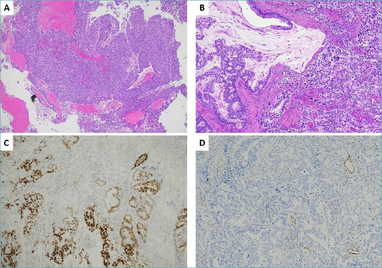
Figure 1.

Histological and immunohistochemical findings observed in the biopsy sample. (A) Low power view (x5) showing endometrial bioptic samples diffusely involved by a neoplastic proliferation with solid and glandular pattern of growth. (B) On higher magnification (x10), irregulary shaped, infiltrative mucinous glands were observed. (C) Neoplastic glands showed diffuse staining for CDX2. (D) Estrogen receptors was negative.
