Abstract
Mammalian vocalizations are critical for communication and are produced through the process of phonation, in which expiratory muscles force air through the tensed vocal folds of the larynx, which vibrate to produce sound. Despite the importance of phonation, the motor circuits in the brain that control it remain poorly understood. In this study, we identified a subpopulation of ~160 neuropeptide precursor Nts (neurotensin)-expressing neurons in the mouse brainstem nucleus retroambiguus (RAm) that are robustly activated during both neonatal isolation cries and adult social vocalizations. The activity of these neurons is necessary and sufficient for vocalization and bidirectionally controls sound volume. RAm Nts neurons project to all brainstem and spinal cord motor centers involved in phonation and activate laryngeal and expiratory muscles essential for phonation and volume control. Thus, RAm Nts neurons form the core of a brain circuit for making sound and controlling its volume, which are two foundations of vocal communication.
Subject terms: Neural circuits, Molecular neuroscience, Motor control, Social behaviour
The authors identify a cluster of ~160 peptidergic neurons in the mouse brainstem whose activity is necessary and sufficient for producing sound and controlling sound volume. These neurons form the final common pathway for vocalization.
Main
Animals communicate through a range of vocalizations. Many of these are innate, such as a laugh, a baby’s cry, a dog’s bark or a rodent’s distinct calls for its mother or a mate. A small number of bird and mammalian species can also produce learned vocalizations, such as human speech1. All types of vocalizations in mammals, both innate and learned, are generated by the larynx, an airway structure containing two vocal folds that, when tensed, vibrate and generate sound as air is moved across them. The vocal folds are brought together (adducted) and tensed by laryngeal muscles, and air is moved across them by abdominal muscles, which generate expiratory force during vocalization. This process of laryngeal sound production (phonation) is central to all mammalian vocalizations, including speech. As sound is produced, fine modulations of expiratory force, laryngeal muscle tension and timing of muscle contractions generate acoustic features, such as loudness (volume), pitch, syllable structure and syntax, which convey meaning to the listener. Despite the importance of phonation, little is known about the neuronal cell types and motor circuits controlling phonation or individual acoustic features.
The motor neurons controlling the laryngeal muscles reside in the nucleus ambiguus (Amb) in the medulla, whereas motor neurons controlling abdominal expiratory muscles reside in the thoracic spinal cord (Extended Data Fig. 1). Motor centers controlling other key aspects of phonation are located across the pons and medulla, in the trigeminal motor nucleus (mouth opening), in the ventral respiratory column (control of inspiration and expiration) and in the hypoglossal nucleus (tongue positioning). How are these disparate and distant motor output centers intricately coordinated during vocalization? Classical anatomical and physiological studies in cats and primates identified the midbrain periaqueductal gray (PAG) as a key center controlling innate vocalization2. PAG stimulation results in natural-sounding vocalizations, whereas PAG lesions cause mutism2. PAG vocalization-active neurons innervate many targets in the lower brainstem, including the nucleus retroambiguus (RAm)3, located caudal to Amb. Medullary transections at the level of RAm abolish PAG-driven vocalization in decerebrate cats4, suggesting that RAm is a critical relay node for innate vocalization. Chemical stimulation of RAm with a glutamate analog produces artificial-sounding vocalizations4, and bulk anterograde tracing studies from RAm found projections to the brainstem and spinal cord regions implicated in phonation5. Although these classical studies indicate an important role for RAm in vocalization, the neuronal cell types in this region and their circuitry and specific contributions to vocalization are unknown.
Extended Data Fig. 1. Schematic of brainstem regions involved in innate vocalization.

Innate vocalizations are generated (gated) by the midbrain periaqueductal gray (PAG) in the brainstem, which sends dense axonal projections to the nucleus retroambiguus (RAm, red) in the caudal medulla. Motor neuron pools (black) innervate the jaw opening muscles (trigeminal motor nucleus, MoV), laryngeal muscles (nucleus ambiguus, Amb), tongue muscles (hypoglossal motor nucleus, nXII), and abdominal expiratory muscles (thoracic ventral horn), which are all involved in vocalization. The ventral respiratory column (VRC, gray) contains inspiratory and expiratory control centers. RAm is hypothesized to control these distant motor pools and breathing centers to generate vocalization.
In this study, we employed neural activity mapping, optogenetics, behavioral experiments and neuroanatomical tracing in mice to genetically identify and characterize a RAm vocalization cell type. Although most mammals produce vocalizations that are audible to humans (<20 kHz), rodents can also produce ultrasonic vocalizations (>20 kHz) via a laryngeal whistle mechanism6. Because both audible7 and ultrasonic8 vocalizations are produced by using expiratory force to push air through the adducted larynx, we will use the term ‘phonation’ when referring to either human-audible or ultrasonic sound production. Here we show that a subpopulation of ~160 vocalization-activated RAm neurons express the neuropeptide precursor gene Nts (neurotensin). RAm Nts neurons are an excitatory subpopulation activated by both neonatal isolation cries and adult social vocalizations. Genetically targeted gain-of-function and loss-of-function studies show that RAm Nts neurons are necessary for adult social vocalization, sufficient for phonation in the audible and ultrasonic ranges, and their neural activity level determines the volume of the produced sound. RAm Nts neurons project to all motor pools involved in phonation and engage key laryngeal and expiratory muscles when activated. Our studies indicate that RAm Nts neurons define a critical brain circuit for phonation and volume control.
Results
Nts marks a subset of vocalization-activated neurons in RAm
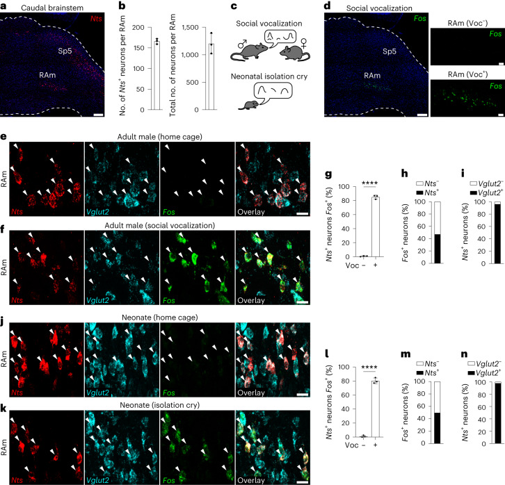
To identify genetic markers of vocalization neurons, we searched the Allen Mouse Brain Atlas9 for genes selectively expressed in RAm. One of the identified genes was Nts, which encodes the preproprotein for neuropeptides neurotensin and neuromedin N (ref. 10). Single-molecule fluorescence in situ hybridization (smFISH) (Fig. 1a) confirmed Nts expression in RAm as well as in the spinal trigeminal nucleus and a sparse population of motor neurons in the Amb9. Cell counts showed ~160 Nts-expressing neurons in RAm, with the Nts-expressing neurons clustered but intermingled with ~1,100 non-expressing neurons (Fig. 1b). To determine if RAm Nts neurons are activated during vocalization, we placed adult male mice with a female mouse to induce social vocalizations3 (Fig. 1c, top) and then examined activity of Nts and other RAm neurons with the neural activity marker Fos, allowing 90 min for Fos transcripts to accumulate. There were few, if any, Fos-expressing (active) neurons in RAm or surrounding regions in home cage male control mice, but there was robust induction of Fos in the RAm of vocalizing male mice (Fig. 1d). Probing of RAm sections for both Nts and Fos showed that RAm Nts neurons are activated during vocalization: ~80% of RAm Nts+ neurons expressed Fos in the vocalizing mice, whereas less than 1% expressed Fos in control mice (Fig. 1e–g). Nts+ neurons comprised ~45% of the Fos+ neurons in RAm, indicating that there is at least one other population of vocalization-activated RAm neurons, which is intermingled with and surrounds the Nts population (Fig. 1f,h). Over 95% of RAm Nts neurons expressed the excitatory marker Vglut2 (vesicular glutamate transporter 2) (Fig. 1e,f,i).
Fig. 1. Nts marks a vocalization-activated RAm subpopulation.
a, Caudal brainstem sagittal section of adult mouse with Nts mRNA labeled by smFISH (red). Note Nts expression in RAm and spinal trigeminal nucleus (Sp5). Scale bar, 200 µm. b, Left, number of Nts+ neurons in RAm (n = 3 mice). Right, number of neurons in RAm (n = 3 mice). c, Top, adult social vocalization. A female mouse placed with a male drives ultrasonic vocalizations by the male. Bottom, neonatal isolation cry. Removing a neonatal mouse from home cage drives ultrasonic isolation cries. d, Left, Fos mRNA labeling by smFISH (green, neural activity marker) in an adult male that vocalized to a female for 90 min. Note Fos expression restricted to RAm (magnified in right lower panel). Right upper panel, Fos labeling in RAm of adult male home cage control mouse that did not vocalize. Left scale bar, 200 µm. Right panel scale bars, 50 µm. e, smFISH labeling for Nts, Vglut2 and Fos in RAm of an adult male home cage control mouse. Note all RAm Nts+ neurons (arrowheads) express Vglut2 but do not express Fos. Scale bar, 15 µm. f, Identical smFISH labeling in RAm of adult male mouse that vocalized to a female. Note induction of Fos in RAm Nts+ neurons. Scale bar, 15 µm. g, Percent of RAm Nts+ neurons that express Fos in adult male mice under home cage control (Voc−) and vocalization (Voc+) conditions (P = 9 × 10⁻⁷, n = 3 mice per condition and 739 total scored Nts+ neurons). h, Percent of RAm Fos+ neurons in vocalizing adult male mice that also express Nts (n = 3 mice and 710 total scored Fos+ neurons). i, Percent of RAm Nts+ neurons that express Vglut2 in adult male mice (n = 3 mice and 739 total scored Nts+ neurons). j–n, Experimental scheme as in c–i except performed in neonatal mice that emitted isolation cries or home cage controls (P = 6 × 10⁻⁶, n = 3 mice per condition, 1,164 total scored Nts+ neurons and 1,115 total scored Fos+ neurons). Scale bars, 15 µm. Data are shown as mean ± s.d. ****P < 0.0001 by unpaired two-tailed t-test.
To determine if RAm Nts+ neurons are activated during other types of vocalization, we examined the activity of Nts neurons in neonatal mice induced to produce isolation calls by removal from their home cage litter11 (Fig. 1c, bottom). In control pups that remained in their home cage, only ~1% of RAm Nts neurons expressed Fos (Fig. 1j,l). However, in vocalizing pups removed from the home cage, ~80% of RAm Nts neurons expressed Fos (Fig. 1k,l). As during adult social vocalizations, Nts neurons comprised ~50% of the total Fos+ neurons in RAm (Fig. 1m), and more than 95% of the Nts neurons were Vglut2+ (Fig. 1n).
To test whether RAm Nts neurons are innervated by PAG neurons, we GFP labeled the caudolateral PAG with AAV-GFP in NtsCre:Ai14 adult male mice in which Nts neurons are labeled with tdTomato (Extended Data Fig. 2a,b). The adult male mice were induced to vocalize as above, and RAm was co-stained for c-Fos along with GFP and tdTomato. RAm Nts neurons (~80%) were innervated by PAG, showing that they receive direct input from this vocalization gating region (Extended Data Fig. 2c–e). c-Fos+Nts− neurons (~50%) were also innervated by PAG, showing that at least one other vocalization-active neuronal population (c-Fos+Nts−) in RAm is also directly engaged by PAG.
Extended Data Fig. 2. PAG innervation of RAm Nts+ and Nts- neurons.
a, Experimental schematic: AAV-GFP was injected into the caudolateral PAG of male mice to label projections of vocalization-gating neurons, and 3 weeks later male mice were induced to vocalize towards a female for 90 minutes before GFP and c-Fos immunostaining. b, Immunostaining for GFP and c-Fos in coronal brain slice containing caudal PAG following injection of AAV-GFP into left caudolateral PAG and subsequent vocalization induction. Note c-Fos expression across the PAG, and injection site targeting the left caudolateral PAG. Scale bar, 200 µm. c, Quantification of PAG innervation of RAm c-Fos+ neurons (n = 4 mice). d, Representative immunostaining image of RAm showing three c-Fos+ neurons (arrowheads) also labeled with Nissl (neuron-filling stain), tdTomato (NtsCre+ neurons), and GFP (PAG innervation). Note right NtsCre + /c-Fos+ neuron receiving PAG innervation (puncta), and two left NtsCre-/c-Fos+ neurons also receiving PAG innervation. Scale bar, 10 µm. e, Additional representative immunostaining image showing four c-Fos+ neurons (arrowheads). Note three top neurons are NtsCre + /c-Fos+ and receive PAG innervation, whereas bottom-right neuron is NtsCre-/c-Fos+ and does not receive PAG innervation. Scale bar, 10 µm. All data shown as mean ± S.D.
We conclude that Nts neurons are an excitatory subpopulation of RAm neurons that are robustly activated during both neonatal isolation cries and adult social vocalization, and they are directly innervated by PAG neurons.
Ablation of RAm Nts neurons abolishes social vocalizations
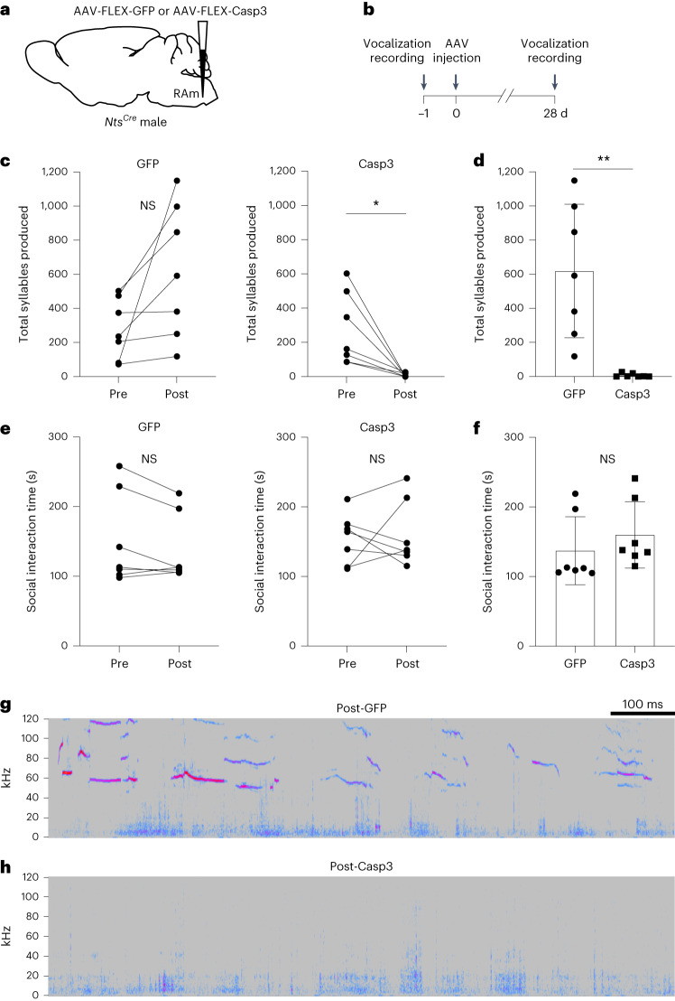
To test for function of RAm Nts neurons in vocalization, we genetically ablated them and examined the effect on social vocalizations. Adult male NtsCre mice were bilaterally injected in RAm with a Cre-dependent adeno-associated virus (AAV) vector encoding Caspase-3 (Flex-Casp3) (Fig. 2a,b), which causes Cre-dependent apoptosis12. To avoid targeting the sparse population of Nts-expressing motor neurons in the adjacent Amb, we used AAV serotype 8, which transduces interneurons but has poor tropism for motor neurons13. Comparing the number of residual RAm Nts+ neurons in Flex-Casp3-injected mice to that of Flex-GFP (mock ablation) controls indicated that this approach ablated nearly all (~90%) RAm Nts neurons, and ablation was specific for RAm Nts neurons (Extended Data Fig. 3). Comparison of social vocalizations in the same animals before and after RAm Nts ablation showed that their ablation almost completely abolished vocalization (Fig. 2c,d,h), whereas mock ablation (Flex-GFP injection) had no effect (Fig. 2c,d,g). The effect of ablation on vocalization was not due to decreased activity or interest in the female, because Flex-Casp3 mice spent the same amount of time interacting with the female (Fig. 2e,f) and in a similar manner (Supplementary Videos 1 and 2) before and after RAm Nts ablation and compared to Flex-GFP control mice. We conclude that RAm Nts neurons are required to produce adult male-to-female social vocalizations, and their role is specific for vocalization.
Fig. 2. Effect of RAm Nts neuron ablation on social vocalizations.
a, RAm Nts neurons were ablated by injecting Cre-dependent AAV encoding GFP (AAV-FLEX-GFP, mock ablation control) or apoptosis inducer Casp3 (AAV-FLEX-Casp3, ablation condition) in adult male NtsCre mice. b, Experimental paradigm. Female-induced vocalizations from male NtsCre mice were recorded in a 5-min trial. One day later (day 0), AAV encoding Cre-dependent GFP or Casp3 was bilaterally injected into RAm of male NtsCre mice. After 28 d (to allow protein expression and cell ablation), the NtsCre mice were induced to vocalize during an identical 5-min trial with a female. c, Syllables produced during 5-min female encounters before (Pre) and after (Post) injection of Cre-dependent GFP (left) or Casp3 (right) (n = 7 mice per group). Note abolishment of vocalizations in Casp3 (ablation) (P = 0.018) condition. Trend toward increase in syllables in GFP (control) condition (P = 0.053) could be due to increased motivation to vocalize after 28-d social isolation between trials. d, Total syllables produced after GFP or Casp3 expression in RAm Nts neurons (P = 0.0014, n = 7 mice per group). e, Social interaction time during 5-min female encounters before (Pre) and after (Post) injection of Cre-dependent GFP (left) or Casp3 (right) (n = 7 mice per group). f, Social interaction time after GFP or Casp3 expression in RAm Nts neurons (P = 0.4, n = 7 mice per group). g, Sonogram of female-induced ultrasonic vocalizations by control NtsCre mouse 28 d after AAV-FLEX-GFP injection into RAm. y axis, frequency (kHz). Warmer colors indicate increased sound amplitude at given frequency. Note complex ultrasonic (>20 kHz) syllables. Audible sounds (<20 kHz) are background noises from mouse movement. h, Sonogram of female-induced ultrasonic vocalizations emitted by an NtsCre mouse 28 d after AAV-FLEX-Casp3 injection into RAm. Note absence of ultrasonic vocalizations. Data are shown as mean ± s.d. *P < 0.05, **P < 0.01 and NS, not significant, by paired (Pre versus Post) or unpaired (GFP versus Casp3) two-tailed t-test. Mouse brain schematic here and those in Figs. 3, 4, 5 and 6 and Extended Data Figs. 1, 2 and 5 are reproduced from ref. 33.
Extended Data Fig. 3. Casp3-mediated ablation of RAm Nts neurons.
a, b, smFISH labeling of Nts in brainstem of NtsCre mice injected in RAm with AAV-FLEX-GFP (a, mock ablation) or AAV-FLEX-Casp3 (b, ablation condition) for RAm Nts neuron ablation experiments (see Fig. 2). Insets, magnified view of Sp5 and RAm regions. Note ablation of RAm Nts+ neurons in Casp3 mice with sparing of Sp5. Scale bars, 200 µm. Inset scale bars, 50 µm. c, Percent of RAm Nts+ neurons remaining following AAV-FLEX-Casp3 injection relative to GFP mock ablation controls. n = 868 total RAm Nts neurons scored in 4 mice per condition. d, e, smFISH of Nts+ subpopulation of Amb motor neurons in GFP (d) and Casp3 (e) conditions above. Red arrowheads, Nts+ Amb motor neurons, marked by expression of Vacht (vesicular acetylcholine transporter). White arrowheads, Nts- Amb motor neurons. Note sparing of Amb Nts+ neurons in Casp3 condition, indicating AAV8 approach was selective for RAm interneurons and avoided nearby motor neurons. Scale bars, 25 µm. All data shown as mean ± S.D.
Activation of RAm Nts neurons produces vocalization
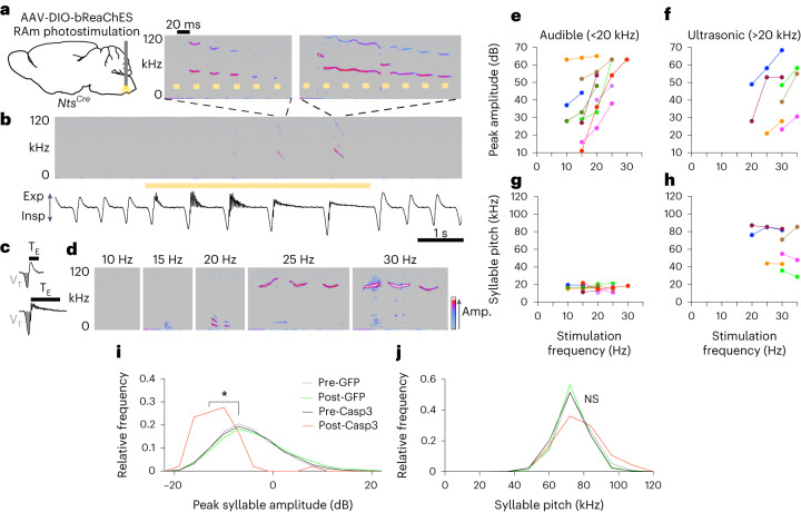
To determine if RAm Nts neuronal activity is sufficient to produce vocalization, we optogenetically activated them in anesthetized mice. A Cre-dependent AAV8 vector encoding the channelrhodopsin bReaChES was bilaterally delivered to the RAm of adult NtsCre mice, and fiber-optic cannulas were implanted above the injection sites (Fig. 3a). After AAV injection into RAm, ~70% of Nts+ neurons in the region expressed bReaChES-eYFP, and this labeling was selective for RAm Nts+ interneurons (Extended Data Fig. 4a–d). Two to four weeks after optical fiber implantation, mice were anesthetized with isoflurane and placed in a nose cone fitted with a spirometer and an ultrasonic microphone to simultaneously measure breathing and vocalization. Upon optogenetic stimulation of RAm Nts neurons, the anesthetized mice emitted vocalizations (Fig. 3b). They also showed related changes in breathing, as detailed below, but otherwise remained fully quiescent. During a single stimulation train, the inspiratory volume and expiratory time progressively increased with each breath (Fig. 3b,c and Extended Data Fig. 4e). Simultaneously, the airflow trace began oscillating rapidly but only during expiration, suggestive of airflow instabilities6 or vocal fold vibration (Fig. 3b and Extended Data Fig. 4f). During the largest breaths in the stimulation train, vocalization was produced. Although these optogenetically induced vocalizations occupied a similar fundamental frequency range as natural mouse ultrasonic vocalizations (30–110 kHz)11, their spectrographic form did not resemble that of any natural vocalization. Rather, the spectrogram reflected features of the laser pulses, with repeating spectral motifs time-locked to each pulse (Fig. 3b). Optogenetic stimulation did not alter heart rate (Extended Data Fig. 4g), which is powerfully regulated by neurons intermingled with RAm14,15, confirming that RAm Nts neurons had been specifically targeted. Optogenetic stimulation in awake, freely moving mice also induced vocalizations (Extended Data Fig. 5a,b). We conclude that optogenetic stimulation of RAm Nts neurons is sufficient to generate artificial vocalizations.
Fig. 3. Optogenetic activation of RAm Nts neurons generates vocalizations and controls sound volume.
a, RAm Nts neuron optogenetic stimulation by bilateral injection of Cre-dependent AAV encoding bReaChES in NtsCre mice, with laser light delivered via optical fiber. b, Respiratory airflow (bottom) and sonogram (top) during optogenetic stimulation of RAm Nts neurons (yellow bar, 30-Hz laser) in an anesthetized mouse. Inspiration (Insp), downward deflections on airflow trace; expiration (Exp), upward deflections. Note ultrasonic vocalizations of increasing volume and duration during optogenetic stimulation, accompanied by breathing changes. Insets (above), induced vocalizations expanded to millisecond timescale. Note ultrasonic vocalizations with motifs that repeat at the same rate as stimulus pulse. c, Airflow trace of single breaths before (top) and during (bottom) optogenetic stimulation of RAm Nts neurons. Note that tidal volume (VT, shaded area) and expiration time (TE) increase during optogenetic stimulation. d, Effect of pulse frequency on vocalizations generated by optogenetic stimulation of RAm Nts neurons in an anesthetized NtsCre mouse. Note lack of vocalization with 10-Hz stimulation, audible (<20 kHz) vocalizations generated with increasing volume from 15 Hz to 20 Hz and ultrasonic (>20 kHz) vocalizations generated with increasing volume from 25 Hz to 30 Hz. Amp., sound amplitude. e,f, Peak syllable amplitude of optogenetically driven vocalizations (n = 9 mice, colored lines) in audible (e) and ultrasonic (f) frequency ranges. Note monotonic increase in peak syllable amplitude of audible vocalizations with increasing stimulation frequency in all mice and similar monotonic increase in ultrasonic vocalization amplitude in a subset of mice that produced ultrasonic vocalizations. g,h, Syllable pitch for the same optogenetic stimulation trials as e,f. Note that syllable pitch does not change. i, Histograms of peak syllable amplitude of all vocalizations generated during RAm Nts neuron ablation experiments (Fig. 2) (n = 7 mice per condition and n = 8,310 total syllables analyzed). Note decrease in peak syllable amplitude of residual syllables after Casp3-mediated ablation of RAm Nts neurons (P = 0.02, Pre-Casp3 versus Post-Casp3). j, Histograms of syllable pitch for the same syllables as i. Note that syllable pitch does not change after RAm Nts ablation (P = 0.7 Pre-Casp3 versus Post-Casp3, n = 7 mice per condition and n = 8,310 total syllables analyzed). *P < 0.05 and NS, not significant, by paired two-tailed t-test.
Extended Data Fig. 4. Expression of bReaChES-eYFP in RAm Nts neurons and stimulation-induced changes in respiration and heart rate.
a, Sagittal section immunostaining of NtsCre;tdTomato mouse caudal medulla following AAV-DIO-bReaChES-eYFP injection into RAm and fiber optic implantation. Note eYFP expression in RAm but not Sp5. tdTomato expression in brain vasculature (bright red streaks) was only observed after lineage labeling, presumably from transient embryonic expression since postnatal vascular Nts expression was not observed with Nts smFISH (Fig. 1) or by in situ hybridization in the Allen Brain Atlas9. Scale bar, 100 µm. b, Quantification of percent of RAm Nts neurons targeted by bReaChES-eYFP (n = 384 total Nts+ neurons scored in 3 mice). c, Immunostaining of RAm Nts neurons expressing bReaChES-eYFP (arrowheads) in NtsCre;tdTomato mouse. Scale bar, 25 µm. d, Immunostaining of Amb Nts+ neurons in NtsCre;tdTomato mouse injected with AAV-DIO-bReaChES-eYFP. Note lack of eYFP expression in ChAT+ (Amb) neurons (arrowheads) or in vasculature (bright red), indicating AAV8 approach was selective for RAm interneurons and avoided nearby motor neurons. Scale bar, 25 µm. e, Respiratory effects of RAm Nts optogenetic stimulation. Note progressive increase in tidal volume and expiratory time with increasing optogenetic stimulation frequency. f.c., fold change from pre-stimulation baseline (n = 3 mice). f, Magnified view of airflow oscillations and their temporal relationship to ultrasonic vocalizations elicited by RAm Nts optogenetic stimulation. Note expiratory airflow oscillations during ultrasonic vocalization production, and dampening of oscillations as syllable volume progressively increases. g, Respiration airflow (top) and heart rate (HR, bottom) traces during optogenetic stimulation (yellow bar, 30 Hz) of RAm Nts neurons as in Fig. 3. Note lack of heart rate response during optogenetic stimulation, indicating AAV8 approach and NtsCre driver was selective to RAm interneurons and avoided nearby cardiac control neurons.
Extended Data Fig. 5. Optogenetic stimulation of RAm Nts neurons produces vocalization in awake mice.

a, Schematic of optogenetic stimulation paradigm. b, Sonogram during optogenetic stimulation of RAm Nts neurons (yellow bar, 20 Hz laser light) in awake mouse. Note ultrasonic vocalizations produced within stimulation period (top, magnification). c, d, Quantification of peak syllable amplitude of optogenetically-driven vocalizations (n = 4 mice) in the audible and ultrasonic frequency ranges. Colored lines, individual mice. Note monotonic increase in all mice in peak syllable amplitude of audible vocalizations (left panel) with increasing stimulation frequency, and similar monotonic increase in ultrasonic vocalization amplitude (right panel) for mice that transitioned to ultrasonic. e, f, Quantification of syllable pitch for same audible and ultrasonic optogenetic stimulation trials as c,d. Note lack of change in syllable pitch for individual mice with increasing stimulation frequency.
RAm Nts neuronal activity controls sound volume
To investigate acoustic features regulated by RAm Nts neuronal activity, we systematically altered the laser pulse frequency of RAm Nts optogenetic activation in anesthetized mice. A distinct minimum pulse frequency was required to elicit vocalization in different mice. At this threshold, low-intensity vocalizations were detected in the audible frequency range (<20 kHz fundamental frequency) (Fig. 3d,e). As pulse frequency was increased, the loudness of the audible vocalizations increased monotonically in all mice (Fig. 3e and Extended Data Fig. 6a), whereas pitch remained unchanged (Fig. 3g and Extended Data Fig. 6b). Then, above a second threshold of pulse frequency, the induced vocalizations transformed from audible to ultrasonic (>20 kHz fundamental frequency) (Fig. 3d,f and Extended Data Fig. 6e). When these ultrasonic vocalizations could be elicited with multiple pulse frequencies, their loudness also increased with pulse frequency (Fig. 3f and Extended Data Fig. 6c), and, likewise, there was no consistent change in pitch (Fig. 3h and Extended Data Fig. 6d). Similar effects were observed when laser power was increased with a constant pulse frequency (Extended Data Fig. 6f,g). Increasing the stimulation rate in awake, freely moving mice also increased loudness without altering pitch (Extended Data Fig. 5c–f). Thus, the neural activity level of RAm Nts neurons controls the loudness of vocalizations as well as the transition from audible to ultrasonic, but it does not regulate pitch within the audible or ultrasonic regime.
Extended Data Fig. 6. Additional quantification of relationship between optogenetic stimulation frequency and acoustic features in anesthetized mice.
a, Relationship between RAm Nts optogenetic stimulation frequency and audible vocalization syllable amplitude. Data from Fig. 3e was aligned to the vocalization threshold (‘0’ on X axis indicates lowest stimulation frequency at which vocalization was elicited). Note progressive increase in peak amplitude with increasing stimulation frequency. b, Relationship between RAm Nts optogenetic stimulation frequency and audible vocalization syllable pitch for same syllables as panel a. Note lack of change in syllable pitch with increasing stimulation frequency. c, Relationship between RAm Nts optogenetic stimulation frequency and ultrasonic vocalization syllable amplitude. Data from Fig. 3f was aligned to the vocalization threshold. Note progressive increase in peak amplitude with increasing stimulation frequency. d, Relationship between RAm Nts optogenetic stimulation frequency and ultrasonic vocalization syllable pitch for same syllables as panel c. Note lack of change in syllable pitch with increasing stimulation frequency. e, Ultrasonic transition points of mice that transitioned from audible to ultrasonic vocalization with RAm Nts optogenetic stimulation. y axis indicates percent of optogenetically-driven vocalizations in the ultrasonic range (>20 kHz) at each stimulation frequency. Colored lines, individual mice (n = 6 mice). Black line, mean across all mice. Note most mice transitioned from audible to ultrasonic vocalization between 20 Hz and 30 Hz optogenetic stimulation. f, g, Quantification of peak syllable amplitude (left) and syllable pitch (right) of audible vocalizations elicited by RAm Nts optogenetic stimulation at different laser powers, while maintaining a constant pulse rate (25 Hz). Note increase in syllable loudness as laser power was increased, but lack of change in syllable pitch. Colored lines, individual mice (n = 4 mice). All r2 and p values in panels a-d calculated from simple linear regression. *: p < 0.05, **: p < 0.01, n.s.: not significant.
To further investigate the relationship between RAm Nts neurons and sound volume, we examined the few remaining syllables in the RAm Nts neuron-ablated animals described above. Here, too, we observed the same relationship between RAm Nts neural activity and sound volume: ablation of RAm Nts neurons greatly reduced the loudness of the remaining syllables relative to that of control, pre-ablation syllables (Fig. 3i), whereas syllable pitch remained unchanged (Fig. 3j). Thus, both gain-of-function and loss-of-function studies support that RAm Nts neurons control sound volume, and the relationship is monotonic and spans the full range from silence to loud social vocalizations.
RAm Nts neurons control laryngeal and expiratory muscles
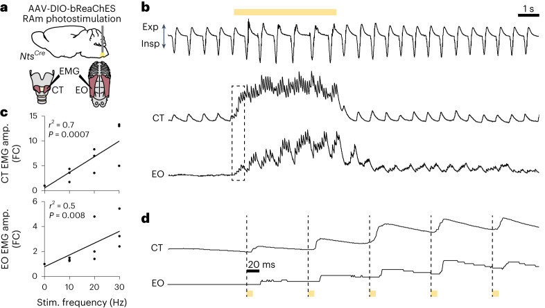
To investigate the mechanism by which RAm Nts neurons produce sound and control its volume, we monitored the activity of two key vocalization muscles by electromyogram (EMG) recording while optogenetically stimulating RAm Nts neurons. One of the muscles was the cricothyroid (CT), a key laryngeal muscle contributing to laryngeal adduction, vocal fold tension and vocal pitch16. The other was the external oblique (EO), a major abdominal muscle that generates expiratory force during vocalization and contributes to sound volume17. RAm Nts neurons were optogenetically stimulated under isoflurane-anesthetized conditions as above while EMG was simultaneously recorded from both muscles (Fig. 4a). During eupneic breathing, the CT demonstrated a low level of rhythmic inspiratory activity, consistent with previous EMG studies18, whereas the EO remained silent, also consistent with previous work19. Optogenetic stimulation of RAm Nts neurons drove rapid and stimulation rate-dependent increases in EMG amplitude of both CT and EO (Fig. 4b,c), indicating coordinated contraction of both muscles. CT muscle activity rapidly returned to baseline levels after cessation of stimulation, whereas the EO muscle had slower off-kinetics, gradually returning to baseline voltage over several seconds (Fig. 4b). Examining the EMG traces at the millisecond timescale (Fig. 4d) revealed that single laser pulses elicited coordinated EMG spikes in the CT and EO muscles with high fidelity and low latency. However, CT had a slightly shorter latency (~8 ms) than EO (~20 ms), a difference that we explore further below. Thus, optogenetic activation of RAm Nts neurons drives coordinated contraction of key laryngeal and expiratory muscles involved in phonation and volume control.
Fig. 4. Optogenetic activation of RAm Nts neurons drives high-fidelity, short-latency spikes in laryngeal and expiratory muscles.
a, Schematic of optogenetic stimulation with EMG recording. NtsCre mice were bilaterally injected with AAV-DIO-bReaChES into RAm. An optical fiber delivered yellow laser light into RAm while EMG was simultaneously recorded from laryngeal (CT) and expiratory (EO) muscles and airflow by a spirometer. b, Airflow trace (top), integrated amplitude EMG trace for CT (middle) and EO (bottom) during optogenetic stimulation of RAm Nts neurons (yellow bar, 10 Hz). Note rhythmic inspiratory activity of CT muscle and lack of EO activity during eupneic (normal) breathing before stimulus and then increases in CT and EO EMG activity during optogenetic stimulation, followed by gradual return to pre-stimulation breathing and EMG patterns. c, Quantification of integrated EMG amplitude (amp.) fold change (FC) from pre-stimulation period for CT (top) and EO (bottom) muscles during 0-Hz, 10-Hz, 20-Hz and 30-Hz optogenetic stimulation (n = 3 mice). Note progressive increase in EMG amplitude of both muscles with increasing stimulation frequency. CT: P = 0.0007 and EO: P = 0.008 by linear regression. d, Magnified traces from dashed boxed region in b during optogenetic stimulation pulses (yellow bars). Note short-latency, high-fidelity EMG spikes in both muscles after an optogenetic stimulation pulse but slightly longer latency of EO (~20 ms) versus CT (~8 ms) activation.
RAm Nts neurons innervate laryngeal and respiratory neurons
To determine if RAm Nts neurons innervate CT motor neurons, we labeled and mapped their axons and synapses. NtsCre mice were injected with a Cre-dependent AAV encoding mGFP and a synaptophysin–mRuby fusion protein, which labeled RAm Nts axons with GFP and putative pre-synaptic terminals with mRuby (Fig. 5a). CT motor neurons were retrogradely labeled by cholera toxin B (CTB) (Fig. 5a). RAm Nts neurons bilaterally and directly innervated almost all CT motor neurons (Fig. 5b,c), consistent with the short-latency activation observed for this muscle after optogenetic stimulation.
Fig. 5. Projection targets of RAm Nts neurons.
a, RAm Nts neuron projection mapping. NtsCre mice were injected in RAm with the AAV indicated for Cre-dependent labeling of axons (mGFP) and putative synapses (mRuby). After AAV injection, retrograde tracer CTB was injected into CT and EO muscles to label their motor neurons. b, Immunostaining of retrogradely labeled (CTB+) CT motor neurons in Amb (top) and EO motor neurons in thoracic ventral horn (VH) (bottom) in NtsCre mice injected in RAm with the AAV. RAm Nts neurons (mGFP+ and mRuby+) directly innervate (red puncta) CT (arrowheads) but not EO motor neurons. Scale bars, 20 μm. c, CT and EO motor neuron innervation by ipsilateral (left bars) and contralateral (right bars) RAm Nts neurons (CT: n = 3 mice; EO: n = 4 mice). d, Sagittal section immunostaining of contralateral ventral respiratory column (VRC) after mGFP labeling of RAm Nts fibers as above. Note selective projection of RAm Nts neurons to VRC, comprising RTN/parafacial respiratory group, BötC, pre-BötC and rVRG, plus sparse innervation of pontine reticular formation (RF). Scale bar, 100 µm. e, Sagittal section immunostaining of contralateral trigeminal motor nucleus (MoV) as above. Note innervation of a subset of MoV, with dense innervation of supratrigeminal nucleus (SupV) and parabrachial (PB)/Kӧlliker–Fuse nuclei. Inset, boxed region of MoV. Scale bar, 100 µm. f, Sagittal section immunostaining of contralateral hypoglossal motor nucleus (nXII) as above. Note innervation of nXII. 10N, dorsal motor nucleus of vagus; Sol, nucleus of the solitary tract. Scale bar, 100 µm. g,h, Quantification of mGFP+ innervation density of RAm Nts neurons across brainstem (g, n = 3 mice) and spinal cord (h, n = 3 mice) regions. DDH, deep dorsal horn; IZ, intermediate zone; SDH, superficial dorsal horn. i, Transverse section immunostaining of cervical, thoracic, lumbar and sacral spinal cord after mGFP labeling of RAm Nts axons. Note RAm axons descending contralaterally through the dorsal corticospinal tract (middle dashed outline) and lateral spinal nucleus (right dashed outline), with extensive arborization in cervical and thoracic DDH, IZ and VH. Scale bar, 500 µm. Data are shown as mean ± s.d. Contra, contralateral; Ipsi, ipsilateral.
In addition to their innervation of CT motor neurons, ascending projections from RAm Nts neurons densely innervated breathing control regions across the brainstem, including the ventral respiratory column (retrotrapezoid nucleus (RTN)/parafacial respiratory group, Bötzinger complex (BӧtC), pre-Bӧtzinger complex (pre-BӧtC) and rostral ventral respiratory group (rVRG)) as well as the parabrachial/Kӧlliker–Fuse nuclei (Fig. 5d,e,g). RAm Nts neurons also innervated a subpopulation of trigeminal (jaw) motor neurons (Fig. 5e,g) and hypoglossal (tongue) motor neurons (Fig. 5f,g) but spared brainstem nuclei not known to be involved in vocalization, such as the nucleus ambiguus compact formation (AmbC, esophageal motor neurons) and the dorsal motor nucleus of vagus (10N) (Fig. 5d,f). Thus, ascending projections from RAm Nts neurons directly innervate the breathing and orofacial motor regions involved in phonation.
RAm Nts neurons project to spinal cord expiratory centers
To determine the spinal cord projection targets of RAm Nts neurons, we labeled their axons and synapses with mGFP and mRuby as above and mapped their projections in the spinal cord. RAm Nts axons primarily descended contralaterally along two distinct spinal cord tracts: the dorsal corticospinal tract and a second tract in the lateral spinal nucleus (Fig. 5i). At the cervical and thoracic levels, which control breathing muscles, axons arborized and innervated spinal gray matter across the deep dorsal horn, intermediate zone and ventral horn (Fig. 5h,i), a distribution similar to abdominal muscle pre-motor neuron distributions20. Unlike classical studies of RAm innervation using bulk tracing methods5, we found that RAm Nts innervation declined precipitously at the lumbar and sacral levels (Fig. 5h,i), which control hindlimb and pelvic muscles.
To determine if RAm Nts neurons directly innervate EO motor neurons, we retrogradely labeled EO motor neurons with CTB. In contrast to CT motor neurons, RAm Nts neurons rarely innervated EO motor neurons (<10%) (Fig. 5b,c). However, they extensively innervated nearby interneurons, suggesting indirect innervation involving local microcircuits, consistent with the longer latency activation and slower off-kinetics observed above for the EO muscle after RAm Nts optogenetic stimulation. We conclude that RAm Nts neurons send descending projections to spinal cord segments controlling expiratory muscles, but, unlike their direct projection to CT motor neurons, they project indirectly to EO motor neurons.
Discussion
We identified and characterized RAm Nts neurons, revealing a neural circuit for phonation (Fig. 6). RAm Nts neurons are an excitatory subpopulation of ~160 neurons located near the spinomedullary junction, an ideal position to coordinate phonation. They are robustly activated by both neonatal isolation cries (aversive context) and adult social vocalizations (appetitive context), showing that they are involved in a broad range of, and potentially all, vocalizations. Ablation of RAm Nts neurons in adult mice abolishes adult male-to-female social vocalizations, whereas optogenetic activation generates artificial vocalizations that reflect features of the stimulus. RAm Nts neurons produce sound by recruiting two key vocalization muscles: the CT, which adducts/tenses the vocal folds and is recruited through direct RAm Nts projections, and the EO, which generates expiratory force and is recruited through indirect projections. The indirect projection to EO provides a ~12-ms delay in activation compared to CT, ensuring that the vocal folds are fully adducted and tensed before subglottal pressure is generated by the EO. In addition to the CT and EO projections, RAm Nts neurons project to all breathing and orofacial nuclei that generate the other key components of phonation: mouth opening (MoV), tongue positioning (nXII), increased tidal volume (pre-BӧtC, rVRG and cervical spinal cord) and increased expiration time (BӧtC and RTN). Thus, the RAm Nts circuit appears to be the core neural circuit for phonation.
Fig. 6. Summary of RAm Nts neuron innervation and function.

RAm Nts neurons (red) innervate the trigeminal motor nucleus (MoV), which contains jaw opening motor neurons; the ventral respiratory column (VRC) and cervical spinal cord, which increase inspiratory (tidal) volume and expiratory time; Amb CT motor neurons, which adduct and tense the vocal folds; and the hypoglossal nucleus (nXII), which positions the tongue. RAm Nts neurons generate expiratory force through their indirect innervation of EO motor neurons in the spinal cord, possibly through a local interneuron (gray). All the above functions combine to produce sound (phonation). In addition to generating phonation, the RAm Nts neuronal activity level (red gradient triangle) determines the volume of the produced sound (black gradient triangle) by adjusting abdominal expiratory pressure via the EO muscle. RAm Nts neurons receive input from the midbrain periaqueductal gray and probably from other brain regions (not shown) to initiate phonation and adjust volume during diverse vocalizations.
In addition to phonation, RAm Nts neurons also control a key acoustic feature of vocalization: sound volume. Gain-of-function and loss-of-function studies show that RAm Nts neural activity monotonically determines the volume of the produced sound at audible and ultrasonic frequencies, spanning the full range from silence to loud social vocalizations. Our respiration and EMG data suggest a simple mechanism by which RAm Nts neurons increase sound volume. Increasing RAm Nts neuronal activity increases tidal volume, which increases the elastic recoil pressure of the lungs and vocal loudness17. Increasing RAm Nts neuronal activity also increases activity of the EO muscle, a muscle known to increase loudness by increasing expiratory force and subglottal pressure17. Our results are in agreement with a recent single-unit recording study in the rat RAm, which identified a subpopulation of RAm neurons with a tonic activity pattern that was highly correlated with vocal loudness21. Our study also complements a recent study that showed that hypothalamic lateral preoptic area neurons expressing estrogen receptor 1 can scale the volume and bout length of adult male social vocalizations22. RAm Nts neurons may provide a downstream or parallel mechanism to adjust sound volume, because they are activated in multiple vocalization contexts.
RAm Nts neuronal activity also controls the transition from audible to ultrasonic vocalization. Rodent ultrasonic vocalizations are thought to be produced by a whistle mechanism generated by a glottal air jet impinging on the thyroid inner wall23. Critical levels of laryngeal adduction and subglottal pressure are necessary to generate ultrasonic vocalization through this mechanism23. Our data suggest that a critical level of RAm Nts neuronal activity is necessary to reach threshold laryngeal adduction and subglottal pressure levels and convert audible vocalizations to ultrasonic.
How does the brain control the other acoustic features of innate vocalization, such as pitch, syllable structure and syntax? These features are thought to be controlled within the pons and medulla, where neurons tuned to some of these features have been identified in multiple regions, including RAm24,25. Artificial stimulation of the PAG generates natural-sounding vocalizations3, consistent with acoustic features being controlled by downstream structures. In contrast, RAm Nts neuron stimulation produced artificial vocalizations, suggesting that cell types downstream of PAG in addition to RAm Nts neurons are required to generate natural vocalizations. Notably, RAm Nts neurons comprise only ~45% of the vocalization-activated RAm neurons, and some of the other neurons are also directly innervated by PAG. Thus, there is at least one other RAm vocalization subpopulation. These might control acoustic features other than volume and may be necessary along with RAm Nts neurons to create natural-sounding vocalizations.
The function of RAm appears to be conserved across vocalizing vertebrates4,24,26, suggesting that the RAm Nts circuit may also be conserved. RAm is present in the avian brain and is thought to be the final common pathway for both innate calls and learned songs27. The avian RAm possesses a remarkably similar circuit architecture to the mammalian RAm Nts circuit, containing pre-motor neurons that innervate the vocal motor neurons, abdominal expiratory neurons and breathing control nuclei28. Additionally, RAm is present in all mammalian species examined, including rodents, non-human primates24 and humans29. As was found in our study, studies in primates have found that RAm contains a dense concentration of neurons activated by diverse innate vocalizations24. Hence, it is possible that RAm mediates human innate vocalizations, such as laughter and crying, and it will be important to determine if it harbors a homolog of the mouse Nts circuit that provides the core phonation drive for these vocalizations. Projections from human motor cortex innervate the RAm region as well as Amb30, suggesting that RAm may be engaged to generate phonation during human speech while parallel human-specific projections to Amb31 generate fine pitch adjustments. Similar circuit architectures that use brainstem pre-motor modules have been identified in other skilled motor control circuits32. Engagement of the RAm Nts module to generate the basic phonation drive for diverse vocalizations while controlling their volume would explain the strong conservation of RAm across vocalizing species.
Methods
Mice
Wild-type C57BL/6NCrl mice (Charles River Laboratories, strain 027), NtsCre knock-in mice34 (Jackson Laboratory, strain 017525) and Ai9 or Ai14 tdTomato mice35 (Jackson Laboratory, strain 007909) were housed and bred in the animal facility at Stanford University in accordance with Institutional Animal Care and Use Committee (IACUC) guidance and were maintained on a 12-h light/dark cycle at temperature 70–75 °F and humidity 35–60%, with food and water provided ad libitum. Male mice of age 6–8 weeks were used for all adult social vocalization studies. Both male and female mice of age 6–8 weeks were used for all optogenetic stimulation and viral tracing studies. Neonatal mice were postnatal day 7 and were not sexed. All mouse experiments were approved by the Stanford University IACUC.
Vocalization induction for neural activity monitoring by Fos labeling
For adult social vocalization induction, adult (6–8 weeks old) male wild-type C57BL/6NCrl mice were individually housed 1 d before experiments. The next day, an adult wild-type female was placed in the male’s cage, and an ultrasonic microphone (Avisoft Bioacoustics, CM16/CMPA) was used to verify vocalization production. After 90 min, the male was immediately killed by CO2 inhalation and transcardial perfusion for smFISH studies.
For neonatal isolation cry induction, postnatal day 7 wild-type mice were removed from their home cage and placed in a large plastic box while an ultrasonic microphone was used to verify vocalization production. After 90 min, the pup was immediately killed by saturating vapors of isoflurane and transcardial perfusion for smFISH studies.
AAV injection and optical fiber implantation
Adult NtsCre mice (6–8 weeks old) were anesthetized with isoflurane (3% for induction and 1–2% for maintenance) for AAV injections. Anesthetized mice were placed in a stereotactic instrument (David Kopf Instruments, model 940) with body temperature maintained at 37 °C using a feedback-controlled heating pad (Physitemp Instruments, TCAT-2LV). Immediately before surgery, mice were given analgesic (carprofen 5 mg kg−1 and buprenorphine SR 0.5–1.0 mg kg−1, subcutaneous). The following AAV vectors were used: for caudolateral PAG labeling, AAVDJ-CAG-GFP (9.3 × 10¹² genome copies per milliliter (GC ml−1), Stanford Gene Vector and Virus Core); for cell ablation, AAV8-Ef1a-FLEX-taCasp3-TEVp12 (8.8 × 10¹³ GC ml−1, Addgene, 45580, Janelia Viral Tools facility); for mock ablation, AAV8-CAG-FLEX-GFP (UNC Vector Core); for optogenetic stimulation, AAV8-Ef1a-DIO-bReaChES-TS-eYFP (2.9 × 10¹² GC ml−1, Stanford Gene Vector and Virus Core); and for projection mapping, AAVDJ-hSyn-FLEX-mGFP-2A-Synaptophysin-mRuby36 (1.2 × 10¹³ GC ml−1, Stanford Gene Vector and Virus Core). To target the caudolateral PAG, 50 nl of AAV-GFP was unilaterally injected on the left side at the following stereotaxic coordinates: −4.5 mm caudal to bregma, −0.7 mm lateral and −2.0 mm ventral to the surface of the brain. To target RAm in NtsCre mice, 500–700 nl of the GFP, Casp3 or bReaChES AAV vector was bilaterally injected at the following stereotactic coordinates: 3.4 mm caudal to lambda, ±1.25 mm lateral to lambda and 6.3 mm ventral to lambda. Immediately after bilateral AAV-DIO-bReaChES injection, fiber-optic cannulas (Doric Lenses, MFC_200/230-0.37_6mm_ZF1.25_FLT) were bilaterally implanted 350 μm above the injection site and secured to the skull with dental cement (Parkell, C&B Metabond). For all experiments, mice in which each injection did not target RAm on histology were excluded from analysis. For projection mapping experiments, 100 nl of the Syp-mRuby AAV vector was injected unilaterally into the left RAm. Mice recovered for 4 weeks for ablation experiments and 3–8 weeks for optogenetic or projection mapping experiments.
Adult vocalization recordings in neural ablation experiments
For vocalization recording before ablation, adult male NtsCre mice (6–8 weeks old) were individually housed 1 d before recording. On the day of recording, the male’s cage was placed in a black plastic box with an ultrasonic microphone (Avisoft Bioacoustics, CM16/CMPA) and a video camera mounted above the cage. After 15 min of acclimation time, an adult wild-type female was placed in the male’s cage for 5 min, and the encounter was recorded. Female mice were verified to be in estrus37 on the day of recording to maintain consistency between trials. The next day, male NtsCre mice were randomized to ablation or control groups, and AAV8-Ef1a-FLEX-taCasp3-TEVp or AAV8-CAG-FLEX-GFP (mock ablation control) was bilaterally injected into RAm. After a 4-week recovery to allow for protein expression and cell ablation, vocalization was recorded as above in a 5-min encounter with a wild-type female in estrus. The male was then killed by transcardial perfusion. Vocalizations were assumed to be produced by the male because males produce the majority of vocalizations while in the presence of a female38.
Ultrasonic vocalization and behavior analysis
Adult social vocalization sound files were analyzed using MATLAB (MathWorks) with MUPET39. To extract syllables, sound files were processed using default MUPET parameters, with the exception of the following: minimum-syllable-duration, 8; minimum-syllable-total-energy, −40; minimum-syllable-peak-amplitude, −40; minimum-syllable-distance, 10; and minimum-usv-frequency, 50,000. All extracted syllables were manually examined by a blinded experimenter, and any falsely detected syllables due to noise from audible mouse movement were excluded from analysis. Peak syllable amplitude and syllable pitch (‘mean frequency’ in MUPET) were extracted directly from the MUPET output file. To calculate social interaction time, videos were manually scored by counting the number of seconds during the 5-min trial in which the male mouse’s nose or forelimb was in contact with the female.
Optogenetic stimulation with recording of vocalization
NtsCre mice injected with AAV8-Ef1a-DIO-bReaChES-TS-eYFP and recovered as above were anesthetized with isoflurane (3% induction and 1–2% maintenance), and body temperature was maintained at 37 °C. Respiration was recorded using a spirometer (ADInstruments) connected to a plastic nose cone that also delivered maintenance isoflurane. Vocalizations were recorded with an ultrasonic microphone (Avisoft Bioacoustics, 40007) attached to the nose cone and connected to a Avisoft Bioacoustics UltraSoundGate 116H recording interface. Single-lead ECG was recorded using needle electrodes (ADInstruments, MLA1203), an ADInstruments Octal Bio Amp and an ADInstruments PowerLab data acquisition system. The implanted fiber-optic cannulas were connected via fiber-optic cable (Doric Lenses, SBP(2)_200/220/900-0.37_1m_SMA-2xZF1.25) to a 577-nm laser (CNI Laser), and laser light was delivered using the following parameters: 10–15-mW power from the fiber tip, 10-ms pulse width and 5-s stimulation train duration. The interval between stimulation trains was 5 min, and two stimulation trains were performed at each pulse rate. Pulse width was not varied. Laser power and pulse frequency were varied as indicated in each figure. Because of the high spike fidelity of bReaChES40, we assume that increasing pulse frequency increased the firing rate of RAm Nts neurons.
For optogenetic stimulation in awake mice, stimulation was performed as above while mice were placed in a black plastic box with an ultrasonic microphone (Avisoft Bioacoustics, CM16/CMPA) and a video camera mounted above the cage. Lower laser powers and stimulus durations were sufficient to elicit vocalization in awake mice, so the stimulation parameters were modified to a stimulus train duration of 500 ms and laser power of 5 mW.
To calculate peak syllable amplitude and syllable pitch of optogenetically driven vocalizations, sound files were analyzed using Audacity (https://www.audacityteam.org/). The onset and offset time of each syllable was manually annotated. The ‘plot spectrum’ function was applied to each syllable to calculate the fast Fourier transform. To calculate peak syllable amplitude, the peak amplitude of each syllable in dB was subtracted from the dB of quiet background noise. The frequency of the syllable at its peak amplitude was reported as the syllable pitch. When multiple syllables were recorded at a given stimulation parameter set, the loudest syllable was used to calculate the peak syllable amplitude and syllable pitch.
Optogenetic stimulation with EO and CT muscle EMG recording
NtsCre mice injected with AAV8-Ef1a-DIO-bReaChES-TS-eYFP and recovered as above were anesthetized with isoflurane (3% induction and 1–2% maintenance), and body temperature was maintained at 37 °C. Respiration was recorded using a spirometer (ADInstruments) connected to a plastic nose cone that also delivered maintenance isoflurane. The mouse was placed in the supine position, and the skin overlying the EO muscle was aseptically prepared. A 1-cm skin incision was made to expose the EO muscle, and a two-lead needle electrode (ADInstruments, MLA1203) was inserted into the muscle. The CT muscle was similarly exposed with a 1-cm ventral neck skin incision, followed by dissection of the overlying strap muscles, and two 76.2-μm-diameter silver wires (A-M Systems) were inserted into the CT muscle. Electrodes were connected to an ADInstruments Octal Bio Amp and an ADInstruments PowerLab data acquisition system that recorded EMG and respiration at a sampling rate of 1 kHz. EMG signals were high-pass filtered at 100 Hz and then integrated using LabChart parameters: Integral, absolute value; Time constant decay, 0.2 s. Fold change of integrated EMG amplitude was calculated by dividing the peak integrated EMG amplitude during laser stimulation by the integrated EMG amplitude immediately before laser stimulation. Laser light (577 nm) was delivered as above using the following parameters: 10–15-mW power from the fiber tip and 10-ms pulse width.
CT and EO muscle injections for RAm Nts neuron projection mapping
For CT and EO muscle injections, adult (age 6–8 weeks) NtsCre mice were used that had been previously injected as described above with AAVDJ-hSyn-FLEX-mGFP-2A-Synaptophysin-mRuby into RAm 8 weeks before the muscle injections. Mice were anesthetized with isoflurane (3% for induction and 1–2% for maintenance) and then pre-treated with analgesic (carprofen 5 mg kg−1 and buprenorphine SR 0.5–1.0 mg kg−1, subcutaneous). For CT injections, a 1-cm incision was made in the ventral neck, and the CT muscles were exposed by dissection of the overlying strap muscles. A pulled glass micropipette (Drummond Scientific, 5-000-2005) was then used to inject 200–300 nl of 1% CTB solution (Sigma-Aldrich, C9903, diluted in PBS + 0.05% Fast Green dye) into the left and right CT muscles. The overlying skin was sutured, and the mouse was placed in a heated recovery cage. For EO injections, 400–1,000 nl of 1% CTB was similarly injected into the left and right EO muscles through an incision in the overlying skin that was sutured after injection. Mice recovered for 3 d (CT) or 7 d (EO) before perfusion and immunostaining.
smFISH and immunostaining
Mice were killed with CO2 and transcardially perfused with 4% paraformaldehyde (PFA), and tissues were post-fixed in 4% PFA overnight at 4 °C. Brains and spinal cords were cryoprotected in 30% sucrose at 4 °C overnight. Cryoprotected tissue was embedded in optimal cutting temperature (OCT) compound and sectioned on a Leica CM3050S cryostat at 20 μm for smFISH and 25 μm for immunostaining.
For smFISH, sections were processed with an RNAscope Multiplex Fluorescent Assay v2 kit according to manufacturer instructions and using the following probes: Mm-Fos (316921), Mm-Nts-C2 (420441-C2), Mm-Slc17a6-C3 (319171-C3) and Mm-Slc18a3-C3 (448771-C3).
For immunostaining, sections were permeabilized in PBS + 0.3% Triton X-100, blocked for 1 h in block buffer (PBS + 0.3% Triton + 10% normal donkey serum) and incubated with primary antibodies in block buffer at 4 °C overnight. Slides were washed three times, incubated in secondary antibodies in block buffer for 1 h at room temperature and washed three times, and a coverslip was applied with ProLong Gold Antifade Reagent. Primary antibodies included: chicken anti-GFP (Aves Labs, GFP-1010, 1:1,000), rabbit anti-c-Fos (Synaptic Systems, 226 003, 1:5,000), goat anti-ChAT (Millipore, AB144P, 1:100) and goat anti-CTB (List Labs, 703, 1:1,000). Species-specific donkey secondary antibodies conjugated to Alexa Fluor 488, 568 or 647 were obtained from Life Technologies or Jackson ImmunoResearch and used at a 1:500 dilution.
To determine the total number of RAm neurons, the cluster of c-Fos+ neurons after vocalization was used to define the boundaries of the region, and NeuroTrace (Invitrogen, N21479) was used to count the number of neurons within those boundaries. To count RAm Nts neurons in AAV experiments, the Nts smFISH probe or an NtsCre-driven tdTomato allele was used. NeuroTrace was used to differentiate Nts+ neurons from lineage-labeled vasculature in NtsCre;tdTomato mice. Stained neurons were counted manually from z-stacks acquired on a Zeiss LSM 780 confocal microscope. To quantify innervated RAm neurons or CT/EO motor neurons, a neuron was scored as innervated if it had at least two GFP+ (PAG experiment) or mRuby+ (CT/EO experiment) puncta directly abutting the cell soma.
To quantify RAm Nts innervation of brainstem and spinal cord nuclei, mGFP+ fibers were quantified in ImageJ (Fiji). ROIs were drawn around the brain regions, and the GFP channel was converted to a mask and then binarized. ‘Area fraction’ was then quantified for each ROI to calculate the percent area innervated.
Data collection and statistics
All results are presented as mean ± s.d. with all data points displayed. All statistical analyses were performed with GraphPad Prism. All statistical tests used are listed in the figure legends, with statistical significance set at P < 0.05. Statistical tests were not used to pre-determine sample size, but sample sizes are similar to those in previous publications3,22. Data distribution was assumed to be normal, but this was not formally tested. Fos labeling and social interaction time were quantified by a blinded experimenter. Syllable count, syllable amplitude and EMG amplitude were quantified using the same automated approaches for all mice, so blinding was not relevant. Wild-type mice were randomized into control or vocalization groups for Fos labeling. Male NtsCre mice were randomized to ablation or control groups.
Reporting summary
Further information on research design is available in the Nature Portfolio Reporting Summary linked to this article.
Online content
Any methods, additional references, Nature Portfolio reporting summaries, source data, extended data, supplementary information, acknowledgements, peer review information; details of author contributions and competing interests; and statements of data and code availability are available at 10.1038/s41593-023-01478-2.
Supplementary information
Male NtsCre mouse behavior toward female after bilateral AAV-FLEX-GFP injection into RAm (mock ablation). Note male (larger mouse) chasing female with frequent olfactory investigation.
Male NtsCre mouse behavior toward female after bilateral AAV-FLEX-Casp3 injection into RAm (Nts neuron ablation). Note male (larger mouse) chasing female with frequent olfactory investigation, similarly to GFP mock ablation condition.
Source data
Statistical source data.
Statistical source data.
Statistical source data.
Statistical source data.
Statistical source data.
Statistical source data.
Statistical source data.
Statistical source data.
Statistical source data.
Statistical source data.
Acknowledgements
A.V. was supported by the Stanford Medical Scientist Training Program and a Lubert Stryer Bio-X Stanford Interdisciplinary Graduate Fellowship. J.P.H. was supported by a National Science Foundation Graduate Research Fellowship and a Stanford Graduate Fellowship. M.A.K. is an investigator of the Howard Hughes Medical Institute, which supported this work. The funders had no role in study design, data collection and analysis, decision to publish or preparation of the manuscript.
Extended data
Author contributions
A.V. and M.A.K. conceived the project. A.V. performed experiments. J.P.H. performed external oblique muscle injections and spinal cord immunostaining. A.V., J.P.H. and M.A.K. analyzed data and wrote the manuscript.
Peer review
Peer review information
Nature Neuroscience thanks Arkarup Banerjee and the other, anonymous, reviewer(s) for their contribution to the peer review of this work.
Data availability
Source data are provided with this paper.
Code availability
This study did not use custom software code.
Competing interests
The authors declare no competing interests.
Footnotes
Publisher’s note Springer Nature remains neutral with regard to jurisdictional claims in published maps and institutional affiliations.
Extended data
is available for this paper at 10.1038/s41593-023-01478-2.
Supplementary information
The online version contains supplementary material available at 10.1038/s41593-023-01478-2.
References
- 1.Jarvis ED. Evolution of vocal learning and spoken language. Science. 2019;366:50–54. doi: 10.1126/science.aax0287. [DOI] [PubMed] [Google Scholar]
- 2.Jurgens U. The neural control of vocalization in mammals: a review. J. Voice. 2009;23:1–10. doi: 10.1016/j.jvoice.2007.07.005. [DOI] [PubMed] [Google Scholar]
- 3.Tschida K, et al. A specialized neural circuit gates social vocalizations in the mouse. Neuron. 2019;103:459–472. doi: 10.1016/j.neuron.2019.05.025. [DOI] [PMC free article] [PubMed] [Google Scholar]
- 4.Zhang SP, Bandler R, Davis PJ. Brain stem integration of vocalization: role of the nucleus retroambigualis. J. Neurophysiol. 1995;74:2500–2512. doi: 10.1152/jn.1995.74.6.2500. [DOI] [PubMed] [Google Scholar]
- 5.Holstege G. Anatomical study of the final common pathway for vocalization in the cat. J. Comp. Neurol. 1989;284:242–252. doi: 10.1002/cne.902840208. [DOI] [PubMed] [Google Scholar]
- 6.Roberts LH. The rodent ultrasound production mechanism. Ultrasonics. 1975;13:83–88. doi: 10.1016/0041-624X(75)90052-9. [DOI] [PubMed] [Google Scholar]
- 7.Titze I, Riede T, Mau T. Predicting achievable fundamental frequency ranges in vocalization across species. PLoS Comput. Biol. 2016;12:e1004907. doi: 10.1371/journal.pcbi.1004907. [DOI] [PMC free article] [PubMed] [Google Scholar]
- 8.Mahrt E, Agarwal A, Perkel D, Portfors C, Elemans CP. Mice produce ultrasonic vocalizations by intra-laryngeal planar impinging jets. Curr. Biol. 2016;26:R880–R881. doi: 10.1016/j.cub.2016.08.032. [DOI] [PubMed] [Google Scholar]
- 9.Lein ES, et al. Genome-wide atlas of gene expression in the adult mouse brain. Nature. 2007;445:168–176. doi: 10.1038/nature05453. [DOI] [PubMed] [Google Scholar]
- 10.Vincent JP, Mazella J, Kitabgi P. Neurotensin and neurotensin receptors. Trends Pharmacol. Sci. 1999;20:302–309. doi: 10.1016/S0165-6147(99)01357-7. [DOI] [PubMed] [Google Scholar]
- 11.Portfors CV. Types and functions of ultrasonic vocalizations in laboratory rats and mice. J. Am. Assoc. Lab. Anim. Sci. 2007;46:28–34. [PubMed] [Google Scholar]
- 12.Yang CF, et al. Sexually dimorphic neurons in the ventromedial hypothalamus govern mating in both sexes and aggression in males. Cell. 2013;153:896–909. doi: 10.1016/j.cell.2013.04.017. [DOI] [PMC free article] [PubMed] [Google Scholar]
- 13.Snyder BR, et al. Comparison of adeno-associated viral vector serotypes for spinal cord and motor neuron gene delivery. Hum. Gene Ther. 2011;22:1129–1135. doi: 10.1089/hum.2011.008. [DOI] [PubMed] [Google Scholar]
- 14.Zhang SP, Davis PJ, Carrive P, Bandler R. Vocalization and marked pressor effect evoked from the region of the nucleus retroambigualis in the caudal ventrolateral medulla of the cat. Neurosci. Lett. 1992;140:103–107. doi: 10.1016/0304-3940(92)90692-Z. [DOI] [PubMed] [Google Scholar]
- 15.Veerakumar A, Yung AR, Liu Y, Krasnow MA. Molecularly defined circuits for cardiovascular and cardiopulmonary control. Nature. 2022;606:739–746. doi: 10.1038/s41586-022-04760-8. [DOI] [PMC free article] [PubMed] [Google Scholar]
- 16.Arnold GE. Physiology and pathology of the cricothyroid muscle. Laryngoscope. 1961;71:687–753. doi: 10.1288/00005537-196107000-00002. [DOI] [PubMed] [Google Scholar]
- 17.Hoit JD, Plassman BL, Lansing RW, Hixon TJ. Abdominal muscle activity during speech production. J. Appl. Physiol. 1988;65:2656–2664. doi: 10.1152/jappl.1988.65.6.2656. [DOI] [PubMed] [Google Scholar]
- 18.Mathew OP, Sant’Ambrogio FB, Woodson GE, Sant’Ambrogio G. Respiratory activity of the cricothyroid muscle. Ann. Otol. Rhinol. Laryngol. 1988;97:680–687. doi: 10.1177/000348948809700619. [DOI] [PubMed] [Google Scholar]
- 19.Kera T, Maruyama H. The effect of posture on respiratory activity of the abdominal muscles. J. Physiol. Anthropol. Appl. Hum. Sci. 2005;24:259–265. doi: 10.2114/jpa.24.259. [DOI] [PubMed] [Google Scholar]
- 20.Goetz C, Pivetta C, Arber S. Distinct limb and trunk premotor circuits establish laterality in the spinal cord. Neuron. 2015;85:131–144. doi: 10.1016/j.neuron.2014.11.024. [DOI] [PubMed] [Google Scholar]
- 21.Concha-Miranda M, Tang W, Hartmann K, Brecht M. Large-scale mapping of vocalization-related activity in the functionally diverse nuclei in rat posterior brainstem. J. Neurosci. 2022;42:8252–8261. doi: 10.1523/JNEUROSCI.0813-22.2022. [DOI] [PMC free article] [PubMed] [Google Scholar]
- 22.Chen J, et al. Flexible scaling and persistence of social vocal communication. Nature. 2021;593:108–113. doi: 10.1038/s41586-021-03403-8. [DOI] [PMC free article] [PubMed] [Google Scholar]
- 23.Hakansson J, et al. Aerodynamics and motor control of ultrasonic vocalizations for social communication in mice and rats. BMC Biol. 2022;20:3. doi: 10.1186/s12915-021-01185-z. [DOI] [PMC free article] [PubMed] [Google Scholar]
- 24.Luthe L, Hausler U, Jurgens U. Neuronal activity in the medulla oblongata during vocalization. A single-unit recording study in the squirrel monkey. Behav. Brain Res. 2000;116:197–210. doi: 10.1016/S0166-4328(00)00272-2. [DOI] [PubMed] [Google Scholar]
- 25.Wei XP, Collie M, Dempsey B, Fortin G, Yackle K. A novel reticular node in the brainstem synchronizes neonatal mouse crying with breathing. Neuron. 2022;110:644–657. doi: 10.1016/j.neuron.2021.12.014. [DOI] [PMC free article] [PubMed] [Google Scholar]
- 26.Schmidt MF, Wild JM. The respiratory-vocal system of songbirds: anatomy, physiology, and neural control. Prog. Brain Res. 2014;212:297–335. doi: 10.1016/B978-0-444-63488-7.00015-X. [DOI] [PMC free article] [PubMed] [Google Scholar]
- 27.Wild JM. The avian nucleus retroambigualis: a nucleus for breathing, singing and calling. Brain Res. 1993;606:319–324. doi: 10.1016/0006-8993(93)91001-9. [DOI] [PubMed] [Google Scholar]
- 28.Wild JM, Kubke MF, Mooney R. Avian nucleus retroambigualis: cell types and projections to other respiratory-vocal nuclei in the brain of the zebra finch (Taeniopygia guttata) J. Comp. Neurol. 2009;512:768–783. doi: 10.1002/cne.21932. [DOI] [PMC free article] [PubMed] [Google Scholar]
- 29.Holstege G, Subramanian HH. Two different motor systems are needed to generate human speech. J. Comp. Neurol. 2016;524:1558–1577. doi: 10.1002/cne.23898. [DOI] [PubMed] [Google Scholar]
- 30.Kuypers HG. Corticobular connexions to the pons and lower brain-stem in man: an anatomical study. Brain. 1958;81:364–388. doi: 10.1093/brain/81.3.364. [DOI] [PubMed] [Google Scholar]
- 31.Simonyan K. The laryngeal motor cortex: its organization and connectivity. Curr. Opin. Neurobiol. 2014;28:15–21. doi: 10.1016/j.conb.2014.05.006. [DOI] [PMC free article] [PubMed] [Google Scholar]
- 32.Arber S, Costa RM. Connecting neuronal circuits for movement. Science. 2018;360:1403–1404. doi: 10.1126/science.aat5994. [DOI] [PubMed] [Google Scholar]
- 33.Paxinos, G. & Franklin, K. B. J. The Mouse Brain in Stereotaxic Coordinates 2nd edn (Academic Press, 2001).
- 34.Leinninger GM, et al. Leptin action via neurotensin neurons controls orexin, the mesolimbic dopamine system and energy balance. Cell Metab. 2011;14:313–323. doi: 10.1016/j.cmet.2011.06.016. [DOI] [PMC free article] [PubMed] [Google Scholar]
- 35.Madisen L, et al. A robust and high-throughput Cre reporting and characterization system for the whole mouse brain. Nat. Neurosci. 2010;13:133–140. doi: 10.1038/nn.2467. [DOI] [PMC free article] [PubMed] [Google Scholar]
- 36.Beier KT, et al. Circuit architecture of VTA dopamine neurons revealed by systematic input-output mapping. Cell. 2015;162:622–634. doi: 10.1016/j.cell.2015.07.015. [DOI] [PMC free article] [PubMed] [Google Scholar]
- 37.Byers SL, Wiles MV, Dunn SL, Taft RA. Mouse estrous cycle identification tool and images. PLoS ONE. 2012;7:e35538. doi: 10.1371/journal.pone.0035538. [DOI] [PMC free article] [PubMed] [Google Scholar]
- 38.Neunuebel JP, Taylor AL, Arthur BJ, Egnor SE. Female mice ultrasonically interact with males during courtship displays. eLife. 2015;4:e06203. doi: 10.7554/eLife.06203. [DOI] [PMC free article] [PubMed] [Google Scholar]
- 39.Van Segbroeck M, Knoll AT, Levitt P, Narayanan S. MUPET-mouse ultrasonic profile extraction: a signal processing tool for rapid and unsupervised analysis of ultrasonic vocalizations. Neuron. 2017;94:465–485. doi: 10.1016/j.neuron.2017.04.005. [DOI] [PMC free article] [PubMed] [Google Scholar]
- 40.Rajasethupathy P, et al. Projections from neocortex mediate top-down control of memory retrieval. Nature. 2015;526:653–659. doi: 10.1038/nature15389. [DOI] [PMC free article] [PubMed] [Google Scholar]
Associated Data
This section collects any data citations, data availability statements, or supplementary materials included in this article.
Supplementary Materials
Male NtsCre mouse behavior toward female after bilateral AAV-FLEX-GFP injection into RAm (mock ablation). Note male (larger mouse) chasing female with frequent olfactory investigation.
Male NtsCre mouse behavior toward female after bilateral AAV-FLEX-Casp3 injection into RAm (Nts neuron ablation). Note male (larger mouse) chasing female with frequent olfactory investigation, similarly to GFP mock ablation condition.
Statistical source data.
Statistical source data.
Statistical source data.
Statistical source data.
Statistical source data.
Statistical source data.
Statistical source data.
Statistical source data.
Statistical source data.
Statistical source data.
Data Availability Statement
Source data are provided with this paper.
This study did not use custom software code.


