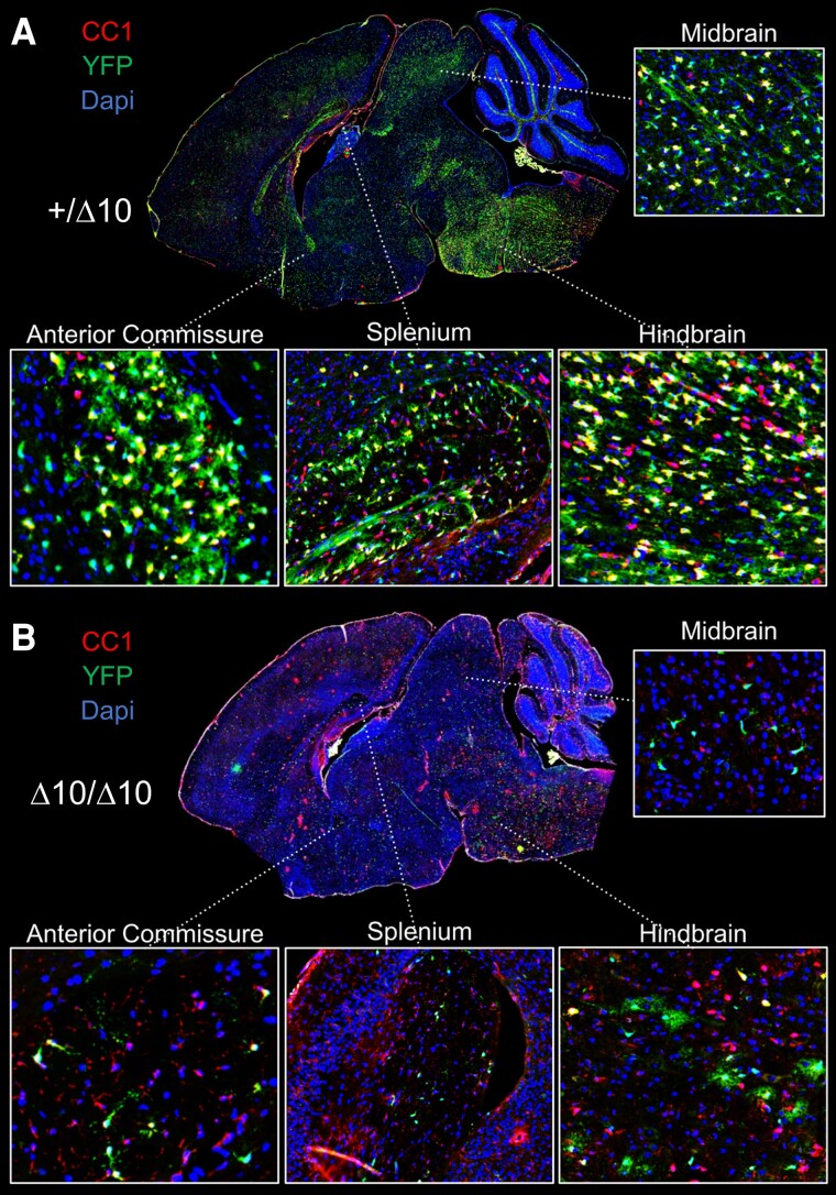
Figure 7.
Depletion of YFP+ cells from the parenchyma in Δ10 homozygous mice. Comparison of +/Δ10 (A) and Δ10/Δ10 (B) demonstrated widespread depletion of yellow fluorescent protein positive (YFP+) cells in the parenchyma of Δ10/Δ10. Representative images shown. Among remaining YFP+ cells, Δ10/Δ10 had cells with shorter processes and more immature morphology. Inset pictures show the hindbrain, midbrain, splenium of the corpus callosum, and anterior commissure. Δ10/Δ10 = tamoxifen-treated transgenic mice carrying a Pdgfrα-CreERT allele and homozygous Polr3bfl alleles; +/Δ10 = tamoxifen-treated transgenic mice carrying a Pdgfrα-CreERT allele and heterozygous Polr3bfl alleles; CTRL = tamoxifen-treated transgenic mice carrying no Pdgfrα-CreERT allele(s); CC1 = clone CC1 of anti-adenomatous polyposis coli (also: Anti-Quaking 7).

