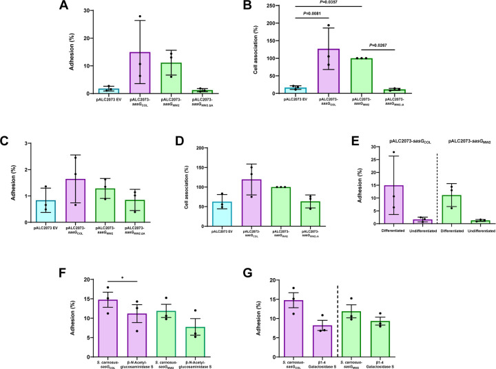
Figure 6. SasG-I and SasG-II-mediated adhesion to differentiated N/TERT keratinocytes following treatment with glycosidases suggests complex N-linked glycans and core 2 O-glycans may be important for SasG-I and SasG-II binding.
S. carnosus-pALC2073 EV, SasG-I-expressing S. carnosus-sasGCOL and SasG-II-expressing S. carnosus-sasGMW2, and SasG-II-expressing S. carnosus with the A-domain deleted (S. carnosus-sasGMW2∆A) at an MOI of 5 were tested for adhesion to either differentiated (A and B) or undifferentiated (C and D) N/TERT keratinocytes. (A) Adhesion to terminally differentiated cells as shown by overall percent adhesion. (B) Adhesion to terminally differentiated cells as shown by percent cell association to pALC2073-sasGMW2 input inoculum. Both SasG-expressing strains adhered more to differentiated cells than the EV and A domain mutant controls. (C) Adhesion to a monolayer of undifferentiated cells as shown by overall percent adhesion. (D) Adhesion to a monolayer of undifferentiated cells as shown by percent cell association to pALC2073-sasGMW2 input inoculum. There were no significant differences in adhesion between the EV and A domain mutant controls and the SasG-expressing strains. (A-D) The CFU/mL of three independent experiments (n = 3) were calculated and analyzed for statistical significance in GraphPad Prism using ordinary one-way ANOVA. (E) Data from panels A and C displaying differences in adhesion between differentiated and undifferentiated N/TERT keratinocytes for SasG-I-expressing S. carnosus-sasGCOL and SasG-II-expressing S. carnosus-sasGMW2. Both strains adhered well to differentiated N/TERT keratinocytes, and did not adhere well to undifferentiated N/TERT keratinocytes. No statistical analyses were performed for Panel E. SasG-I-expressing S. carnosus-sasGCOL and SasG-II-expressing S. carnosus-sasGMW2 were tested for overall percent adhesion to differentiated N/TERT keratinocytes following treatment with (F) β-N-Acetylglucosaminidase S and (G) β1–4 Galactosidase S. β1–4 Galactosidase S reduced adhesion of both strains, while β-N-Acetylglucosaminidase S resulted in a greater reduction in adhesion of S. carnosus-sasGMW2. The CFU/mL of three independent experiments (n = 3) were calculated and analyzed for statistical significance in GraphPad Prism using an unpaired t-test. *P=0.0500.

