The completion of the human genome sequence, in conjunction with newer, cheaper, and more reliable methods of gene expression analysis has the potential to revolutionize cancer diagnosis and treatment. By providing a molecular portrait of an individual cancer, this technology will allow clinicians to determine the origin of the cancer, its potential for metastasis, its specific drug responsiveness, and the probability of its recurrence.
Currently, when cancer is diagnosed it is generally classified according to the gross morphological appearance of the cells and surrounding tissue. This traditional classification scheme is limited by a number of factors. First, it relies on a subjective review of the tissue that is dependent on the knowledge and experience of a pathologist, and therefore may not be reproducible.1 The classification is discrete, rather than continuous, meaning that patients are classified into broad treatment groups (e.g., low, medium, or high probability of recurrence) with limited ability to determine the individual recurrence risk. In addition, current pathology reports either lack or offer very little information regarding the potential drug treatment regime to which a cancer will respond. While current pathology does help determine treatment that leads to better outcomes, tumors with identical pathology may have different origins and respond differently to treatment.2
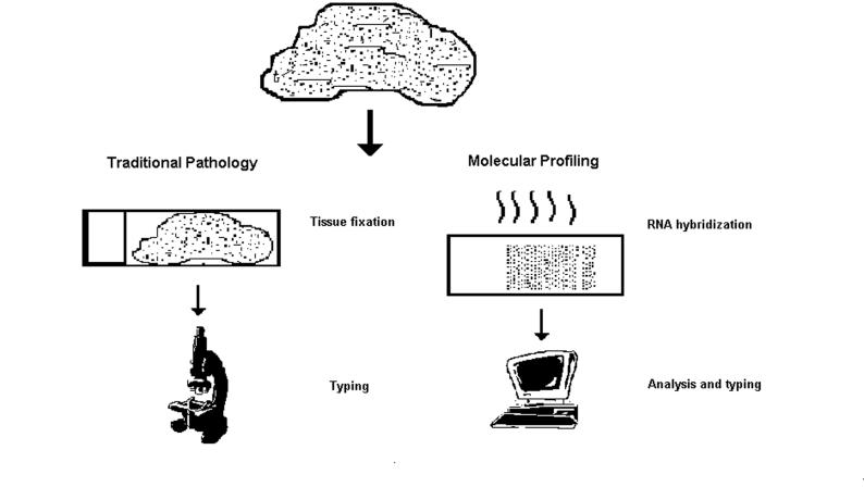
Classification of cancerous tissue based on its molecular profile overcomes these limitations. A molecular profile determines the level of gene expression within the cancer by hybridizing the cellular RNA with known genes. Currently this is done using microarray technology to provide information on thousands of genes simultaneously. Once the gene expression pattern is determined, this information is compared to the expression profiles of cancers with known outcomes using a predetermined algorithm. The algorithm then places the cancer into an outcome class based on similar gene expression patterns, or it will return a survival probability (figure 1). The potential of molecular profiling is illustrated in the following two examples: diffuse large B-cell lymphoma, a cancer with a 50% or less 5-year survival rate,3 and breast cancer which has a much higher 5-year survival rate (80% average), but affects far more individuals (1 in 8 females).4 Using examples from such disparate cancers highlights the limitations of classical cancer classifications and the potential of molecular profiling.
Figure 1.
Schematic of traditional cancer typing versus cancer typing by molecular profiling.
The current classification scheme for diffuse large B-cell lymphoma begins with distinguishing this type of non-Hodgkin's lymphoma using characteristics of the cellular morphology from the biopsy specimens. The tumor is ranked according to stage and grade depending on the extent of spread throughout the tissue and the degree of cellular differentiation, respectively. This information along with the age of the patient and lactate dehydrogenase concentration is used in the International Prognostic Index to determine if the cancer has a low, intermediate, or high risk of recurrence.3 Unfortunately, even with good prognostic indicators for diffuse large B-cell lymphoma, 36% do not respond to treatment.3 With molecular profiling using Lymphochip, an expression array designed for lymphomas, one algorithm successfully distinguished patients with two subtypes of lymphoma originating from different progenitor cells, one with a 76% response rate to chemotherapy and the other with a 16% chemotherapy response rate.5 This was a strong predictor of survival even in patients classified in the low risk group according to standard tumor typing methods. By identifying patients who are unlikely to respond to standard treatment, more aggressive alternatives can be sought earlier in the course of therapy. Another classification algorithm was developed using a subset of the gene expression data that provided a continuous rather than discrete survival probability.6 In the future, this information could be used to make individual patient decisions. These two examples of molecular profiling highlight the advantages over traditional typing and prognosticating.
Molecular profiling can be of benefit even in cancers that, traditionally, are highly curable. Standard treatment regimes for breast cancer rely on the grade and stage of the tumor, as well as estrogen receptor and HER2/neu expression status.7 However, all estrogen receptor positive breast cancers are not the same. Molecular profiling of a variety of breast cancers separated the tumors into five different classes; estrogen receptor positive tumors fell into two distinct classes with different survival profiles.8 An additional problem with breast cancer treatment is over-treatment with chemotherapy. Using the current pathology based methods of determining chemotherapy for breast cancer patients, only 3% of those afflicted show a survival benefit related to chemotherapy. Approximately 83% of these individuals would have remained breast cancer-free without treatment, representing a large population of unnecessarily treated patients. Fourteen percent would die despite receiving chemotherapy, representing a population that would benefit from early identification for aggressive or experimental treatments.9 One molecular profiling algorithm developed for breast cancer decreases the number of patients placed in the high-risk population by 33% to 38%, thus significantly reducing the number of patients undergoing needless chemotherapy.10 In addition, a large proportion of patients classified as low risk with current pathology classifications were found to have poor prognosis via molecular profiling.10 These candidates might benefit from early, aggressive treatments or experimental treatments.
Despite the promise demonstrated with molecular profiling, several barriers must be overcome prior to routine diagnostic implementation for patient intervention. One barrier is the cost of microarray technology for determining the molecular profile of the tumor. This technology is expensive, requires special handling procedures, and lacks standardization within the research community. A single microarray analysis can cost more than $900 in materials alone (cost estimate averaged from several microarray facilities using the Affymetrix human microarray 133 chip set). To obtain the RNA necessary for molecular profiling of a tumor, a tissue sample must be snap frozen in liquid nitrogen at the time of excision adding a step to the standard excision and formalin fixation procedure. In addition, standardized microarrays, necessary for clinical application and interpretation of results, have not been created. Of the 409 genes used in one study to form a clinical profile for breast cancer, only 67% and 37%, respectively, were used in two other breast cancer tumor studies, limiting the usefulness of the profile because of differences in the microarray used.11 In addition to these technical hurdles, a large data set of existing microarray analyses of cancer with known outcomes is necessary to build a robust algorithm for determining outcome in the malignancy to be profiled. These algorithms often consist of analyses of hundreds of genes simultaneously, with only some of the genes contributing to the prognostic value of the test.
Efforts are underway to reduce the problems associated with molecular profiling in order to bring this technology from bench to bedside. Ipsogen, a French biotechnology company, is commercializing a 900-gene microarray for breast cancer. If this chip proves to have diagnostic merit, it would standardize outcomes for comparison. This technology is expected to be available within the next year.12
In an effort to overcome the cost limitation of microarray technology, a small number of genes taken from a larger expression data set can be tested for clinical relevance. Recently, clinically relevant outcomes have been predicted for diffuse large B-cell lymphoma using quantitative reverse transcription-polymerase chain reaction (RT-PCR: a technology routinely used for laboratory testing) of only 6 genes. RT-PCR can be performed on formalin fixed tissue and can be designed to examine multiple genes per reaction, making it much cheaper than a microarray analysis.13
Another interesting application being investigated is attempting to determine a handful of genes that are differentially expressed in aggressive tumors of various cellular origins. A single gene expression pattern, representing an activated wound healing phenotype, was a significant indicator of metastasis and death in breast, lung, and gastric cancer.14 This gene expression pattern, known as the core serum response, consisted of 512 genes, including genes that were involved in the cell cycle (e.g., FOXM1, E2F1), with cell motility, extracellular remodeling (e.g., PLOD2, LOXL2, PLAUR), cell-cell signaling, and myofibroblast phenotype development (e.g., MYL6, AGLN).
In the post-genomic era, a cancer patient would not only know the grade and stage of the tumor or malignancy, but also the relevant gene expression pattern. This information will become a tool for selecting the most promising drug regime, predicting the metastatic potential of the cancer and allowing the patients and physicians to weigh the relative merits of aggressive treatment earlier in the course of disease. This potential, while not yet commercially available, will quickly become a reality. In the case of diffuse large B-cell lymphoma, this information can be obtained with a 6-gene RT-PCR assay,13 that can be easily standardized in a clinical laboratory.
The potential of molecular profiling is not limited to lymphoma and breast cancer; progress is being made with molecular profiling of lung and prostate cancer, as well as acute leukemia.15–20 Selected papers detailing the progress of molecular profiling for these cancers are summarized in table 1. The potential of molecular profiling is clear and the hurdles for the implementation of this powerful new tool are rapidly being overcome.
Table 1.
Examples of microarray analyses of cancers other than lymphoma and breast.
| Cancer | Summary statement | Reference |
| Acute myeloid leukemia | A 13,000-gene array separated acute myeloid leukemia patients into 16 classes, including 1 class with a particularly poor clinical outcome. | 15 |
| A 133-gene predictor classified patients into clinically relevant subtypes. | 16 | |
| Prostate | Prostate tumors separated into three distinct classes using microarrays of 26,000 genes. | 17 |
| A 4-gene model predicated relapse of cancer independent of stage and grade. | 18 | |
| Lung | Review of molecular profiling for non-small cell lung cancer. | 19 |
| Biopsy specimens were separated with a 99-gene profile and a 42-gene profile associated with increased risk of death. | 20 |
Acknowledgments
The authors thank Marshfield Clinic Research Foundation for its support through the assistance of Linda Weis and Alice Stargardt in the preparation of this manuscript.
Contributor Information
Deanna Cross, Center for Personalized Medicine, Marshfield Clinic Research Foundation, Marshfield, Wisconsin.
James K. Burmester, Center for Personalized Medicine, Marshfield Clinic Research Foundation, Marshfield, Wisconsin.
References
- 1.Boiesen P, Bendahl PO, Anagnostaki L, Domanski H, Holm E, Idvall I, Johansson S, Ljungberg O, Ringberg A, Ostberg G, Ferno M. Histologic grading in breast cancer—reproducibility between seven pathologic departments. South Sweden Breast Cancer Group. Acta Oncol. 2000;39:41–45. doi: 10.1080/028418600430950. [DOI] [PubMed] [Google Scholar]
- 2.Isaacs C, Stearns V, Hayes DF. New prognostic factors for breast cancer recurrence. Semin Oncol. 2001;28:53–67. doi: 10.1016/s0093-7754(01)90045-4. [DOI] [PubMed] [Google Scholar]
- 3.The International Non-Hodgkin's Lymphoma Prognostic Factors Project. A predictive model for aggressive non-Hodgkin's lymphoma. N Engl J Med. 1993;329:987–994. doi: 10.1056/NEJM199309303291402. [DOI] [PubMed] [Google Scholar]
- 4.Jemal A, Tiwari RC, Murray T, Ghafoor A, Samuels A, Ward E, Feuer EJ, Thun MJ. American Cancer Society. Cancer statistics, 2004. CA Cancer J Clin. 2004;54:8–29. doi: 10.3322/canjclin.54.1.8. [DOI] [PubMed] [Google Scholar]
- 5.Alizadeh AA, Eisen MB, Davis RE, Ma C, Lossos IS, Rosenwald A, Boldrick JC, Sabet H, Tran T, Yu X, Powell JI, Yang L, Marti GE, Moore T, Hudson J, Jr, Lu L, Lewis DB, Tibshirani R, Sherlock G, Chan WC, Greiner TC, Weisenburger DD, Armitage JO, Warnke R, Levy R, Wilson W, Grever MR, Byrd JC, Botstein D, Brown PO, Staudt LM. Distinct types of diffuse large B-cell lymphoma identified by gene expression profiling. Nature. 2000;403:503–511. doi: 10.1038/35000501. [DOI] [PubMed] [Google Scholar]
- 6.Bair E, Tibshirani R. Semi-supervised methods to predict patient survival from gene expression data. PLoS Biol. 2004;2:E108. doi: 10.1371/journal.pbio.0020108. [DOI] [PMC free article] [PubMed] [Google Scholar]
- 7.Hlupic L, Jakic-Razumovic J, Bozikov J, Coric M, Belev B, Vrbanec D. Prognostic value of different factors in breast carcinoma. Tumori. 2004;90:112–119. doi: 10.1177/030089160409000123. [DOI] [PubMed] [Google Scholar]
- 8.Sorlie T, Perou CM, Tibshirani R, Aas T, Geisler S, Johnsen H, Hastie T, Eisen MB, van de Rijn M, Jeffrey SS, Thorsen T, Quist H, Matese JC, Brown PO, Botstein D, Eystein Lonning P, Borresen-Dale AL. Gene expression patterns of breast carcinomas distinguish tumor subclasses with clinical implications. Proc Natl Acad Sci USA. 2001;98:10869–10874. doi: 10.1073/pnas.191367098. [DOI] [PMC free article] [PubMed] [Google Scholar]
- 9.Cleator S, Ashworth A. Molecular profiling of breast cancer: clinical implications. Br J Cancer. 2004;90:1120–1124. doi: 10.1038/sj.bjc.6601667. [DOI] [PMC free article] [PubMed] [Google Scholar]
- 10.van de Vijver MJ, He YD, van't Veer LJ, Dai H, Hart AA, Voskuil DW, Schreiber GJ, Peterse JL, Roberts C, Marton MJ, Parrish M, Atsma D, Witteveen A, Glas A, Delahaye L, van der Velde T, Bartelink H, Rodenhuis S, Rutgers ET, Friend SH, Bernards R. A gene-expression signature as a predictor of survival in breast cancer. N Engl J Med. 2002;347:1999–2009. doi: 10.1056/NEJMoa021967. [DOI] [PubMed] [Google Scholar]
- 11.Yu K, Lee CH, Tan PH, Hong GS, Wee SB, Wong CY, Tan P. A molecular signature of the Nottingham prognostic index in breast cancer. Cancer Res. 2004;64:2962–2968. doi: 10.1158/0008-5472.can-03-2430. [DOI] [PubMed] [Google Scholar]
- 12.Borie N, Birnbaum D, Viens P, Jacquemier J, Bachelot T, Deraco S, Debono S, Hermitte F, Fert V, Koki A. Breast Cancer ProfileChip: from large scale gene expression profiling to oncodiagnostic device. EJC Supplements. 2004;2((3)):99. [Google Scholar]
- 13.Lossos IS, Czerwinski DK, Alizadeh AA, Wechser MA, Tibshirani R, Botstein D, Levy R. Prediction of survival in diffuse large-B-cell lymphoma based on the expression of six genes. N Engl J Med. 2004;350:1828–1837. doi: 10.1056/NEJMoa032520. [DOI] [PubMed] [Google Scholar]
- 14.Chang HY, Sneddon JB, Alizadeh AA, Sood R, West RB, Montgomery K, Chi JT, Rijn Mv M, Botstein D, Brown PO. Gene expression signature of fibroblast serum response predicts human cancer progression: similarities between tumors and wounds. PLoS Biol. 2004;2:E7. doi: 10.1371/journal.pbio.0020007. [DOI] [PMC free article] [PubMed] [Google Scholar]
- 15.Valk PJ, Verhaak RG, Beijen MA, Erpelinck CA, Barjesteh van Waalwijk van Doorn-Khosrovani S, Boer JM, Beverloo HB, Moorhouse MJ, van der Spek PJ, Lowenberg B, Delwel R. Prognostically useful gene-expression profiles in acute myeloid leukemia. N Engl J Med. 2004;350:1617–1628. doi: 10.1056/NEJMoa040465. [DOI] [PubMed] [Google Scholar]
- 16.Bullinger L, Dohner K, Bair E, Frohling S, Schlenk RF, Tibshirani R, Dohner H, Pollack JR. Use of gene-expression profiling to identify prognostic subclasses in adult acute myeloid leukemia. N Engl J Med. 2004;350:1605–1616. doi: 10.1056/NEJMoa031046. [DOI] [PubMed] [Google Scholar]
- 17.Lapointe J, Li C, Higgins JP, van de Rijn M, Bair E, Montgomery K, Ferrari M, Egevad L, Rayford W, Bergerheim U, Ekman P, DeMarzo AM, Tibshirani R, Botstein D, Brown PO, Brooks JD, Pollack JR. Gene expression profiling identifies clinically relevant subtypes of prostate cancer. Proc Natl Acad Sci USA. 2004;101:811–816. doi: 10.1073/pnas.0304146101. [DOI] [PMC free article] [PubMed] [Google Scholar]
- 18.Latil A, Bieche I, Chene L, Laurendeau I, Berthon P, Cussenot O, Vidaud M. Gene expression profiling in clinically localized prostate cancer: a four-gene expression model predicts clinical behavior. Clin Cancer Res. 2003;9:5477–5485. [PubMed] [Google Scholar]
- 19.Petty RD, Nicolson MC, Kerr KM, Collie-Duguid E, Murray GI. Gene expression profiling in non-small cell lung cancer: from molecular mechanisms to clinical application. Clin Cancer Res. 2004;10:3237–3248. doi: 10.1158/1078-0432.CCR-03-0503. [DOI] [PubMed] [Google Scholar]
- 20.Borczuk AC, Shah L, Pearson GD, Walter KL, Wang L, Austin JH, Friedman RA, Powell CA. Molecular signatures in biopsy specimens of lung cancer. Am J Respir Crit Care Med. 2004;170:167–174. doi: 10.1164/rccm.200401-066OC. [DOI] [PubMed] [Google Scholar]


