Abstract
Objectives
The aim of this study was to examine the use of hand or rotary files by pre-graduation (fourth- and fifth-year) and postgraduate students in endodontic treatments and to determine the incidence of file fracture and the management of cases with broken instruments.
Materials and Methods
A total of 2,168 teeth undergoing primary endodontic treatment were included in this study. It was determined that 79 of these teeth resulted in broken tools. In the case of broken tools, the education level of the treating clinician, the tooth that was being treated, the canal and fracture level, the curvature of the tooth and the management of the broken instrument were recorded. Periapical radiographs of the patients were used to calculate curvature following the Schneider method.
Results
There was no significant difference in the incidence of broken tools according to education level (p > 0.05). The incidence of file fracture in molar teeth (73.4%) was higher than in other teeth (p < 0.05). More files were broken in the mandibular molar MB canal (20.25%) and in the apical third of the canals (72.1%). The risk of instrument fracture was high in teeth with moderate (44.3%) and severe (38%) curvature canals. The management of apically broken (80%) files mostly involved lefting (p < 0.05).
Conclusions
There was no statistically significant difference between fourth-year students, fifth-year students and postgraduate students in terms of instrument fracture.
Keywords: Endodontic file, File fracture, File fracture management, Separate file
INTRODUCTION
In endodontic practice, various procedural errors may occur at any stage of root canal treatment [1]. Intracanal file fracture, which poses a great challenge for routine root canal treatment, is one of the most common procedural errors that occurs during root canal treatment [2,3,4]. Endodontic appliances are made of various materials, including nickel-titanium (NiTi), stainless steel and carbon steel. The fracture of endodontic instruments can occur for a variety of reasons, including excessive strain or instrument fatigue from use as well as operators who are not fully trained [4,5,6].
Stainless steel hand file fracture usually occurs after visible deformation of the instrument, whereas the fracture of NiTi instruments can occur without prior visible warning to the clinician [7]. It has been reported that the fracture prevalence of stainless steel hand files varies between 2% and 6%, while the fracture prevalence of NiTi rotary files varies between 1.04% and 13.54% [7,8,9,10,11]. Although many factors lead to instrument fracture, the most important factor is the experience of the clinician. In fact, the main problem that occurs when the biomechanical preparation process is performed by inexperienced clinicians is instrument fracture [12]. To reduce this possibility, it is important to ensure that clinicians receive adequate training [13]. Theoretical and practical training in endodontics is an important component of the undergraduate dental curriculum [14]. This training process should enable dental students on graduation to be able to manage uncomplicated root canal treatments for both single-rooted and multi-rooted teeth [15].
The education levels of fourth- and fifth-year students performing endodontic treatment at the faculty of dentistry and postgraduate students in the endodontics program are different from each other. Research exploring endodontic practices among undergraduate students revealed findings regarding the number of root fillings performed. Specifically, the study observed that third-year students displayed minimal root fillings (averaging only 0.71) in the laboratory setting, without any clinical experience. However, as the students progressed to the fourth and fifth years, a remarkable increase in root fillings was noted, with fourth-year students averaging 7.40 and fifth-year students averaging 7.47 root fillings, respectively. Moreover, the fifth-year students, on average, did a higher number of root fillings compared to their third and fourth-year students [15]. In addition, a systematic review showed that postgraduate students outperformed undergraduate students in primary endodontic treatment [16]. In this case, it is clear that the students’ ability to manage endodontic treatments and possible complications differ according to their education level. Therefore, the aim of this study was to investigate the prevalence of hand file and rotary instrument fracture during root canal preparation among fourth- and fifth-year undergraduate and postgraduate endodontics students. The null hypothesis of the study is that fourth- and fifth-year students break more files than postgraduate students.
MATERIALS AND METHODS
Ethical approval (No. 2022/12-29) for this study was obtained from the Ethics Committee of Fırat University. According to 95% confidence (1-α), 95% test power (1-β), and 2-way hypothesis, the total number of cases that should be included in the study was 124 [7].
Patients who underwent endodontic treatment between October 2022 and February 2023 were prospectively analyzed. The study included all maxillary and mandibular permanent incisors, premolars and molars. Retreatments and primary tooth endodontic treatments were excluded from this study. During this semester, the number of fourth-year students who treated patients in the endodontic clinic was 66, the number of fifth-year students was 72, and the number of postgraduate students was 16. A total of 2,168 teeth were included in the study. 1,205 teeth were treated by postgraduate students, 373 teeth were treated by fourth-year students, and 590 teeth were treated by fifth-year students.
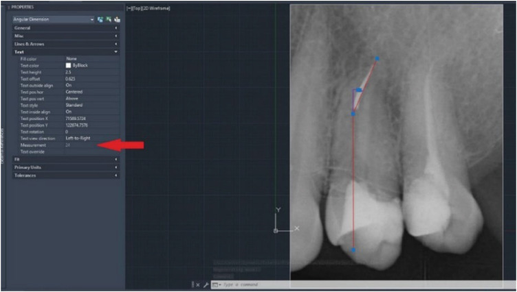
Data were recorded for the patients, including the person performing the treatment (fourth-year, fifth-year, or postgraduate student), tooth type (molar, premolar, and incisor), type of instrument (hand or rotary file), location of the broken instrument (apical, middle, or coronal), and broken file length information. All clinicians used a K file (Mani Inc., Tochigi, Japan) as a hand file and a reciprocating file (Endoart Expert Gold, İnci Dental, Istanbul, Turkey) as a rotary file for biomechanical preparation. Each clinician was informed, and immediately after the file was broken, the length of the file was measured, the amount of separation of the file segmented was recorded, and a periapical radiograph (Planmeca Oy, Helsinki, Finland) image was taken. The length of the broken file was found by measuring the difference between the length of the file before and after separation with a ruler. Canal curvature angles were calculated following the Schneider method using [17], AutoCAD software (Autodesk, San Rafael, CA, USA) (Figure 1). The cases were divided into 4 groups based on canal curvature: 1) mild (curvature < 10); 2) moderate (curvature ≥ 10 and < 25); 3) severe (curvature ≥ 25 and < 45); and 4) ultra-severe (curvature ≥ 45) [18].
Figure 1. Calculation of curvature angle with AutoCAD software.
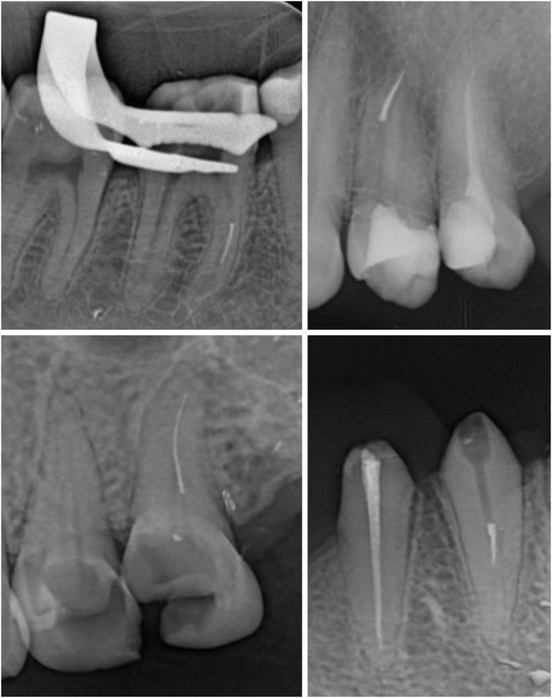
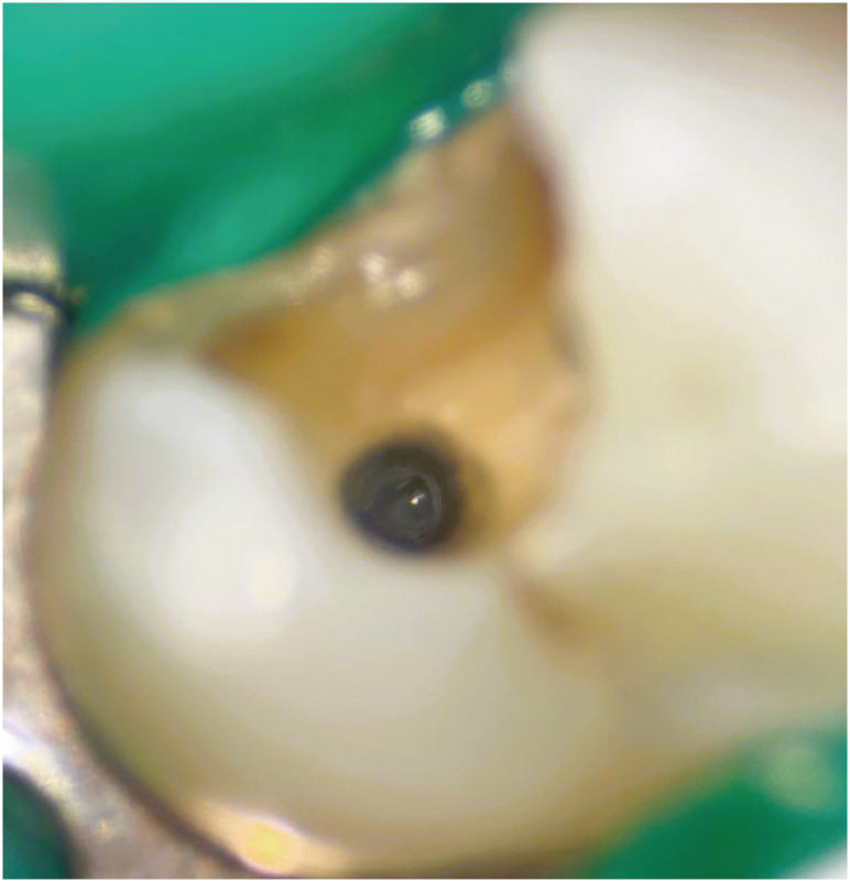
Periapical radiograph images (Planmeca Oy) of hand files and rotary instrument files broken at various levels in different teeth are shown in Figure 2. The localization of the broken files was determined under a dental operating microscope (Zumax Medical, Suzhou, China) (Figure 3). Ultrasonic tips (Guilin Woodpecker Medical Instrument, Guilin, Guangxi, China) were used to retrieve these instruments.
Figure 2. Periapical X-ray images of broken files.
Figure 3. Dental operation microscope image of the broken instrument.
Statistical analysis
Data were analyzed with SPSS (v23; IBM, Armonk, NY, USA). Pearson’s χ2 test was used to compare categorical data, and multiple comparisons were made with Bonferroni correction. The analysis results were presented as frequencies (percentages). The level of significance was set as p < 0.05.
RESULTS
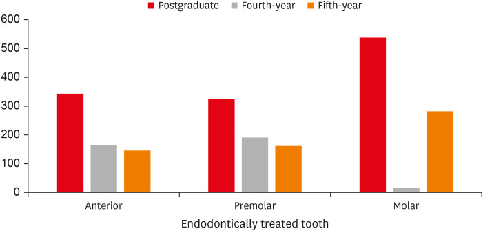
The distribution of teeth treated by postgraduate, fourth-, and fifth-year students according to tooth type is presented in Figure 4. The number of molar teeth treated by postgraduate students was 538, the number of premolars was 324, and the number of incisor teeth was 343. The number of molar teeth treated by fourth-year students was 17, the number of premolars was 191, and the number of incisor teeth was 165. The number of molar teeth treated by fifth-year students was 282, the number of premolars was 162, and the number of incisor teeth was 146.
Figure 4. Distribution of endodontically treated tooth type according to students.
While NiTi reciprocating files were broken in 59 cases, hand files were broken in 20 cases. In the treatments performed by fourth- and fifth-year students, 20 cases with broken instruments were referred to the specialist clinic. In these cases, the procedures of removing, leaving or bypassing the broken instrument were performed by the specialist physician. In addition, the average length of the broken file was 2.42 mm in incisor teeth, 3.4 mm in premolars, and 3.63 mm in posterior teeth.
There was no statistically significant difference between the distribution of file fracture occurrence by education level (p = 0.051). For postgraduate students, the rate of fracture occurrence in the treatments was 3.2%. In comparison, it was 2.4% for the fourth-year students and 5.4% for the fifth-year students (Table 1). There was no statistically significant difference between the fracture frequency of endodontic files in the maxillary and mandibular teeth (p = 0.922) (Table 1). The incidence of fractures in incisor teeth was 0.9%, whereas it was 2.2% in premolars, and there was no significant difference in the presence of fractures in these 2 tooth groups (p > 0.05). There were significantly more file fractures in molar teeth (6.9%) than in other tooth groups (p < 0.001) (Table 1).
Table 1. Comparison of the fracture presence/absence distribution by education level, jaw, and tooth type.
| Variables | Fractured instrument | p * | ||
|---|---|---|---|---|
| Presence | Absent | |||
| Education level | 0.051 | |||
| Postgraduate student | 38 (3.2) | 1,167 (96.8) | ||
| Fourth-year student | 9 (2.4) | 364 (97.6) | ||
| Fifth-year student | 32 (5.4) | 558 (94.6) | ||
| Arch | 0.922 | |||
| Mandibula | 40 (3.7) | 1,046 (96.3) | ||
| Maxilla | 39 (3.6) | 1,043 (96.4) | ||
| Tooth type | < 0.001 | |||
| Incisor | 6 (0.9)a | 648 (99.1) | ||
| Premolar | 15 (2.2)a | 662 (97.8) | ||
| Molar | 58 (6.9)b | 779 (93.1) | ||
Values are presented as number of patients (%).
*Pearson χ2 test; a,bThere is no difference between rows with the same letter.
While the bypassed files did not differ by level (p > 0.05), there was a significant difference by level in the leaved and removed files (p < 0.001) (Table 2). While none of the files broken at the apical level could be removed, 5.3% of files broken at the middle level and 66.7% of files at the coronal level could be removed. The rate of left files was 80% at the apical level and 20% at the middle level, while no files were left at the coronal level. In terms of left files, the rate at the coronal level was significantly lower than at the apical level (p < 0.05).
Table 2. Comparison of file management by fracture level.
| Management of file | Fractured file level | p * | ||
|---|---|---|---|---|
| Apical | Middle | Coronal | ||
| Retrieval | 0 (0.0)a | 1 (5.3)a | 2 (66.7)b | < 0.001 |
| By-passed | 9 (15.8) | 6 (31.6) | 1 (33.3) | |
| Left | 48 (84.2)a | 12 (63.2)ab | 0 (0.0)b | |
Values are presented as number of patients (%).
*Pearson χ2 test; a,bThere is no difference between rows with the same letter.
There was no statistically significant difference between the distribution of the file removal-release status by the canal of the fracture (p = 0.125) (Table 3).
Table 3. Comparison of file management by fracture canal.
| Management of file | Fractured file location | p * | |||||||
|---|---|---|---|---|---|---|---|---|---|
| Man-MB | Max-MB | MB2 | D | P | B | ML | OR | ||
| Retrieval | 1 (4.8) | 0 (0.0) | 1 (16.7) | 1 (14.3) | 0 (0.0) | 0 (0.0) | 0 (0.0) | 0 (0.0) | 0.125 |
| By-passed | 4 (19.0) | 4 (33.3) | 0 (0.0) | 4 (57.1) | 1 (16.7) | 1 (11.1) | 0 (0.0) | 2 (33.3) | |
| Left | 16 (76.2) | 8 (66.7) | 5 (83.3) | 2 (28.6) | 5 (83.3) | 8 (88.9) | 12 (100.0) | 4 (66.7) | |
Values are presented as number of patients (%).
Man-MB, mandibular molar mesiobuccal canal; Max-MB, maxillary molar mesiobuccal canal; MB2, mesiobuccal 2; D, distal; P, palatinal; B; buccal; ML, mesiolingual; OR, one root.
*Pearson χ2 test.
According to the curvature classification, most fractures were seen in the root canals with the most moderate curvature. This was followed by severe, mild, and ultra-severe curvatures, respectively (Table 4). A statistically significant difference was found between the distribution of the removal-left status of the file according to curvature classification (p = 0.028) (Table 4). The rate of bypassed files was 41.7% in canals with mild curvature, 28.6% in canals with moderate curvature, and 3.3% in canals with severe curvature. There were no bypassed files in canals with ultra-severe curvature. The bypass rate of files in canals with severe curvature was significantly lower than the rate in those with mild and moderate curvature (p < 0.05). The proportion of files left in canals with mild curvature was 50%, while it was 65.7% in those with moderate curvature, 96.7% in those with severe curvature, and 100% in those with ultra-severe curvature. The drop rate of files in canals with severe curvature was significantly higher than the rate in canals with mild and moderate curvature (p < 0.05).
Table 4. Comparison of the distribution of the removal-left status of the file by curvature classification.
| Management of file | Canal curvature classification | p * | |||
|---|---|---|---|---|---|
| Mild | Moderate | Severe | Ultra-severe | ||
| Retrieval | 1 (8.3) | 2 (5.7) | 0 (0.0) | 0 (0.0) | 0.028 |
| By-passed | 5 (41.7)a | 10 (28.6)a | 1 (3.3)b | 0 (0.0)ab | |
| Left | 6 (50.0)a | 23 (65.7)a | 29 (96.7)b | 2 (100.0)ab | |
| Total | 12 (100.0) | 35 (100.0) | 30 (100.0) | 2 (100.0) | |
Values are presented as number of patients (%).
*Pearson χ2 test; a,bThere is no difference between rows with the same letter.
DISCUSSION
Endodontic treatment depends on the quality of the cleaning and shaping of the root canal system, and the fracture of the files in the root canal during these procedures is often caused by operator negligence [19]. File fractures are considered to be one of the most troublesome hazards that jeopardize endodontic treatment and can affect prognosis [20]. The results of this study show that the fracture rate of NiTi reciprocating files is 3 times higher than the fracture rate of stainless steel hand files. The length of the broken files in this study ranged from 1–14 mm, with an average length of 3.63 ± 1.15 mm.
Our study aimed to determine the fracture prevalence of hand and NiTi rotary files during root canal treatment performed by fourth-year, fifth-year, and postgraduate students in a dentistry faculty. According to the findings of this study, the fracture frequency of NiTi rotary files was higher than that of hand files. This finding is also consistent with the results of other studies [7]. However, it was observed that the prevalence of broken instruments was higher than in similar studies [7,10,19]. While NiTi rotary files usually break as a result of flexural or torsional loading, it has been reported that fractures in hand files occur due to excessive apical pressure and turning of the instrument [20,21]. In addition, many parameters are involved in the breaking of files [22]. For this reason, the fracture rate of NiTi files is higher than that of hand files, but it does not mean that NiTi files should break more easily than hand files [7].
Various studies have stated that the most important factor in the occurrence of file fracture is the ability of the clinician [23]. In our study, it was found that the prevalence of fractured instruments during root canal treatments was 2.4% for fourth-year students, 5.4% for fifth-year students, and 3.2% for postgraduate students. The null hypothesis was rejected because there was no statistical difference between the groups. In our study, postgraduate students treated an average of 75.31 cases per person during the semester in which the data were collected, while this number was 8.19 for fifth-year students and 5.65 for fourth-year students. Although the difference between the postgraduate and fifth-year students was not statistically significant, the lower prevalence of broken instruments supports this situation. However, with regard to the postgraduate and fifth-year students, the fact that they performed fewer treatments and that the teeth requiring endodontic treatment were chosen from relatively simple teeth may have led to this finding.
The biomechanical preparation in root canal treatment procedures performed by postgraduate students in our faculty of dentistry was as follows: After creating the glide path with a size 10 or 15 K-files, Endoart Expert Gold with a size 25 apical diameter was completed with a reciprocating file. In the biomechanical preparation process, the Endoart WISMY endomotor (İnci Dental) was used, and the standard treatment protocol was followed in accordance with the instructions provided by the manufacturers’ instructions. In this study, the Endoart Expert Gold reciprocating file used for root canal preparation has a fixed 0.06 taper, while manual hand files have a 0.02 taper. In addition, the Endoart Expert Gold file has an S cross-section. The manufacturer claims that Endoart Expert Gold files have high cutting efficiency and high fracture strength due to their heat treatment technology. In the present study, the balanced force technique was used for the manual preparation of the root canals. For manual preparation, standard hand K-files were used, as they have a cutting tip and rectangular cross sections.
The preparation of different root canal anatomies was left to the discretion of the clinician. In the biomechanical preparation procedures of fourth- and fifth-year students, after the apical preparation, was made up to a size 20 K-file, it was completed with a size 25 apical Endoart Expert Gold reciprocating file. The postgraduate students performed manual glide-path creation before using the NiTi rotary instrument, which may have resulted in fewer file fractures, as it reduced the stress on the NiTi rotary file system. In addition, the rotary files used had a fixed angle performing the reciprocating motion, but the use of files with different kinematics or variable taper may alter the incidence of fracture.
Molar teeth have more roots and usually more curvature canals than premolars and incisor teeth [24]. It has been reported that this leads to more frequent instrument fractures in molar teeth [19,25]. In our study, hand files and NiTi rotary files were broken more frequently in molars than in premolars and incisor teeth. In addition, it has been reported that the apical parts of the mesiobuccal roots of the maxillary and mandibular first molars are narrow and the curvatures are greater [24,25,26]. In this study, the most common location of broken files was in the apical part of the molar teeth, which is consistent with the results of similar studies [7,25,27]. We found more instrument fractures in teeth with a moderate curvature. In a similar study evaluating the Mtwo file (VDW, Munich, Germany), fracture was found to be more common in teeth with ultra-severe curvature [18]. It is possible that this difference may be caused by the different file designs, the kinematics of the file, and the clinician’s experience.
Periapical rontgen images of the hand or NiTi rotary files were taken after they were broken, and they were referred to the specialist clinic. Attempts to remove or bypass these files using various methods were attempted under the dental operating microscope (Zumax Medical). While the most frequently extracted files were located in the coronal part of the root canal system, the most frequently bypassed root canal system was found to have mild curvature. The files in the coronal part are more accessible and the curvature is < 10°, making it easier to remove or bypass the broken instruments. This finding is consistent with the results of similar studies [7,18]
Although many studies have stated that the files should be examined before use for the risk of instrument fracture, that deformed files should not be used, and that they should not be used after various numbers, they mainly focused on the way in which the files were used. In these studies, it was stated that NiTi files were more broken than hand files [28,29,30,31,32]. However, anatomical difficulties should not be ignored. According to the results of our study, the probability of instrument breakage in the apical part of the mesiobuccal canals is higher than in the other canals. Undergraduate and postgraduate students should have sufficient knowledge of the root canal anatomy of all teeth, especially molars, and pay attention to the biomechanical preparation of the mesiobuccal canals of the molars.
CONCLUSIONS
Although there is no statistical difference in the percentage of file fractures, when looking at the number of patients treated, it is clear that students treat far fewer cases than postgraduate students. Undergraduate students’ involvement in more cases may increase their experience managing root canal endodontic treatments and file fractures. Graduation of students with the knowledge and skills to perform root canal treatment at a high standard will increase the quality of endodontic treatment applied to patients and will reduce the risk of file fracture.
Footnotes
Conflict of Interest: No potential conflict of interest relevant to this article was reported.
- Conceptualization: Eskibağlar M, Ocak MS.
- Data curation: Eskibağlar M, Öztekin F.
- Formal analysis: Eskibağlar M, Özata MY.
- Investigation: Özata MY.
- Methodology: Özata MY, Ocak MS.
- Project administration: Eskibağlar M.
- Resources: Ocak MS, Öztekin F.
- Software: Eskibağlar M.
- Supervision: Eskibağlar M, Özata MY.
- Validation: Özata MY.
- Visualization: Özata MY.
- Writing - original draft: Eskibağlar M, Öztekin F.
- Writing - review & editing: Eskibağlar M, Özata MY, Ocak MS.
References
- 1.Patnana AK, Chugh A, Chugh VK, Kumar P. The incidence of nickel-titanium endodontic hand file fractures: a 7-year retrospective study in a tertiary care hospital. J Conserv Dent. 2020;23:21–25. doi: 10.4103/JCD.JCD_254_20. [DOI] [PMC free article] [PubMed] [Google Scholar]
- 2.Grossman LI. Guidelines for the prevention of fracture of root canal instruments. Oral Surg Oral Med Oral Pathol. 1969;28:746–752. doi: 10.1016/0030-4220(69)90423-x. [DOI] [PubMed] [Google Scholar]
- 3.Parashos P, Messer HH. Rotary NiTi instrument fracture and its consequences. J Endod. 2006;32:1031–1043. doi: 10.1016/j.joen.2006.06.008. [DOI] [PubMed] [Google Scholar]
- 4.Gambarini G. Cyclic fatigue of ProFile rotary instruments after prolonged clinical use. Int Endod J. 2001;34:386–389. doi: 10.1046/j.1365-2591.2001.00259.x. [DOI] [PubMed] [Google Scholar]
- 5.Ankrum MT, Hartwell GR, Truitt JE. K3 Endo, ProTaper, and ProFile systems: breakage and distortion in severely curved roots of molars. J Endod. 2004;30:234–237. doi: 10.1097/00004770-200404000-00013. [DOI] [PubMed] [Google Scholar]
- 6.Sattapan B, Nervo GJ, Palamara JE, Messer HH. Defects in rotary nickel-titanium files after clinical use. J Endod. 2000;26:161–165. doi: 10.1097/00004770-200003000-00008. [DOI] [PubMed] [Google Scholar]
- 7.Tzanetakis GN, Kontakiotis EG, Maurikou DV, Marzelou MP. Prevalence and management of instrument fracture in the postgraduate endodontic program at the Dental School of Athens: a five-year retrospective clinical study. J Endod. 2008;34:675–678. doi: 10.1016/j.joen.2008.02.039. [DOI] [PubMed] [Google Scholar]
- 8.Abu-Tahun I, Al-Rabab’ah MA, Hammad M, Khraisat A. Technical quality of root canal treatment of posterior teeth after rotary or hand preparation by fifth year undergraduate students, The University of Jordan. Aust Endod J. 2014;40:123–130. doi: 10.1111/aej.12069. [DOI] [PubMed] [Google Scholar]
- 9.Yılmaz A, Gökyay SS, Dağlaroğlu R, Küçükay IK. Evaluation of deformation and fracture rates for nickel-titanium rotary instruments according to the frequency of clinical use. Eur Oral Res. 2018;52:89–93. doi: 10.26650/eor.2018.461. [DOI] [PMC free article] [PubMed] [Google Scholar]
- 10.Iqbal MK, Kohli MR, Kim JS. A retrospective clinical study of incidence of root canal instrument separation in an endodontics graduate program: a PennEndo database study. J Endod. 2006;32:1048–1052. doi: 10.1016/j.joen.2006.03.001. [DOI] [PubMed] [Google Scholar]
- 11.Cheung GS, Bian Z, Shen Y, Peng B, Darvell BW. Comparison of defects in ProTaper hand-operated and engine-driven instruments after clinical use. Int Endod J. 2007;40:169–178. doi: 10.1111/j.1365-2591.2006.01200.x. [DOI] [PubMed] [Google Scholar]
- 12.Sonntag D, Delschen S, Stachniss V. Root-canal shaping with manual and rotary Ni-Ti files performed by students. Int Endod J. 2003;36:715–723. doi: 10.1046/j.1365-2591.2003.00703.x. [DOI] [PubMed] [Google Scholar]
- 13.Deepika G, Mitthra S, Anuradha B, Karthick A. Separated instruments—a mind-set between hard and rock: a review. J Evolut Med Dent Sci. 2017;6:6077–6080. [Google Scholar]
- 14.Field J, Cowpe J, Walmsley D. The Profile of Undergraduate Dental Education in Europe. Dublin: Association for Dental Education in Europe; 2017. [Google Scholar]
- 15.Davey J, Bryant ST, Dummer PM. The confidence of undergraduate dental students when performing root canal treatment and their perception of the quality of endodontic education. Eur J Dent Educ. 2015;19:229–234. doi: 10.1111/eje.12130. [DOI] [PubMed] [Google Scholar]
- 16.Ng YL, Mann V, Rahbaran S, Lewsey J, Gulabivala K. Outcome of primary root canal treatment: systematic review of the literature - Part 1. Effects of study characteristics on probability of success. Int Endod J. 2007;40:921–939. doi: 10.1111/j.1365-2591.2007.01322.x. [DOI] [PubMed] [Google Scholar]
- 17.Sonntag D, Stachniss-Carp S, Stachniss V. Determination of root canal curvatures before and after canal preparation (part 1): a literature review. Aust Endod J. 2005;31:89–93. doi: 10.1111/j.1747-4477.2005.tb00311.x. [DOI] [PubMed] [Google Scholar]
- 18.Wang NN, Ge JY, Xie SJ, Chen G, Zhu M. Analysis of Mtwo rotary instrument separation during endodontic therapy: a retrospective clinical study. Cell Biochem Biophys. 2014;70:1091–1095. doi: 10.1007/s12013-014-0027-0. [DOI] [PubMed] [Google Scholar]
- 19.Siqueira JF., Jr Aetiology of root canal treatment failure: why well-treated teeth can fail. Int Endod J. 2001;34:1–10. doi: 10.1046/j.1365-2591.2001.00396.x. [DOI] [PubMed] [Google Scholar]
- 20.Panitvisai P, Parunnit P, Sathorn C, Messer HH. Impact of a retained instrument on treatment outcome: a systematic review and meta-analysis. J Endod. 2010;36:775–780. doi: 10.1016/j.joen.2009.12.029. [DOI] [PubMed] [Google Scholar]
- 21.Ungerechts C, Bårdsen A, Fristad I. Instrument fracture in root canals - where, why, when and what? A study from a student clinic. Int Endod J. 2014;47:183–190. doi: 10.1111/iej.12131. [DOI] [PubMed] [Google Scholar]
- 22.Gomes MS, Vieira RM, Böttcher DE, Plotino G, Celeste RK, Rossi-Fedele G. Clinical fracture incidence of rotary and reciprocating NiTi files: a systematic review and meta-regression. Aust Endod J. 2021;47:372–385. doi: 10.1111/aej.12484. [DOI] [PubMed] [Google Scholar]
- 23.Madarati AA, Watts DC, Qualtrough AJ. Factors contributing to the separation of endodontic files. Br Dent J. 2008;204:241–245. doi: 10.1038/bdj.2008.152. [DOI] [PubMed] [Google Scholar]
- 24.McGuigan MB, Louca C, Duncan HF. Endodontic instrument fracture: causes and prevention. Br Dent J. 2013;214:341–348. doi: 10.1038/sj.bdj.2013.324. [DOI] [PubMed] [Google Scholar]
- 25.Shen Y, Coil JM, McLean AG, Hemerling DL, Haapasalo M. Defects in nickel-titanium instruments after clinical use. Part 5: Single use from endodontic specialty practices. J Endod. 2009;35:1363–1367. doi: 10.1016/j.joen.2009.07.004. [DOI] [PubMed] [Google Scholar]
- 26.Cunningham CJ, Senia ES. A three-dimensional study of canal curvatures in the mesial roots of mandibular molars. J Endod. 1992;18:294–300. doi: 10.1016/s0099-2399(06)80957-x. [DOI] [PubMed] [Google Scholar]
- 27.Wu J, Lei G, Yan M, Yu Y, Yu J, Zhang G. Instrument separation analysis of multi-used ProTaper Universal rotary system during root canal therapy. J Endod. 2011;37:758–763. doi: 10.1016/j.joen.2011.02.021. [DOI] [PubMed] [Google Scholar]
- 28.Kosti E, Zinelis S, Molyvdas I, Lambrianidis T. Effect of root canal curvature on the failure incidence of ProFile rotary Ni-Ti endodontic instruments. Int Endod J. 2011;44:917–925. doi: 10.1111/j.1365-2591.2011.01900.x. [DOI] [PubMed] [Google Scholar]
- 29.Suter B, Lussi A, Sequeira P. Probability of removing fractured instruments from root canals. Int Endod J. 2005;38:112–123. doi: 10.1111/j.1365-2591.2004.00916.x. [DOI] [PubMed] [Google Scholar]
- 30.Parashos P, Gordon I, Messer HH. Factors influencing defects of rotary nickel-titanium endodontic instruments after clinical use. J Endod. 2004;30:722–725. doi: 10.1097/01.don.0000129963.42882.c9. [DOI] [PubMed] [Google Scholar]
- 31.Regan JD, Sherriff M, Meredith N, Gulabivala K. A survey of interfacial forces used during filing of root canals. Endod Dent Traumatol. 2000;16:101–106. doi: 10.1034/j.1600-9657.2000.016003101.x. [DOI] [PubMed] [Google Scholar]
- 32.Yared GM, Bou Dagher FE, Machtou P. Failure of ProFile instruments used with high and low torque motors. Int Endod J. 2001;34:471–475. doi: 10.1046/j.1365-2591.2001.00420.x. [DOI] [PubMed] [Google Scholar]


