



Abstract
Purpose of Review
To review the role of the immune cells and their interaction with cells found in gingiva, periodontal ligament, and bone that leads to net bone loss in periodontitis or bone remodeling in orthodontic tooth movement.
Recent Findings
Periodontal disease is one of the most common oral diseases causing inflammation in the soft and hard tissues of the periodontium and is initiated by bacteria that induce a host response. Although the innate and adaptive immune response function cooperatively to prevent bacterial dissemination, they also play a major role in gingival inflammation and destruction of the connective tissue, periodontal ligament, and alveolar bone characteristic of periodontitis. The inflammatory response is triggered by bacteria or their products that bind to pattern recognition receptors that induce transcription factor activity to stimulate cytokine and chemokine expression. Epithelial, fibroblast/stromal, and resident leukocytes play a key role in initiating the host response and contribute to periodontal disease. Single-cell RNA-seq (scRNA-seq) experiments have added new insight into the roles of various cell types in the response to bacterial challenge. This response is modified by systemic conditions such as diabetes and smoking. In contrast to periodontitis, orthodontic tooth movement (OTM) is a sterile inflammatory response induced by mechanical force. Orthodontic force application stimulates acute inflammatory responses in the periodontal ligament and alveolar bone stimulated by cytokines and chemokines that produce bone resorption on the compression side. On the tension side, orthodontic forces induce the production of osteogenic factors, stimulating new bone formation. A number of different cell types, cytokines, and signaling/pathways are involved in this complex process.
Summary
Inflammatory and mechanical force-induced bone remodeling involves bone resorption and bone formation. The interaction of leukocytes with host stromal cells and osteoblastic cells plays a key role in both initiating the inflammatory events as well as inducing a cellular cascade that results in remodeling in orthodontic tooth movement or in tissue destruction in periodontitis.
Keywords: Gingivitis, Innate immune response, Adaptive immune response, RANKL, Lymphocyte, NF-kB, Neutrophil
Osteoimmunology in Periodontitis
Periodontal disease results from inflammation triggered by a bacterial dysbiosis [1]. Periodontal disease, which includes gingivitis and periodontitis, is the sixth most common non-communicable disease worldwide [2]. Gingivitis represents a reversible inflammation of the gingiva that may subsequently lead to periodontitis. In contrast, periodontitis involves inflammation that leads to irreversible loss of bone and connective tissue that attaches to the tooth root or surrounding bone. The host response to microbial dysbiosis is a key contributing factor that leads to tissue destruction [3]. Bone loss occurs through osteoclast activity and inhibition of bone coupling [4.••, 5].
Bacteria form on the oral mucosal surfaces and teeth. Defense against microorganisms comes from salivary macromolecules including mucins, antimicrobial peptides, salivary lectins, secretory IgA, and other molecules [6]. Microbes or their products bind to pattern recognition receptors found on keratinocytes, stromal cells (e.g., fibroblasts and mesenchymal progenitor cells), vascular cells, osteocytes, and a wide range of leukocytes. They include toll-like receptors, nucleotide-binding oligomerization domain (NOD) proteins, complement receptor 3 (CR3), and a number of scavenger receptors [7]. Pattern recognition, cytokine, and chemokine receptors activate pro-inflammatory signaling pathways and transcription factors in the cell types mentioned above to stimulate a response to microbial infection or injury-induced tissue perturbation. Clinical studies with antibacterial agents provide concrete evidence that bacteria play a critical role in the initiation and progression of periodontal disease [8]. Studies with germ-free mice confirmed these results by establishing cause-and-effect relationships: the transfer of bacteria from mice with periodontitis to healthy mice leads to the establishment of a dysbiotic environment and the initiation of the diseased phenotype [9•, 10••].
Innate and adaptive immune cells work together with resident epithelial cells and stromal cells to limit the invasion of periodontal tissues by microbes [11]. Innate immune cells produce chemokines, cytokines, and co-stimulatory factors. They also present antigens that upregulate the activation of adaptive immune cells and produce chemokines needed for homing to lymph nodes or sites of infection [12]. Both the innate and adaptive immune response have a role in protecting the host; however, they can also promote destructive inflammation [13].
The activation or upregulation of pro-inflammatory transcription factors induces the expression of cytokines, chemokines, enzymes that produce reactive oxygen species, prostaglandins, and degradative enzymes. Some of these pro-inflammatory transcription factors include nuclear factor kappa-light-chain-enhancer of activated B cells (NF-κB), interferon regulatory factors (IRF), signal transducers and activators of transcription (STAT), activator protein-1 (AP-1), CCAAT Enhancer Binding Protein Beta (CEBPb), and forkhead box O1 (FOXO1) [5,14–16]. These transcription factors can induce the expression of proteins that produce prostaglandins or cytokines such as tumor necrosis factor (TNF), interleukin-1 (IL-1), IL-6, IL-17, and receptor activator of nuclear factor kappa-B ligand (RANKL) [14–16]. Each of these has been shown to contribute to periodontal disease through cause-and-effect studies using specific inhibitors or gene deletion in animal models [5, 17].
In addition to specific cell types contributing to periodontal disease, factors present in the interstitial fluid found in the gingiva can also play an important role. It is well known that proteases are important in the periodontal breakdown as described previously [18]. Similarly, it has been established that lipid-based mediators such as prostaglandins and leukotrienes contribute to periodontal breakdown, while other lipid mediators such as lipoxins and resolvins play an important protective role in resolving inflammation [14, 19]. Complement, which is activated by multiple pathways, is also a key contributor to periodontal inflammation and periodontal breakdown [20••]. A recent publication has identified genetic defects that lead to plasmin deficiency and an overabundance of fibrin as a contributor to abnormal inflammatory responses [21••]. Insufficient fibrinolysis leads to excess fibrin accumulation that activates neutrophils and upregulates oral mucosal inflammation and periodontitis.
Epithelial Barrier
The epithelial barrier is an essential line of defense against bacterial invasion. Epithelial cells produce antibacterial defensins and possess intercellular connections that limit the penetration of bacteria [22, 23]. Epithelial cells release several cytokines including IL-1α, IL-1β, IL-6, IL-8, TNFα, and others, which are involved in the pathogenesis of periodontitis [24]. A homeostatic relationship generally exists between the epithelium and bacteria, which is made possible through the production of antibacterial factors by keratinocytes and the tendency of these cells to not “overreact” to bacteria present [24, 25]. The epithelial barrier is effective, in part, because it is continually shedding; therefore, it limits biofilm maturation. Teeth are a prominent site of bacterial colonization because the tooth surface is non-shedding and does not produce antibacterial factors [26].
One of the first events to occur in periodontal disease is the loss of barrier function. Two recent publications [27••, 28••] report changes in the transcriptomes of individual cell types in inflamed gingiva compared to healthy gingiva utilizing single-cell RNA-seq (scRNA-seq) (Table 1). Sharpe and colleagues identified ten subpopulations of epithelial cells from gingiva that presented as healthy [28••]. The subpopulations included two basal cell clusters, three epithelial clusters expressing high transcript levels of cell cycle genes (indicating a high rate of proliferation), one cluster expressing genes related to extracellular matrix organization and angiogenesis, and four distinct gingival epithelial subpopulations having transcriptomes related to immune regulation. These immune-regulating epithelial cells express factors involved in chronic inflammation, which showed enrichment for B-lymphocyte receptor signaling and expression of factors that regulate neutrophils. In periodontitis, most epithelial subpopulations were depleted, while immunity-related epithelial subpopulations were increased. Moutopolous and colleagues identified four epithelial subtypes in healthy oral mucosa: three types of keratinocytes and one cluster consisting of melanocytes [27••]. The three keratinocyte subpopulations were further categorized: a basal cell layer, a layer enriched in genes involved in cornification, and a layer with a gene expression profile consistent with inflammatory responses. The overall percent of epithelial cells decreased in periodontitis subjects with a shift to a more inflammatory state with upregulated pathways related to antimicrobial responses. Despite the difference in resolution of the epithelial subtypes, both publications agreed that there are epithelial subpopulations in gingiva that feature pro-inflammatory gene signatures and that there is a general loss of non-inflammatory epithelial cells with periodontal disease.
Table 1.
Findings from two recent scRNA-seq publications in healthy and diseased gingiva [27••] and [28••]. Due to differences in resolution, the findings are not identical for each sub-type nor the change in periodontitis. For example, cycling basal cells were reported to be increased in the diseased state by Sharpe and colleagues only, and neutrophils were only detected by Moutopolous and colleagues. Additionally, Moutopolous and colleagues [27••] reported an increase in mast cells, while Sharpe and colleagues [28••] reported the opposite in the diseased state
| Type | Detected in healthy gingiva | Change in periodontitis | |
|---|---|---|---|
| Epithelial cells | Epithelial stem cells | Decrease | |
| Basal cells | Decrease | ||
| Cycling basal cells[28••] | Increase[28••] | ||
| Regulate ECM organization and angiogenesis | Decrease | ||
| Melanocytes[27••] | Decrease[27••] | ||
| Immune modulatory | Increase | ||
| Mesenchymal stromal cells/fibroblasts | Extracellular matrix-producing | Decrease | |
| Myofibroblast | Decrease | ||
| Pericyte | Decrease | ||
| Immune modulatory | Increase | ||
| Innate immune cells | Myeloid | Dendritic cells | Same |
| Mast cells | Increase[27••] | ||
| Decrease[28••] | |||
| Macrophages | Same | ||
| Neutrophils[27••] | Increase[27••] | ||
| Lymphoid | Natural killer cells | Same | |
| T cells | Same[27••] | ||
| Decrease[28••] | |||
| B cells | Significantly expanded | ||
| Plasma cells | Significantly expanded | ||
Contribution of Stromal Cells in Periodontal Disease
Fibroblasts are known to be instrumental in establishing tissue architecture and repair of damaged connective tissue. Fibroblasts are also crucial to the recognition of microbes: they express protease-activated receptors (PARs), nucleotide-binding oligomerization domain (NODs), and extracellular and intracellular toll-like receptors (TLRs) [29]. scRNA-seq analysis has provided a new understanding of the role of fibroblasts/stromal cells in gingival inflammation (Table 1). Sharpe and colleagues identified seven distinct fibroblast/stromal cell subpopulations [28••]. Three subpopulations showed enrichment of genes related to the production of extracellular matrix, one subpopulation of pericytes, and the other of myofibroblasts; two other fibroblast subpopulations had an inflammatory profile. In periodontitis, there was a selective loss of stromal populations with a myofibroblast or pericyte phenotype and an increase in inflammatory fibroblasts. In the publication by Moutopolous and colleagues, five stromal cell subpopulations were identified [27••]. Two of the clusters had a transcriptome profile associated with collagen synthesis and tissue remodeling. Three of the five had gene signatures related to immune function associated with leukocyte proliferation, granulocyte migration, and complement activation. Subjects with periodontitis had an overall decrease in fibroblast subpopulations but a specific increase in inflammatory fibroblast subpopulations. These sub-populations were enriched in transcripts that upregulate antimicrobial responses, cytokine biosynthesis, neutrophil recruitment (CXCL1, CXCL8), and chemokines known to attract other leukocytes. Thus, both papers identified a decrease in fibroblast numbers with periodontal disease, apart from an expansion of pro-inflammatory stromal cells in subjects with periodontitis.
Contribution of Innate Immune Response to Periodontal Inflammation
The innate immune response is mediated by leukocyte activation and the production of soluble antimicrobial factors, such as complement and defensins. Innate immune cells include myeloid innate immune cells (e.g., neutrophils, macrophages, eosinophils, basophils, mast cells, and dendritic cells) and innate lymphocytic cells (e.g., γδT cells, NK cells, NK-T cells, and innate lymphoid cells (ILC) populations ILC1, ILC2, and ILC3). One of the first steps in the activation of the innate immune response is the production of chemokines that stimulate innate immune cells to migrate to the site of infection and upregulate antibacterial defense mechanisms [30]. A report using scRNA-seq analysis identified three myeloid immune cell subpopulations [28••] (Table 1). Dendritic cells (DCs) of myeloid origin, mast cells, and macrophages were all found in distinct clusters. However, there was no clear difference in the dendritic cell compartment during periodontal disease. Single-cell analysis of neutrophils was not achieved due to technical reasons related to their high susceptibility to degradation/apoptosis during tissue digestion [28••]. In another publication, myeloid immune cell populations were identified in four distinct populations: myeloid dendritic cells, macrophages, neutrophils, mast cells, and small populations of plasmacytoid DCs [27••]. In periodontitis, myeloid and granulocytes were present with an expansion of neutrophils.
Neutrophils are abundant in periodontal tissue. The balance between neutrophils and the microbial community is essential to periodontal homeostasis [31]. Neutrophils have multiple functions that play a role in anti-microbial defense: respiratory burst, phagocytosis, degranulation, and formation of neutrophil extracellular traps (NET) [4••, 31]. An imbalance of neutrophil deficiency or excess can lead to periodontal disease [4••]. Transcription factors such as NF-kB and FOXO1 regulate neutrophil function via multiple pathways. Normal neutrophil function is needed to protect the host from bacteria. For instance, there is a reduced ability to clear bacteria or increased periodontitis when neutrophil function is disrupted by deletion of FOXO1 (which is needed for neutrophil chemotaxis and phagocytosis) and when neutrophil recruitment is blocked by leukocyte adhesion deficiency [4, 32].
Macrophages defend against bacterial infection, have antigen presentation properties, and participate in the up- and downregulation of inflammation. The process of resolving inflammation paves the way for tissue repair [33]. Periodontitis is initially associated with enhanced pro-inflammatory M1 macrophage phenotypes followed by the presence of M2 macrophages [34]. Thus, the early development of periodontal tissue infiltration by macrophages is characterized by pro-inflammatory changes. In later phases, the resolution of inflammation is mediated by Th2, Treg lymphocytes, and M2 macrophages [34]. The fact that active periodontal tissue destruction is resolved but leaves a deep pocket supports the concept that deep pocket depth does not indicate an active disease process. Similarly, bacteria obtained from deep pockets are not necessarily indicative of active disease.
Classic antigen-presenting cells (dendritic cells) have important functions in maintaining health in periodontal tissues [35]. Bacteria and their byproducts activate DCs, which in turn modify the host response by stimulating lymphocyte polarization and activity [36]. Activated DCs may promote periodontal disease through activation of Th1 or Th17 responses or reduce inflammation via induction of Treg subsets. Immature DCs may also form osteoclasts [37•]. An increased susceptibility to periodontitis was previously shown with lineage-specific deletion of the transcription factor FOXO1 in DCs as they are involved in the homing of regional lymph nodes and activating lymphocytes [38, 39].
Innate lymphocytes such as natural killer (NK) cells and natural killer T cells (NK-T cells) play important roles in maintaining oral tissue homeostasis and can also initiate a destructive cascade through the production of pro-inflammatory mediators [40, 41•]. NK cells directly kill pathogens through the secretion of perforin and granzymes. Dendritic cell crosstalk with NK cells leads to the secretion of IL-12 by dendritic cells and interferon gamma from NK cells, which can lead to macrophage and T cell activation as well as destructive inflammation [42, 43]. Innate lymphoid cells (ILCs) maintain tissue integrity; during inflammation and pathological conditions, they participate in pathogen clearance and tissue remodeling. ILCs are divided into three major subsets: ILC1, ILC2, and ILC3. These “helper-like” innate lymphocyte subsets are considered to be the counterparts of Th1, Th2, and Th17 cells, respectively, because they are functionally similar to their T-helper counterparts. ILC1s require the transcription factor T-bet for development and produce interferon gamma [44]. ILC2s are similar to Th2 lymphocytes and depend on TGFβ and transcription factor GATA-3 for development and activation [42, 43]. ILC2s secrete IL-4, IL-10, and IL-13. ILC3s require the transcription factor RORγt for induction and produce IL-17A, IL-22, and GM-CSF [45]. GATA-3 is also critical in the induction and maintenance of ILC3s. Both Th2 and ILC2 cells have the plasticity and potential to differentiate into IL-17A-producing Th17 or ILC3-like cells, respectively [46]. ILC1 and ILC3 from periodontitis patients produce more IL-17A and IFN-γ than those from healthy subjects [47]. Understanding the specific mechanisms, function, and plasticity of ILCs is an important future goal of periodontal research.
Contribution of Adaptive Immune Response to Periodontal Inflammation
The adaptive immune system consists of a diverse set of lymphoid cells that maintain homeostasis in the periodontium and respond to microbial perturbation. They include a number of T-helper cells including T-helper subsets, CD4+ T cells, CD8+ T cells, follicular T cells, B cells, and plasma cells [48]. Sharpe and colleagues identified four lymphoid immune cell populations in the gingiva by single-cell RNAseq [28••] (Table 1). B cells were shown in three distinct sub-populations characteristic of follicular, IgG-producing plasma B cells, and memory B cells. T cells were resolved to only one cluster, although multiple T-cell populations exist in the gingiva [36]. In health, low numbers of follicular and plasma B cells and a progressive increase in these B cells were found with periodontal disease. Meanwhile, memory B cells and T cells showed a distinct decrease in disease. Moutopolous and colleagues found that T cells were subclustered into CD4+, Th17, mucosal-associated invariant T cells (MAIT), CD8+, γδT, Treg, and NK/T cells [27••]. They also observed a small population of B and plasma cells in health. In periodontal disease, T cells were the major lymphocyte population, followed by B and plasma cells. Plasma cells were significantly expanded in periodontitis compared to health. Albeit, these two groups reported differences in lymphocyte subsets, they found increases in lymphocytes in specimens obtained from patients with periodontitis compared to health.
T lymphocytes (T cells) can be distinguished by the presence of αβ or γδ T cell receptors on the cell surface. Antigen presentation to αβ CD4+ T-cells drives their differentiation into several subsets, such as Th1, Th2, Th9, Th17, T follicular helper (Tfh), and regulatory T cells (Treg) [48, 49]. Cytokines, such as IL-1 produced by Th1 cells and IL-17A by Th17 cells, stimulate RANKL expression in a number of cell types including periodontal ligament cells and osteocytes [49]. Osteoclastogenesis is regulated by a cytokine network that includes RANKL, IL-6, TNFα, IL-1β, and lipid mediators (e.g., PGE2) [48]. On the other hand, cytokines such as TGF-β and IL-10 are produced by Tregs, to promote resolution and prevent bone destruction in periodontal diseases [48].
B lymphocytes (B cells) are part of the humoral component of the adaptive immune system and are specialized in secreting antibodies. B cells can also present antigens and enhance inflammation through the production of cytokines and opsonization [30, 49]. B cells produce RANKL that stimulates osteoclast formation and bone loss. Therefore, the proinflammatory and pro-osteoclastogenic effects of B cells can increase the RANKL/OPG ratio [30]. On the other hand, the deficiency of B cells in periodontitis murine models results in increased severity of alveolar bone loss, which is associated with upregulated RANKL expression [50]. However, it has also been reported that IgD-deficient mice have reduced numbers of activated B cells with less bone resorption [51•].
Bone and Periodontitis
Bone resorption in normal circumstances is closely followed by osteoblast-mediated bone formation in a process referred to as bone coupling. Ideally, the amount of new bone is equal to the amount of bone resorbed, resulting in no net bone loss. The process of bone formation involves mesenchymal stem cells (MSC), also known as mesenchymal stromal cells, and cell-signaling pathways such as TGF-β1, BMP, IGF-1, PDGF, FGF, and Wnt signaling, which then drives proliferation of MSC and/or differentiation into osteoblasts [52]. When the number or activity of osteoblasts is insufficient, the resorbed bone is incompletely replaced, and uncoupling occurs. This leads to net bone loss and the formation of osteolytic lesions. Thus, a key feature of periodontitis is insufficient bone coupling [53]. The impact of inflammation of the periodontium and bone cells is summarized in (Fig. 1).
Fig. 1.
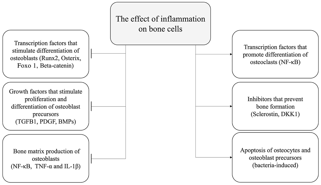
Impact of inflammation of the periodontium. Inflammation inhibits osteogenic transcription factors, growth factors, and bone matrix production of osteoblast. Simultaneously, inflammation stimulates inhibitors which block osteogenic signaling or stimulates apoptosis of bone cells [69–71]
Osteoclasts are multinucleated bone-resorbing cells derived from hematopoietic stem cells and crucial to normal skeletal development and homeostasis [54]. Differentiation to multinucleated osteoclasts requires RANKL, which is encoded by the Tnfsf11 gene [55, 56]. RANKL is expressed by PDL cells, osteoblasts, and osteocytes [57]; lineage-specific deletion of RANKL in these cells inhibits periodontitis [58••]. Bone resorption is reduced by osteoprotegerin (OPG), a RANKL decoy receptor. Treatment of periodontal disease results in a reduced RANKL/OPG ratio, consistent with diminished bone resorption [59].
Osteoblasts are derived from pluripotential precursor cells. Osteoblasts produce osteoid, the organic matrix of bone, which becomes calcified to form bone [60]. Osteocytes are derived from the osteoblasts that are trapped in the bone matrix. Osteocytes account for 90–95% of all bone cells and can regulate bone homeostasis by coordinating the interaction of osteoclasts and osteoblasts [61, 62]. Osteocytes express a number of factors that can promote bone formation. They include chemokines, prostaglandins, and growth factors: BMPs, TGF-β, fibroblast growth factor-2 (FGF-2), and insulin-like growth factor-1 (IGF-1) [63]. Inflammation has a significant effect on osteoblasts and osteocytes. For example, TNF inhibits the bone-forming function of osteoblasts and the differentiation of osteoblasts from precursor cells. TNF can limit bone formation by the production of sclerostin and Dkk1, which inhibits the Wnt pathway [64]. Osteocytes can also contribute to bone resorption through the production of RANKL, TNFα, IL-1α, IL-6, and M-CSF [65]. In experimental periodontitis, RANKL ablation in osteocytes inhibits osteoclastogenesis and bone loss, demonstrating that osteocytes are an important source of RANKL in periodontitis [57].
PDL cells exhibit similarities to tendon cells and immature mesenchymal cells with some osteoblastic characteristics as demonstrated by the expression of a 2.3-kb regulatory unit of Col1α1 [66, 67]. Bacterial dysbiosis in mice stimulates NF-kB expression in PDL fibroblasts, which stimulates the production of chemokines and RANKL to enhance inflammation and bone loss [66]. The periodontal ligament contains MSC that can differentiate into periodontal ligament fibroblasts, cementoblasts, or osteoblasts. TGF-β1 is a key mediator of this differentiation [67]. Thus, MSC in the PDL can differentiate into cells that produce cementum, PDL, or bone. They can also produce mediators that regulate inflammation in a positive or negative direction and can promote bone resorption through the production of RANKL [66]. A recent study examined PDL cells by single-cell RNA-seq [68]. Two transcription factors distinguished different PDL fibroblast populations: Scx (scleraxis) and Mkx (mohawk homeobox), which are found in tendon cells. Scx + PDL cells are located in the central part of the PDL and produce collagen. Mkx+ cells are seen throughout the PDL and produce oxytalan fibers and proteoglycan [68].
The Impact of Diabetes on Periodontal Disease
Diabetes has a significant effect on the periodontium. Diabetes can be classified into type 1 diabetes, which is typically due to autoimmune β-cell destruction leading to insulin deficiency, or type 2 diabetes, which is due to insulin resistance and most often but not always linked to obesity [72]. A positive bidirectional association between periodontal disease and diabetes mellitus has been established—both conditions affect each other [73,74•]; the treatment of one can reduce the risk of the other [74•]. The pathogenesis of the two diseases involves chronic inflammation; diabetes can enhance periodontal inflammation and periodontal dysbiosis, which subsequently leads to periodontal tissue destruction [75, 76]. Diabetes contributes to the formation of a pathogenic oral microbiota. This was demonstrated by a transfer of bacteria from diabetic mice to germ-free recipients compared to a transfer of bacteria from normoglycemic to germ-free mice [10••]. Periodontal tissue destruction is linked to diabetes through several potential pathogenic mechanisms. For example, diabetes increases inflammation, leads to increased expression and activity of proteolytic enzymes, inhibits repair mechanisms, increases death of stromal cells (e.g., MSC, fibroblasts, and osteoblasts), and reduces coupled bone formation [77–80].
Studies in experimental periodontitis have revealed that diabetes enhances RANKL expression in osteocytes and PDL cells, increases periodontal bone resorption, and interferes with the resolution of periodontal inflammation [58••, 66•]. In periodontitis, diabetic animals have difficulty downregulating inflammation, which contributes to prolonged osteoclastogenesis, bone resorption, and interference with coupled bone formation [78, 79]. Diabetes causes a reduction in the number of bone-lining cells, osteoblasts, and PDL cells by increasing their apoptosis [80]. Moreover, TNFα mediates diabetes-enhanced apoptosis of matrix-producing cells and impairs diabetic healing. However, TNF inhibitors, significantly reduce death of matrix producing cells during the resolution of inflammation to reduce periodontitis [81].
The Impact of Smoking on Periodontal Disease
Smoking is a major risk factor for periodontal disease, affecting its prevalence, severity, progression, and treatment [82]. Epidemiological studies consistently show a significantly higher risk for periodontal disease in smokers compared to non-smokers, which is proportional to the duration and amount of smoking [83–85]. Several mechanisms have been proposed. Smoking is thought to alter the oral microbiota, which may accelerate periodontitis [86]. Smokers with periodontitis have increased bacterial anaerobes compared to non-smokers. Tobacco smoke may interfere with the effectiveness of neutrophils [84]. Smoking increases oxidative stress by enhancing the production of radical oxygen species (ROS) [87] and increases the RANKL/OPG ratio [88]. Smoking reduces osteoclast apoptosis, which increases osteoclast numbers and bone resorption [89]. This is mechanistically due to smoking-reduced activation of caspase 3 in osteoclasts or their precursors. In contrast to the effect on osteoclasts, smoking promotes osteoblast and osteoblast precursor apoptosis [88, 90]. This is significant since smoking reduces bone coupling leading to reduced repair of osteolytic lesions, in part, by upregulating sclerostin and DKK1 [88]. This finding is supported by observations that smoking reduces bone formation in fracture healing [90].
Osteoimmunology in Orthodontic Tooth Movement
In contrast to periodontitis, which is a bacteria-induced inflammatory response, orthodontic tooth movement (OTM) is a sterile inflammatory response induced by tissue damage from mechanical force. Orthodontic bone remodeling involves both bone resorption by osteoclasts and new bone formation by osteoblasts in response to compression and tension respectively [91–93]. Upregulation of innate and adaptive immunity in the bone remodeling plays an essential role [94••]. Immune cells that participate in OTM include neutrophils, monocytes/macrophages, NK cells, T cells, and B cells [91–93, 94••, 95]. During OTM, the process of how cells transmit mechanical forces and produce biological responses is important for bone remodeling [96]. However, the precise mechanosensing mechanisms remain unclear.
OTM is mainly divided into three consecutive stages including initial, lag or arrest, acceleration and linear phases [97, 98••, 99]. Initially, mechanical force induces tooth displacement that distorts the interstitial space within the PDL and alveolar bone [95, 99]. Mechanical loading triggers an acute inflammatory response within hours of force application, stimulating PDL cells and immune cells to release inflammatory mediators, recruit leukocytes including osteoclasts precursors, and stimulate osteoclastogenesis. This phase lasts up to 3 days in mice. PDL fibroblasts are mechanosensing cells responsible for the complex immune response associated with the initiation of bone remodeling [100]. Neutrophils are among the first-recruited cells and release chemotactic mediators that recruit other granulocytes, monocytes, and macrophages [101]. Most immune cells are highly active during the initial phase. Due to the occlusion of blood vessels by heavy orthodontic forces, the periodontal tissue becomes necrotic on the compression side during the lag or arrest phase, which occurs between 3 and 7 days after orthodontic loading and lasts from 7 to 14 days or more in mice and up to 30 days in humans [102, 103]. Bone resorption on the compression side is a rate-limiting step of orthodontic tooth movement. Macrophages remove necrotic tissue, facilitating tooth movement [104]. In addition to bone resorption, the new bone formation is also evident during the late stages of the lag phase. During some parts of the lag phase and the acceleration and linear phases, the number of pro-inflammatory innate immune cells decrease, but remain higher than normal, indicating maintained inflammation [105]. At these phases of OTM, the balance between the innate and the adaptive immune system shifts towards the adaptive immune system, which is evident with an increase in T and B cells as well as the upregulation of related pathways. T and B cells produce proinflammatory cytokines and RANKL, facilitating bone resorption [94••, 106]. Following the three stages of active tooth movement, there is a decrease in immune cells. This resolution of inflammation is necessary to return to tissue homeostasis, avoid further tissue damage, and allow for bone formation to persist until bone remodeling is complete. A recent study examined the differentially expressed genes on days 1, 3, 7, and 14 during OTM in mice [94••]. On day 3, genes related to tissue degradation, phagocytosis, and innate and adaptive immune system responses peaked and declined by day 14. In contrast, expression of genes related to cell proliferation and migration, cytoskeletal rearrangement, tissue homeostasis, and angiogenesis initially decreased, but increased at day 14.
Contribution of Innate Immune Response to Orthodontic Tooth Movement
Initiation of the innate immune response is marked by the rapidly infiltrating neutrophils. Their primary function is to secrete various mediators that amplify the inflammatory response and recruit leukocytes such as monocytes and macrophages that clear debris. Neutrophils release myeloperoxidase, which exacerbates the inflammatory response and leads to necrosis of the surrounding tissue [107]. In normal conditions, immune cells present in the PDL, as shown in murine studies, are primarily monocytes and neutrophils [103]. Both cells accumulate in the PDL during OTM. In humans, neutrophils increase in both the gingival crevicular fluid and saliva of orthodontic patients at 2 h after orthodontic force loading [101]. Monocytes are attracted to the injured tissue by chemotactic mediators produced from neutrophils and other cell types [98••]. Monocytic precursors can differentiate into macrophages and dendritic cells, which function in phagocytosis, antigen presentation, and differentiation into osteoclasts [108]. Neutrophils and monocytes are highly active in the initial phase within 3 days after orthodontic loading and maintain upregulated during acute inflammation [98••, 105].
The role of macrophages in OTM has been well studied, reaching their peak on day 7 in OTM animal studies. Their main functions include inflammation (M1 phenotype) and regeneration (M2 phenotype) [98••, [109]. Polarized macrophages exhibit a range of activation states and can be identified by the molecules they express [110]. During inflammation, M1 macrophages promote the proinflammatory phase, while M2 macrophages promote the anti-inflammatory/tissue repair phase of the immune response [110]. During OTM, M1 macrophages are the predominant subtype [110], which promotes bone resorption during the initial stage of OTM [110, 111]. The number of CD68 + iNOS + M1 macrophages significantly increases on day 3 and peaks on day 7 on the compression side. In contrast, the number of CD68 + CD163 + M2 macrophages increases on the compression side 3 days after orthodontic force removal using an orthodontic retention mouse model [111]. The rapid increase of osteoclasts in the PDL is due to the differentiation of macrophages, likely the M1 phenotype, in the presence of RANKL and M-CSF [94••, 108, 112–114]. In rats, M2 macrophages appear in the later stages at 21 days following OTM, signifying their importance in bone formation [109]. Human osteoblasts co-cultured with either M1 or M2 macrophages for 2 weeks show that M2 macrophages promote osteogenesis, compared to M1 macrophages which downregulate osteogenesis [115]. Deletion of monocyte/macrophages significantly blocks OTM and reduces osteoclast formation with decreased expression of TNFα [110]. Systemic transfusion of M1 macrophages promotes bone resorption and OTM, whereas M2 macrophage transfusion has no significant effect on OTM [110]. Activators of M1 macrophages, such as TNFα and IFNγ, are released by monocytes, macrophages, NK cells, and Th1 cells [113]. Although IFNγ directly inhibits osteoclastogenesis, it indirectly stimulates osteoclast formation by increasing the production of proinflammatory cytokine and RANKL [116–118].
NK cells control tumors and microbial infections by killing aberrant cells and secreting various cytokines such as IFNγ, TNFα, IL-5, IL-13, and GM-CSF. The number of NK cells significantly increases in the PDL during OTM [94••, 119]. Evidence that NK cells are functionally significant is shown by significantly reduced tooth movement in NK cell-deleted mice compared to WT mice. In addition, the crosstalk between NK cells and DCs is crucial during the early stages of the immune response [94••, 120]. DCs present the antigen to lymphocytes and trigger adaptive immunity. In addition, monocytic DC precursor can differentiate into osteoclasts in the presence of RANKL and M-CSF. In rat OTM, dendritic-like cells are observed in the PDL via an immunohistochemical stain with a CD11b antibody [121].
During the initiation of the innate immune response, the cells typically associated with allergic responses, including mast cells, eosinophils, and basophils, participate early during the initial phase of bone resorption in OTM [94••, 122]. They produce histamine, cause vasodilation, increase permeability for leukocytes, and release the proinflammatory cytokines and RANKL [123], stimulating bone resorption. In particular, the number of mast cells stained with toluidine blue decreases on both compression and tension sides 15 min after orthodontic loading while basophils increase throughout the initial phase [124]. This signifies a quick degranulation in response to mechanical force. In rats, mast cells in the periodontium produce cytokines such as TNFα, IL-6, and IL-β, which are potent inducers of bone resorption [125].
Contribution of Adaptive Immune Response to Orthodontic Tooth Movement
During OTM, classical αβ T lymphocytes are upregulated, as reflected by the proliferation of CD4 + T cells and increased cytokine production [94••, 112]. Th1 cells produce proinflammatory cytokines such as TNFα and IL-1, inducing bone resorption. Orthodontic force–induced alveolar bone remodeling increases the ratio of CD4 + /CD3 + cells and Th1 cells in the local bone marrow in mice [113]. Th17 cells produce IL-17A, a potent stimulator of RANKL expression [126]. IL-6 and IL-17 have a positive-feedback loop, maintaining high amounts of these cytokines, which favors osteoclastogenesis in response to orthodontic forces [127]. T cell-deletion significantly inhibits OTM with less osteoclast formation and reduced expression of TNFα and IFNγ compared with WT mice, which is reversed by T cell transfer [113]. However, another study reports that the deletion of CD4+ and CD8+ T cells has no impact on OTM, questioning the roles of T cells in OTM [128]. A recent study found that γδT cells increased during the lag phase and are essential for OTM [98••, 103]. Deletion of γδT cells in mice reduced IL-17A and RANKL expression, monocyte and neutrophil recruitment, osteoclast formation, and OTM distance [103]. It should be noted that in mice γδT cells are a predominant source of IL-17A in the oral cavity, while in humans Th17 cells are the predominant source. Excessive production of IL-6 and IL-17 could potentially lead to root resorption [129]. Treg cells increase during the lag phase and potentially lead to the resolution of inflammation and bone formation by suppressing the activity of Th1 and Th17 cells [130, 131]. Furthermore, T cells are affected by mechanical forces, which modulate the activation and function of T cells [132].
OTM involves the upregulation of B cell signaling through three phases [91, 94••]. B cells release IL-6, IL-12, GM-CSF, and RANKL, which promote osteoclastogenesis [105, 133]. In mice, B cells are observed as early as 3 days after orthodontic loading and B cell-deletion reduces the OTM rate by 28% [119]. B cells contribute to both bone resorption and anti-inflammation during OTM [94••, 103]. B cells indirectly promote bone resorption by the production of IL-12, which induces Th1 cells to release IL-1 and TNFα [134]. In addition, the B cell numbers significantly increased after 14 days, while other immune cells such as neutrophils, monocytes, and γδT, and CD4+ T cells significantly decreased [103]. The roles of immune cells in OTM are summarized in Table 2.
Table 2.
Functions of immune cells during OTM
| Cell type | Main immune function | Proposed role in OTM | Identifying markers in OTM | Species | Increase of immune cells in OTM | |||
|---|---|---|---|---|---|---|---|---|
| Myeloid | Neutrophils | Secrete myeloperoxidase (MPO) and exacerbate the inflammatory response; and facilitate debris clearance | Recruit monocytes and macrophages via chemoattractants and debris clearance [94••] | Ly6G+; MPO+ [135] | Human | 2 h [101] | ||
| Mouse | 2 h; 3d [94••, 103] | |||||||
| Rat | - | |||||||
| Monocytes | Precursor for Mφ and DCs | Secrete TNFα and differentiate into mononuclear osteoclasts [136] | Ly6C+; CD43+; CD115+; CD11b+ [137] | Human | 14d [138] | |||
| Mouse | 3d; 7d [94••, 103, 139] | |||||||
| Rat | 3d; 7d [121, 140] | |||||||
| Mφ | M1: promote inflammation, M2: reduce inflammation; Phagocytize debris | M1: secrete TNFα and differentiates into osteoclasts [136] M2: secrete TGF-β, IL-10 to promote osteoblastogenesis [141] | CD11b+; CD14+; CD68+; MHC-II+ M1: iNOS+; TNFα+ M2: ARG1+; IL-10+; TGF-β+; [142] | Human | 14d [138] | |||
| Mouse | M1: 7d [143]; M2: - | |||||||
| Rat | M1: 5d 7d; M2: 21d [109, 144] | |||||||
| NK cells | Stimulate lymphoid cells and kill damaged cells | Secrete mediators of osteoclastogenesis (TNFα and IFNγ) and kill damaged cells [94••] | NK1.1+; NKp46+; CD49b+ [145] | Human | - | |||
| Mouse | 3d [94••] | |||||||
| Rat | - | |||||||
| Eosinophils | Associated with allergic reactions; secrete diverse repertoire of major cationic proteins; cytolytic activity | Early release of histamine to stimulate PDL cells to release proinflammatory cytokines [141] | Ly6G−; CD117−; CD125+; CD193+; Siglec-F+ [146, 147] | Human | - | |||
| Mouse | 3d [94••] | |||||||
| Rat | - | |||||||
| Basophils | Release IL-4 to promote Th2 cell differentiation and activate ILC | CD117−; CD123+; CD203c+; CD193+; CD200R3+ [146] | Human | - | ||||
| Mouse | 3d [94••] | |||||||
| Rat | - | |||||||
| Mast cells | Involved in immediate hypersensitivity; regulate adaptive immune response by release of TNFα, IL-6 and IL-1β | CD117+; CD125+; CD200R3+ [146] | Human | - | ||||
| Mouse | - | |||||||
| Rat | 15 min [124] | |||||||
| DCs | Professional APCs; stimulate differentiation of helper T cells | Present antigens to B and T helper cells and differentiate into osteoclasts [94••] | Ly6G−; Ly6C+; CD11c+; MHC-II+; CD103+ or CD11b+ [148] | Human | - | |||
| Mouse | 3d [94••] | |||||||
| Rat | 3d [121] | |||||||
| Lymphoid | γδ T cells | Produce high levels of IL-17A and facilitate neutrophil and monocyte recruitment | Produce the initial secretion of IL-17A and recruit monocytes and neutrophils [103] | Vγ6+ [94••];gdTCR+; IL-17+ [103] | Human | - | ||
| Mouse | 3d in gingiva/7d in PDL [103] | |||||||
| Rat | - | |||||||
| αβ T cells | CD4 +T cells | Tregs | Regulation and suppression of immune response, including Th1, Th17, T-cell related cytokines | Suppress the function of Th1 and Th17 [131] | CD4+; CD25+; FoxP3+; TGF-β+; IL-10+ [149–153] |
Human | - | |
| Mouse | 5d [131] | |||||||
| Rat | 3d [154] | |||||||
| Th1 | Activate Mφ and cytotoxic T cells | Secrete IFNγ and TNFα to promote osteoclastogenesis [131] | CD4+; T-Bet+; IFNγ+ [155, 156] | Human | - | |||
| Mouse | 3-5d [131] | |||||||
| Rat | - | |||||||
| Th17 | Production of IL-17A and recruit neutrophils | Secrete IL-17A and promote the release of osteoclastogenic cytokines [131] | CD4+; RORγt+; IL-17+; IL-22+ [155, 157] | Human | - | |||
| Mouse | 3d [131] | |||||||
| Rat | 14d [154] | |||||||
| CD8+ T cells | Cell-mediated immunity | Dampening the bone remodeling response [158] | CD8+ [159] | Human | - | |||
| Mouse | - | |||||||
| Rat | - | |||||||
| B cells | Humoral immunity | Source of RANKL and OPG [94••] | CD19+ [160] | Human | - | |||
| Mouse | 3d [94••] | |||||||
| Rat | - |
DC, dendritic cells; Mφ, macrophage; NK, natural killer; Th, T helper; -, unknown
Immune Cells and Bone Formation
On the tension side, new bone is formed as a result of applied forces during OTM. Contrary to the multiple roles of immune cells in bone resorption on the compression side, studies related to the function of immune cells in new bone formation are sparse. The anti-inflammatory M2 macrophage is a pivotal immune cell for the cessation of bone resorption and initiation of tissue repair [111]. As an essential regulator of the wound healing process, M2 macrophages produce anti-inflammatory cytokines such as TGF-β and IL-10, promoting osteoblastogenesis [141]. Treg cells reduce progressive inflammatory responses by inhibiting Th1 and Th17 cells and facilitating new bone formation [131]. B cells release IL-10 and TGF-β. IL-10 reduces the production of RANKL, upregulates OPG in osteoblasts, and inhibits osteoclast differentiation. On the other hand, TGF-β attracts osteoblast precursors, induces proliferation and chemotaxis of PDL cells, and upregulates collagen 1 gene and tissue inhibitors of metalloproteinases (TIMPs) [161]. Moreover, B cell-deleted mice present with lower bone formation and less OPG expression, supporting the role of B cell in bone formation [162].
Contribution of PDL Cells, Osteoblasts, and Osteocytes to Bone Resorption in OTM
Periodontal ligament stem cells (PDLSCs) are a subpopulation of mesenchymal stem cells (MSCs) residing in the PDL that have the potential for self-renewal, proliferation, and multilineage differentiation [163, 164]. The roles of PDLSCs in response to mechanical force have been examined in in vitro and in vivo studies, which support their involvement in alveolar bone remodeling [164–166]. Static compression alters the morphology and differentiation of PDLSCs [91, 167]. Upon compression, hPDLSCs express significantly reduced amounts of type I collagen (Col-I), altering formation of the periodontal ligament [168]. After mechanical force withdrawal, reduced collagen expression recovers, which is regulated by TGF-β. Under compression, hPDLSCs present increased osteogenic differentiation at 1 h, but shortly after present significantly increased RANKL/OPG ratio and decreased ALP activity at 12 h [165]. In OTM rat models, the number of PDLSCs, identified by Nestin and PDGFRα, gradually increase until day 3 and then decrease on day 7 on both compression and tension sides, demonstrating their participation in early OTM phases [165]. Interestingly, PDLSC-deletion blocks OTM by 60% and osteoclastogenesis by 80% in transgenic mice [169]. PDLSCs are also mechanosensing cells in the PDL via activation of Yes-associated protein (YAP) [170]. During OTM, YAP expression is significantly increased on the compression and tension side with nuclear translocation [171, 172]. In line with this finding, YAP deletion in PDLSCs significantly decreases OTM and osteoclast formation on the compression side [169].
PDL fibroblasts represent 50–60% of PDL cells, maintain tissue homeostasis, and are the primary mechanosensitive cells in PDL [173–175]. Interestingly, PDL fibroblasts express the alkaline phosphatase, osteocalcin, and positive Von Kossa staining to show bone mineralization in common with osteoblasts [66, 176]. Upon compression, they upregulate the RANKL mRNA and protein expression and regulate the extracellular matrix via the MMP-8/TIMP-1 ratio [177–180]. Expression of MMP-1, −8, and −13, which are the collagenase group of MMP, is upregulated on the compression side in the rat OTM model [181]. MMP-8 is expressed from days 2 to 7 and MMP-13 from days 2 to 4 after orthodontic loading. Both increased expression levels returns to basal levels by day 14. Furthermore, recent studies have supported an important role of PDL fibroblasts and osteoblast lineage cells as a main source of RANKL during OTM via NF-Kb regulation [93, 173].
Osteoblasts are bone-building cells derived from mesenchymal progenitor cells and produce PGE, M-CSF, RANKL, OPG, and BMP, regulating both catabolic and anabolic responses in OTM [182–184]. RANKL expression in osteoblasts is stimulated by PGE2, vitamin D3, PTH, IL-1, IL-6, IL-11, IL-17, and TNFα [185]. Using a mouse OTM organ culture model, Uribe et al. found that the expression of 3.6-kb Col1 and bone sialoprotein (BSP), early osteoblastic markers, decreased on the compression side following 6 h of OTM [186]. Later, at the linear and acceleration phase, osteoblast-related pathways were upregulated [94••]. Levels of IL-1, IL-16, and IFNγ decreased while levels of IL-10 and TGFβ further increased [94••].
Osteocytes are the most abundant cells in bone tissue and individually reside in lacunae [100]. They express M-CSF, RANKL, OPG, and sclerostin, affecting both osteoblasts and osteoclasts during OTM [187–189]. Osteocytes strongly express MMP-13 at days 2 and 4 on the compression side, which returns to normal levels by day 14 in the rat OTM model [181]. Osteocyte damage results in an increased production of M-CSF and RANKL, stimulating osteoclast formation in vitro and in vivo [190, 191]. Microdamage in bone during OTM affects RANKL expression, causing a peak osteocyte apoptosis at 24 h in mice OTM model [192]. RANKL contained in apoptotic bodies can induce osteoclast formation. The role of osteocytes as an important source of RANKL during OTM has been examined using transgenic mice [187–189]. Osteocyte ablation mice present a significant reduction of osteoclast formation and OTM by 60% and 50% respectively [189]. Lineage-specific RANKL deletion in osteocytes blocks OTM by 40% and osteoclastogenesis by 60% compared with control mice [188]. In addition, osteocytes are the only cells to secrete sclerostin, which stimulates bone resorption and inhibits bone formation [193, 194]. Sclerostin also stimulates RANKL expression by osteocytes [195, 196]. Osteocytes are the main mechanosensing cells in bone and have several features that participate in mechanotransduction including cytoskeletons, dendritic processes, primary cilia, focal adhesion, gap junction, ion channels, and cell surface receptors such as integrins [197]. A recent study examined the effect of IFT80 in osteocytes during OTM, which is essential for cilia formation and function [198]. Primary cilia are considered the mechanosensory structure in bone, cartilage, and kidney. Deletion of IFT80 in osteoblasts, chondrocytes, and osteocytes leads to the decreased number of cilia. However, the cilia in osteocytes did not significantly impact the mechanical force-induced bone remodeling, which presents similar OTM distance, osteoclastogenesis, and expression of RANKL or sclerostin in IFT80-deletion mice compared to WT mice [198]. This suggests other possible mechanosensory systems in osteocytes and anatomic limitations to cilia deflection in osteocytes in vivo [199, 200].
Contribution of PDL Cells, Osteoblasts, and Osteocytes to Bone Formation in OTM
Bone formation is delayed during the initial OTM phase due to the predominant proinflammatory markers such as TNFα, which inhibit the expression of Runx2 [185]. On the tension side, PDL fibers are stretched and induce bone deposition. Upon tensional strain in vitro, PDLSCs significantly upregulate the mRNA and protein expression of Runx2, osterix, and Satb2 [201]. The magnitude of tension differentially regulates osteogenic and osteoclastic responses, with excessive tension upregulating PDLSCs to stimulate osteoclast differentiation. Similar to the compression side, expressions of YAP and TAZ are significantly increased on the tension side with nuclear translocation [171, 172]. In addition, YAP deletion in PDLSCs reduces Runx2 expression on the tension side [169].
PDL fibroblasts produce Runx2 and OPG in response to tensional force [178–180]. A recent study investigated the relationship between the platelet-derived growth factor-BB (PDGF-BB) and PDL fibroblasts on the tension side during OTM. PDGF-BB level was significantly enhanced in PDL fibroblasts on the tension side, activating JAK2/STAT3 signals in bone formation during OTM [174]. The inhibitor treatment for PDGFRβ and JAK-STAT signals attenuated OTM with downregulated osteogenic differentiation and bone generation on the tension side. In addition, the expression of the collagenase such as MMP-8 and MMP-13 is upregulated on the tension side in a rat OTM model in vivo and in vitro [181]. In humans, the mRNA expression of MMP-1 and TIMP-1 increases in the PDL on the tension side [202].
Using a mouse OTM organ culture model, Uribe et al. found that the expression of 3.6-kb Col1 and bone sialoprotein (BSP), early osteoblastic markers, increased on the tension side following 6 h of OTM [186]. OTM mice model demonstrated that an expression of alpha-smooth muscle actin (α-SMA), a reliable marker of osteoprogenitor cells, peaked at 2 days while an expression of osteopontin and osteocalcin, markers for osteoid mineralization, peaked at day 4 on the tension side [203]. The levels of TGF-β, responsible for differentiation of osteoblast precursors, increased on the tension side after 3 days in mice OTM model [204]. The expression of MMP-13 mRNA and protein levels was highly expressed in osteoblasts in response to tensile strain in vitro [205, 206].
In response to mechanical strain, osteocytes produce NO and PGE2 within seconds to minutes [207]. Osteocyte-derived NO promotes osteoblast differentiation and bone formation in vitro and in vivo. In addition, sclerostin expression by mature osteocytes was immediately reduced and maintained at low levels on the tension side, nullifying their inhibitory effect on bone formation [193]. Interestingly, lineage-specific RANKL deletion in osteocytes had significantly decreased the osteoblast number on the tension side compared with control mice, suggesting the possible bone coupling mechanism [188]. The expression of MMP-8 in osteocytes increased on days 4 and 7 after orthodontic loading on the tension side using a rat OTM model [181].
The presence of multiple cells on the compression and tension sides during OTM is described in Fig. 2.
Fig. 2.
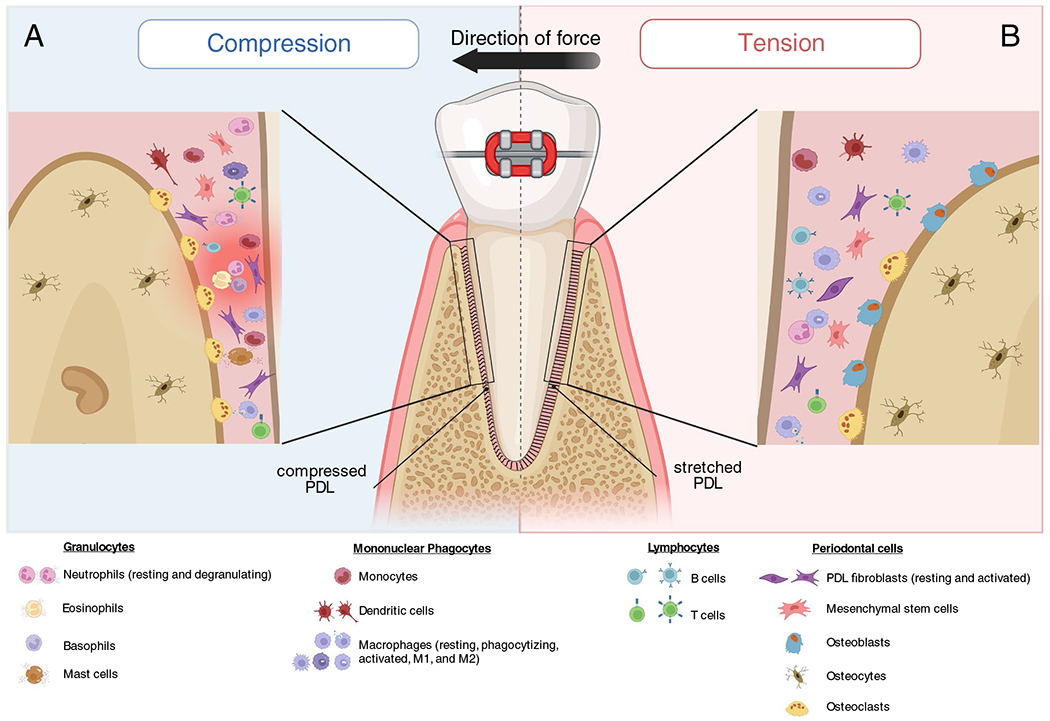
Compression vs. tension side in OTM. A On the compression side, the PDL is infiltrated by a large number of cells the produce inflammatory mediators (activated fibroblasts, mesenchymal stem cells, granulocytes, mononuclear phagocytes, M1 macrophages, osteoclasts, T lymphocytes). Later, anti-inflammatory T and B regulatory lymphocytes reduce the aseptic inflammatory response. B Compared to the compression side, the tension side has fewer proinflammatory leukocytes. The stretched PDL on the tension side exhibits a high degree of fibroblast activation, differentiation of mesenchymal stem cells to osteoblasts, and the presence of M2 macrophages
Conclusion
Inflammatory and mechanical force-induced bone remodeling involves bone resorption and bone formation. A number of cell types are involved in both processes. In particular, the important role of immune cells in bone remodeling developed the concept of “osteoimmunology” or “immunorthodontics.” Periodontal disease is classically destructive to the periodontium by the host response to bacteria. This review thoroughly examined the ways in which the host response contributes to periodontal disease. The host response is evident with the healthy epithelial barrier being armed with immune cells to defend against microbes. When periodontal disease is initiated, this barrier is breached. The inflammatory osteoimmune nature of periodontitis results in affecting the behavior of bone cells driving the bone coupling cycle either towards or away from bone formation/resorption. This relationship is evident in the cause-and-effect studies covered in this review. However, questions regarding how these processes are initiated, why inflammation is causing aggressive host response, and how to control it are still unknown. Understanding the communication between periodontal disease contributing cells and bone cells in both health and disease is vital in order to reveal new insights on how to ultimately treat and prevent periodontal disease. OTM is a highly coordinated process in which various cells such as immune, bone and PDL cells, cytokines, and signals/pathway are involved. To date, a number of OTM studies have focused on bone resorption, which is considered a rate-limiting step. The bone formation itself on the tension side cannot promote the OTM velocity while their effects are more critical in the late phase of OTM. Understanding cellular and molecular targets in OTM can benefit our daily orthodontic practice by applying our knowledge to accelerate tooth movement or prevent orthodontic relapse.
Funding
This work was supported by the Biomedical Research Award from the American Association of Orthodontists Foundation (HHJ) and a grant R01DE017732 and R01DE021921 from the NIDCR (DTG).
Footnotes
Conflict of Interest The authors declare no competing interests.
Human and Animal Rights and Informed Consent This article does not contain any studies with human or animal subjects performed by any of the authors.
References
Papers of particular interest, published recently, have been highlighted as:
• Of importance
•• Of major importance
- 1.Lenartova M, Tesinska B, Janatova T, Hrebicek O, Mysak J, Janata J, Najmanova L. The oral microbiome in periodontal health. Front Cell Infect Microbiol. 2021;11:629723. [DOI] [PMC free article] [PubMed] [Google Scholar]
- 2.Wolf TG, Cagetti MG, Fisher J-M, Seeberger GK, Campus G. Non-communicable diseases and oral health: an overview. Front Oral Health. 2021;2:725460–725460. [DOI] [PMC free article] [PubMed] [Google Scholar]
- 3.Hajishengallis G, Lamont RJ, Graves DT. The enduring importance of animal models in understanding periodontal disease. Virulence. 2015;6:229–35. [DOI] [PMC free article] [PubMed] [Google Scholar]
- 4.••.Hajishengallis G, Lamont RJ. Polymicrobial communities in periodontal disease: their quasi-organismal nature and dialogue with the host. Periodontol. 2021;2000(86):210–30. [DOI] [PMC free article] [PubMed] [Google Scholar]; This review discusses the concept of the ‘polymicrobial synergy and dysbiosis’ (PSD) model of periodontal disease pathogenesis and its effect on dysregulated host inflammatory response.
- 5.Xiao W, Li S, Pacios S, Wang Y, Graves DT. Bone remodeling under pathological conditions. Front Oral Biol. 2016;18:17–27. [DOI] [PMC free article] [PubMed] [Google Scholar]
- 6.Hathaway-Schrader JD, Aartun JD, Poulides NA, Kuhn MB, McCormick BE, Chew ME, Huang E, Darveau RP, Westwater C, Novince CM. Commensal oral microbiota induces osteoimmunomodulatory effects separate from systemic microbiome in mice. JCI Insight. 2022;7:e140738. [DOI] [PMC free article] [PubMed] [Google Scholar]
- 7.Duan T, Du Y, Xing C, Wang HY, Wang RF. Toll-like receptor signaling and its role in cell-mediated immunity. Front Immunol. 2022;13:812774. 10.3389/fimmu.2022.812774. [DOI] [PMC free article] [PubMed] [Google Scholar]
- 8.Curtis MA, Diaz PI, Van Dyke TE. The role of the microbiota in periodontal disease. Periodontol 2000. 2020;83:14–25. [DOI] [PubMed] [Google Scholar]
- 9.•.Payne MA, Hashim A, Alsam A, Joseph S, Aduse-Opoku J, Wade WG, Curtis MA. Horizontal and vertical transfer of oral microbial dysbiosis and periodontal disease. J Dent Res. 2019;98:1503–10. [DOI] [PubMed] [Google Scholar]; This study establishes cause-and-effect evident relationships through the transfer of bacteria from mice with periodontitis to healthy mice, which leads to the establishment of a dysbiotic community and initiation of the diseased phenotype.
- 10.••.Xiao E, Mattos M, Vieira GHA, Chen S, Corrêa JD, Wu Y, Albiero ML, Bittinger K, Graves DT (2017) Diabetes enhances IL-17 expression and alters the oral microbiome to increase its pathogenicity. Cell Host Microbe 22:120–128.e124. [DOI] [PMC free article] [PubMed] [Google Scholar]; This is the first study to demonstrate that diabetes contributes to formation of a pathogenic oral microbiota as demonstrated by transfer of bacteria from diabetic mice to germ-free mice compared to the transfer from normoglycemic to germ-free mice.
- 11.Şenel S. An overview of physical, microbiological and immune barriers of oral mucosa. Int J Mol Sci. 2021;22:7821. [DOI] [PMC free article] [PubMed] [Google Scholar]
- 12.Zimmerman KA, Hopp K, Mrug M. Role of chemokines, innate and adaptive immunity. Cell Signal. 2020;73:109647. [DOI] [PMC free article] [PubMed] [Google Scholar]
- 13.Pan W, Wang Q, Chen Q. The cytokine network involved in the host immune response to periodontitis. Int J Oral Sci. 2019;11:30. [DOI] [PMC free article] [PubMed] [Google Scholar]
- 14.Loos BG, Van Dyke TE. The role of inflammation and genetics in periodontal disease. Periodontol 2000. 2020;83:26–39. [DOI] [PMC free article] [PubMed] [Google Scholar]
- 15.Ito H, Kifune T, Ishiyama M, Iwasa S, Takei H, Hasegawa T, Asano M, Shirakawa T. Effect of hypoxia on the expression of CCAAT/enhancer-binding protein β and receptor activator of nuclear factor kappa-B ligand in periodontal ligament cells. J Oral Sci. 2018;60:544–51. [DOI] [PubMed] [Google Scholar]
- 16.Jurdziński KT, Potempa J, Grabiec AM. Epigenetic regulation of inflammation in periodontitis: cellular mechanisms and therapeutic potential. Clin Epigenetics. 2020;12:186. [DOI] [PMC free article] [PubMed] [Google Scholar]
- 17.Usui M, Onizuka S, Sato T, Kokabu S, Ariyoshi W, Nakashima K. Mechanism of alveolar bone destruction in periodontitis — periodontal bacteria and inflammation. Jpn Dent Sci Rev. 2021;57:201–8. [DOI] [PMC free article] [PubMed] [Google Scholar]
- 18.de Morais EF, Pinheiro JC, Leite RB, Santos PPA, Barboza CAG, Freitas RA. Matrix metalloproteinase-8 levels in periodontal disease patients: a systematic review. J Periodontal Res. 2018;53:156–63. [DOI] [PubMed] [Google Scholar]
- 19.Neupane SP, Virtej A, Myhren LE, Bull VH. Biomarkers common for inflammatory periodontal disease and depression: a systematic review. Brain Behav Immun Health. 2022;21:100450. [DOI] [PMC free article] [PubMed] [Google Scholar]
- 20.••.Hajishengallis G, Hasturk H, Lambris JD; Contributing authors. C3-targeted therapy in periodontal disease: moving closer to the clinic. Trends Immunol 2021;42(10):856–864. 10.1016/j.it.2021.08.001. [DOI] [PMC free article] [PubMed] [Google Scholar]; This clinical trial might represent a novel and transformative host-modulation therapy (C3-targeted intervention) warranting further investigation for the treatment of periodontitis.
- 21.••.Silva LM, Doyle AD, Greenwell-Wild T, et al. (2021) Fibrin is a critical regulator of neutrophil effector function at the oral mucosal barrier. Science 374:eabl5450. [DOI] [PMC free article] [PubMed] [Google Scholar]; This study identifies fibrin as a critical immune regulator of oral mucosal barrier homeostasis, mediated by the local engagement and activation of neutrophils.
- 22.Yamamoto M, Aizawa R. Maintaining a protective state for human periodontal tissue. Periodontol 2000. 2021;86:142–56. [DOI] [PubMed] [Google Scholar]
- 23.Polak D, Zigron A, Eli-Berchoer L, Shapira L, Nussbaum G. Myd88 plays a major role in the keratinocyte response to infection with Porphyromonas gingivalis. J Periodontal Res. 2019;54:396–404. [DOI] [PubMed] [Google Scholar]
- 24.Groeger S, Meyle J (2019) Oral Mucosal Epithelial Cells. Front Immunol 208 [DOI] [PMC free article] [PubMed] [Google Scholar]
- 25.Jiang Y, Tsoi LC, Billi AC, Ward NL, Harms PW, Zeng C, Maverakis E, Kahlenberg JM, Gudjonsson JE. Cytokinocytes: the diverse contribution of keratinocytes to immune responses in skin. JCI Insight. 2020;5(20):e142067. 10.1172/jci.insight.142067. [DOI] [PMC free article] [PubMed] [Google Scholar]
- 26.Mombelli A. Microbial colonization of the periodontal pocket and its significance for periodontal therapy. Periodontol 2000. 2018;76:85–96. [DOI] [PubMed] [Google Scholar]
- 27.••.Williams DW, Greenwell-Wild T, Brenchley L, et al. Human oral mucosa cell atlas reveals a stromal-neutrophil axis regulating tissue immunity. Cell. 2021;184:4090–4104.e4015. [DOI] [PMC free article] [PubMed] [Google Scholar]; This study is a resource characterizing the role of tissue stroma in regulating mucosal tissue homeostasis and disease pathogenesis utilizing single cell RNA-seq (scRNA-seq).
- 28.••.Caetano AJ, Yianni V, Volponi A, Booth V, D’Agostino EM, Sharpe P Defining human mesenchymal and epithelial heterogeneity in response to oral inflammatory disease. eLife 2021;10:e62810. [DOI] [PMC free article] [PubMed] [Google Scholar]; This study establishes an RNA reference map of the human oral mucosa in health and disease.
- 29.Hosokawa I, Hosokawa Y, Ozaki K, Yumoto H, Nakae H, Matsuo T. Proinflammatory effects of muramyldipeptide on human gingival fibroblasts. J Periodontal Res. 2010;45:193–9. [DOI] [PubMed] [Google Scholar]
- 30.Cavalla F, Letra A, Silva RM, Garlet GP. Determinants of periodontal/periapical lesion stability and progression. J Dent Res. 2020;100:29–36. [DOI] [PubMed] [Google Scholar]
- 31.Jiang Q, Zhao Y, Shui Y, Zhou X, Cheng L, Ren B, Chen Z, Li M. Interactions between neutrophils and periodontal pathogens in late-onset periodontitis. Front Cell Infect Microbiol. 2021;11:627328–627328. [DOI] [PMC free article] [PubMed] [Google Scholar]
- 32.Dong G, Song L, Tian C, Wang Y, Miao F, Zheng J, Lu C, Alsadun S, Graves DT. FOXO1 Regulates bacteria-induced neutrophil activity. Front Immunol. 2017;4(8):1088. 10.3389/fimmu.2017.01088. [DOI] [PMC free article] [PubMed] [Google Scholar]
- 33.Ko KI, Syverson AL, Kralik RM, Choi J, DerGarabedian BP, Chen C, Graves DT. Diabetes-induced NF-κB dysregulation in skeletal stem cells prevents resolution of inflammation. Diabetes. 2019;68:2095–106. [DOI] [PMC free article] [PubMed] [Google Scholar]
- 34.Sun X, Gao J, Meng X, Lu X, Zhang L, Chen R. Polarized macrophages in periodontitis: characteristics, function, and molecular signaling. Front Immunol. 2021;12:763334–763334. [DOI] [PMC free article] [PubMed] [Google Scholar]
- 35.Sharawi H, Heyman O, Mizraji G, Horev Y, Laviv A, Shapira L, Yona S, Hovav AH, Wilensky A. The prevalence of gingival dendritic cell subsets in periodontal patients. J Dent Res. 2021;100:1330–6. [DOI] [PubMed] [Google Scholar]
- 36.Song L, Dong G, Guo L, Graves DT. The function of dendritic cells in modulating the host response. Mol Oral Microbiol. 2018;33:13–21. [DOI] [PMC free article] [PubMed] [Google Scholar]
- 37.•.Hovav AH. Dendritic cells of the oral mucosa. Mucosal Immunol. 2014;7:27–37. [DOI] [PubMed] [Google Scholar]; This review demonstrates an overview of the phenotype and distribution of DCs in the oral mucosa is provided.
- 38.Xiao W, Dong G, Pacios S, Alnammary M, Barger LA, Wang Y, Wu Y, Graves DT. FOXO1 deletion reduces dendritic cell function and enhances susceptibility to periodontitis. Am J Pathol. 2015;185:1085–93. [DOI] [PMC free article] [PubMed] [Google Scholar]
- 39.Dong G, Wang Y, Xiao W, Pacios Pujado S, Xu F, Tian C, Xiao E, Choi Y, Graves DT. FOXO1 regulates dendritic cell activity through ICAM-1 and CCR7. J Immunol. 2015;194:3745–55. [DOI] [PMC free article] [PubMed] [Google Scholar]
- 40.Seidel A, Seidel CL, Weider M, Junker R, Gölz L, Schmetzer H. Influence of natural killer cells and natural killer T cells on periodontal disease: a systematic review of the current literature. Int J Mol Sci. 2020;21(24):9766. 10.3390/ijms21249766. [DOI] [PMC free article] [PubMed] [Google Scholar]
- 41.•.Wilensky A, Chaushu S, Shapira L. The role of natural killer cells in periodontitis. Periodontol. 2015;2000(69):128–41. [DOI] [PubMed] [Google Scholar]; This study states the important role of (NK-T) in maintaining oral tissue homeostasis and initiating a destructive cascade through the production of pro-inflammatory mediators.
- 42.Aoki-Nonaka Y, Nakajima T, Miyauchi S, Miyazawa H, Yamada H, Domon H, Tabeta K, Yamazaki K. Natural killer T cells mediate alveolar bone resorption and a systemic inflammatory response in response to oral infection of mice with Porphyromonas gingivalis. J Periodontal Res. 2014;49:69–76. [DOI] [PubMed] [Google Scholar]
- 43.Krämer B, Kebschull M, Nowak M, Demmer RT, Haupt M, Körner C, Perner S, Jepsen S, Nattermann J, Papapanou PN. Role of the NK cell-activating receptor CRACC in periodontitis. Infect Immun. 2013;81:690–6. [DOI] [PMC free article] [PubMed] [Google Scholar]
- 44.Quintino-de-Carvalho IL, Gonçalves-Pereira MH, Faria Ramos M, de Aguiar Milhim BHG, Da Costa ÚL, Santos ÉG, Nogueira ML, Da Costa SH. Type 1 innate lymphoid cell and natural killer cells are sources of interferon-γ and other inflammatory cytokines associated with distinct clinical presentation in early dengue infection. J Infect Dis. 2022;225:84–93. [DOI] [PubMed] [Google Scholar]
- 45.Pearson C, Thornton EE, McKenzie B, et al. ILC3 GM-CSF production and mobilisation orchestrate acute intestinal inflammation. Elife. 2016;5:e10066. [DOI] [PMC free article] [PubMed] [Google Scholar]
- 46.Bernink JH, Ohne Y, Teunissen MBM, et al. c-Kit-positive ILC2s exhibit an ILC3-like signature that may contribute to IL-17-mediated pathologies. Nat Immunol. 2019;20:992–1003. [DOI] [PubMed] [Google Scholar]
- 47.Li C, Liu J, Pan J, Wang Y, Shen L, Xu Y. ILC1s and ILC3s exhibit inflammatory phenotype in periodontal ligament of periodontitis patients. Front Immunol. 2021;12:708678–708678. [DOI] [PMC free article] [PubMed] [Google Scholar]
- 48.Balaji S, Cholan PK, Victor DJ. An emphasis of T-cell subsets as regulators of periodontal health and disease. J Clin Transl Res. 2021;7:648–56. [PMC free article] [PubMed] [Google Scholar]
- 49.Figueredo CM, Lira-Junior R, Love RM. T and B cells in periodontal disease: new functions in a complex scenario. Int J Mol Sci. 2019;20(16):3949. 10.3390/ijms20163949. [DOI] [PMC free article] [PubMed] [Google Scholar]
- 50.Zeng W, Liu G, Luan Q, Yang C, Li S, Yu X, Su L. B-Cell deficiency exacerbates inflammation and bone loss in ligature-induced experimental periodontitis in mice. J Inflamm Res. 2021;14:5367–80. [DOI] [PMC free article] [PubMed] [Google Scholar]
- 51.•.Baker PJ, Boutaugh NR, Tiffany M, Roopenian DC. B Cell IgD deletion prevents alveolar bone loss following murine oral infection. Interdiscip Perspect Infect Dis. 2009;2009:864359. 10.1155/2009/864359. [DOI] [PMC free article] [PubMed] [Google Scholar]; This study suggests that producing IgD B-cells are an important mediator of alveolar bone resorption, possibly through antigen-specific coactivation of B cells and CD4+ T cells.
- 52.Bartold M, Gronthos S, Haynes D, Ivanovski S. Mesenchymal stem cells and biologic factors leading to bone formation. J Clin Periodontol. 2019;46(Suppl 21):12–32. [DOI] [PubMed] [Google Scholar]
- 53.Graves DT, Li J, Cochran DL. Inflammation and uncoupling as mechanisms of periodontal bone loss. J Dent Res. 2011;90:143–53. [DOI] [PMC free article] [PubMed] [Google Scholar]
- 54.Durdan MM, Azaria RD, Weivoda MM. Novel insights into the coupling of osteoclasts and resorption to bone formation. Semin Cell Dev Biol. 2022;123:4–13. [DOI] [PMC free article] [PubMed] [Google Scholar]
- 55.Park JH, Lee NK, Lee SY. Current understanding of RANK signaling in osteoclast differentiation and maturation. Mol Cells. 2017;40:706–13. [DOI] [PMC free article] [PubMed] [Google Scholar]
- 56.Tsukasaki M, Takayanagi H. Osteoimmunology: evolving concepts in bone-immune interactions in health and disease. Nat Rev Immunol. 2019;19:626–42. [DOI] [PubMed] [Google Scholar]
- 57.Pacios S, Xiao W, Mattos M, Lim J, Tarapore RS, Alsadun S, Yu B, Wang CY, Graves DT. Osteoblast lineage cells play an essential role in periodontal bone loss through activation of nuclear factor-kappa B. Sci Rep. 2015;5:16694. [DOI] [PMC free article] [PubMed] [Google Scholar]
- 58.••.Graves DT, Alshabab A, Albiero ML, Mattos M, Corrêa JD, Chen S, Yang Y. Osteocytes play an important role in experimental periodontitis in healthy and diabetic mice through expression of RANKL. J Clin Periodontol. 2018;45:285–92. [DOI] [PMC free article] [PubMed] [Google Scholar]; This study demonstrates, for the first time, the imperative role of osteocytes in bacteria induced periodontal bone loss.
- 59.López Roldán A, García Giménez JL, Alpiste Illueca F. Impact of periodontal treatment on the RANKL/OPG ratio in crevicular fluid. PLoS One. 2020;15:e0227757–e0227757. [DOI] [PMC free article] [PubMed] [Google Scholar]
- 60.Liu Y, Krez A, Stein EM, Chapter 52 - Osteoporosis in organ transplant patients, in Marcus and Feldman’s Osteoporosis (Fifth Edition), Dempster DW, et al. , Editors. 2021, Academic Press. 1281–1307. [Google Scholar]
- 61.Rupp M, Merboth F, Daghma DE, Biehl C, El Khassawna T, Heiβ C. Osteocytes. Z Orthop Unfall. 2019;157:154–63. [DOI] [PubMed] [Google Scholar]
- 62.Robling AG, Bonewald LF. The osteocyte: new insights. Annu Rev Physiol. 2020;82:485–506. [DOI] [PMC free article] [PubMed] [Google Scholar]
- 63.Hienz SA, Paliwal S, Ivanovski S. Mechanisms of bone resorption in periodontitis. J Immunol Res. 2015;2015:615486–615486. [DOI] [PMC free article] [PubMed] [Google Scholar]
- 64.Fukumoto S. Osteocytes and Wnt signaling. Clin Calcium. 2019;29:317–21. [DOI] [PubMed] [Google Scholar]
- 65.Cao W, Helder MN, Bravenboer N, Wu G, Jin J, Ten Bruggenkate CM, Klein-Nulend J, Schulten EAJM. Is there a governing role of osteocytes in bone tissue regeneration? Curr Osteoporos Rep. 2020;18:541–50. [DOI] [PMC free article] [PubMed] [Google Scholar]
- 66.Zheng J, Chen S, Albiero ML, Vieira GHA, Wang J, Feng JQ, Graves DT. Diabetes activates periodontal ligament fibroblasts via NF-κB In Vivo. J Dent Res. 2018;97:580–8. [DOI] [PMC free article] [PubMed] [Google Scholar]
- 67.Lim J-C, Bae S-H, Lee G Ryu Chun Jeih, Jang Young-Joo, Activation of β-catenin by TGF-β1 promotes ligament-fibroblastic differentiation and inhibits cementoblastic differentiation of human periodontal ligament cells. Stem Cells. 2020;38(12):1612–23. 10.1002/stem.3275. [DOI] [PubMed] [Google Scholar]
- 68.Takada K, Chiba T, Miyazaki T, Yagasaki L, Nakamichi R, Iwata T, Moriyama K, Harada H, Asahara H. Single cell RNA sequencing reveals critical functions of Mkx in periodontal ligament homeostasis. Front Cell Dev Biol. 2022;10:795441. 10.3389/fcell.2022.795441. [DOI] [PMC free article] [PubMed] [Google Scholar]
- 69.Yamada S, Tsushima K, Kinoshita M, Sakashita H, Kajikawa T, Fujihara C, Yuan H, Suzuki S, Morisaki T, Murakami S. Mouse model of loeys-dietz syndrome shows elevated susceptibility to periodontitis via alterations in transforming growth factor-beta signaling. Front Physiol. 2021;12:715687–715687. [DOI] [PMC free article] [PubMed] [Google Scholar]
- 70.Eferl R, Hoebertz A, Schilling AF, Rath M, Karreth F, Kenner L, Amling M, Wagner EF. The Fos-related antigen Fra-1 is an activator of bone matrix formation. Embo j. 2004;23:2789–99. [DOI] [PMC free article] [PubMed] [Google Scholar]
- 71.Taichman RS, Hauschka PV. Effects of interleukin-1 beta and tumor necrosis factor-alpha on osteoblastic expression of osteocalcin and mineralized extracellular matrix in vitro. Inflammation. 1992;16:587–601. [DOI] [PubMed] [Google Scholar]
- 72.American Diabetes Association Professional Practice Committee. 2. Classification and diagnosis of diabetes: standards of medical care in diabetes-2022. Diabetes Care. 2022;45(Suppl 1):S17–38. 10.2337/dc22-S002. [DOI] [PubMed] [Google Scholar]
- 73.Stöhr J, Barbaresko J, Neuenschwander M, Schlesinger S. Bidirectional association between periodontal disease and diabetes mellitus: a systematic review and meta-analysis of cohort studies. Sci Rep. 2021;11:13686. [DOI] [PMC free article] [PubMed] [Google Scholar]
- 74.•.Genco RJ, Borgnakke WS. Diabetes as a potential risk for periodontitis: association studies. Periodontol 2000. 2020;83:40–5. [DOI] [PubMed] [Google Scholar]; This review summarizes the established bidirectional association between periodontal disease and diabetes mellitus— both conditions affect each other.
- 75.Graves DT, Ding Z, Yang Y. The impact of diabetes on periodontal diseases. Periodontol 2000. 2020;82:214–24. [DOI] [PubMed] [Google Scholar]
- 76.Barutta F, Bellini S, Durazzo M, Gruden G. Novel insight into the mechanisms of the bidirectional relationship between diabetes and periodontitis. Biomedicines. 2022;10:178. [DOI] [PMC free article] [PubMed] [Google Scholar]
- 77.Kang J, de Brito BB, Pacios S, Andriankaja O, Li Y, Tsiagbe V, Schreiner H, Fine DH, Graves DT. Aggregatibacter actinomycetemcomitans infection enhances apoptosis in vivo through a caspase-3-dependent mechanism in experimental periodontitis. Infect Immun. 2012;80:2247–56. [DOI] [PMC free article] [PubMed] [Google Scholar]
- 78.Andriankaja OM, Galicia J, Dong G, Xiao W, Alawi F, Graves DT. Gene expression dynamics during diabetic periodontitis. J Dent Res. 2012;91:1160–5. [DOI] [PMC free article] [PubMed] [Google Scholar]
- 79.Graves DT, Naguib G, Lu H, Leone C, Hsue H, Krall E. Inflammation is more persistent in type 1 diabetic mice. J Dent Res. 2005;84:324–8. [DOI] [PubMed] [Google Scholar]
- 80.Liu R, Bal HS, Desta T, Krothapalli N, Alyassi M, Luan Q, Graves DT. Diabetes enhances periodontal bone loss through enhanced resorption and diminished bone formation. J Dent Res. 2006;85:510–4. [DOI] [PMC free article] [PubMed] [Google Scholar]
- 81.Liu R, Bal HS, Desta T, Behl Y, Graves DT. Tumor necrosis factor-alpha mediates diabetes-enhanced apoptosis of matrix-producing cells and impairs diabetic healing. Am J Pathol. 2006;168:757–64. [DOI] [PMC free article] [PubMed] [Google Scholar]
- 82.Jiang Y, Zhou X, Cheng L, Li M. The impact of smoking on subgingival microflora: from periodontal health to disease. Front Microbiol. 2020;11:66–66. [DOI] [PMC free article] [PubMed] [Google Scholar]
- 83.Eke PI, Wei L, Thornton-Evans GO, Borrell LN, Borgnakke WS, Dye B, Genco RJ. Risk Indicators for Periodontitis in US Adults: NHANES 2009 to 2012. J Periodontol. 2016;87:1174–85. [DOI] [PMC free article] [PubMed] [Google Scholar]
- 84.White PC, Hirschfeld J, Milward MR, Cooper PR, Wright HJ, Matthews JB, Chapple ILC. Cigarette smoke modifies neutrophil chemotaxis, neutrophil extracellular trap formation and inflammatory response-related gene expression. J Periodontal Res. 2018;53:525–35. [DOI] [PubMed] [Google Scholar]
- 85.Rosa GM, Lucas GQ, Lucas ON. Cigarette smoking and alveolar bone in young adults: a study using digitized radiographs. J Periodontol. 2008;79:232–44. [DOI] [PubMed] [Google Scholar]
- 86.Zhang J, Yu J, Dou J, Hu P, Guo Q. The impact of smoking on subgingival plaque and the development of periodontitis: a literature review. Front Oral Health. 2021;2:751099–751099. [DOI] [PMC free article] [PubMed] [Google Scholar]
- 87.Lee J, Taneja V, Vassallo R. Cigarette smoking and inflammation: cellular and molecular mechanisms. J Dent Res. 2012;91:142–9. [DOI] [PMC free article] [PubMed] [Google Scholar]
- 88.Weng W, Li H, Zhu S. An overlooked bone metabolic disorder: cigarette smoking-induced osteoporosis. Genes (Basel). 2022;13(5):806. 10.3390/genes13050806. [DOI] [PMC free article] [PubMed] [Google Scholar]
- 89.Qin Y, Liu Y, Jiang Y, Mei S, Liu Y, Feng J, Guo L, Du J, Graves DT, Liu Y. Cigarette smoke exposure inhibits osteoclast apoptosis via the mtROS pathway. J Dent Res. 2021;100:1378–86. [DOI] [PMC free article] [PubMed] [Google Scholar]
- 90.Patel RA, Wilson RF, Patel PA, Palmer RM. The effect of smoking on bone healing: a systematic review. Bone Joint Res. 2013;2:102–11. [DOI] [PMC free article] [PubMed] [Google Scholar]
- 91.Krishnan V, Davidovitch Z. Cellular, molecular, and tissue-level reactions to orthodontic force. Am J Orthod Dentofacial Orthop. 2006;129:469.e461–432. [DOI] [PubMed] [Google Scholar]
- 92.Huang H, Williams RC, Kyrkanides S. Accelerated orthodontic tooth movement: molecular mechanisms. Am J Orthod Dentofacial Orthop. 2014;146:620–32. [DOI] [PubMed] [Google Scholar]
- 93.Yang CY, Jeon HH, Alshabab A, Lee YJ, Chung CH, Graves DT. RANKL deletion in periodontal ligament and bone lining cells blocks orthodontic tooth movement. Int J Oral Sci. 2018;10:3. [DOI] [PMC free article] [PubMed] [Google Scholar]
- 94.••.Klein Y, Fleissig O, Polak D, Barenholz Y, Mandelboim O, Chaushu S. Immunorthodontics: in vivo gene expression of orthodontic tooth movement. Sci Rep. 2020;10:8172. [DOI] [PMC free article] [PubMed] [Google Scholar]; This study proposed the concept of “immunorthodontics” to describe the critical roles of immune cells in OTM for the first time.
- 95.Li Y, Zhan Q, Bao M, Yi J, Li Y. Biomechanical and biological responses of periodontium in orthodontic tooth movement: update in a new decade. Int J Oral Sci. 2021;13:20. [DOI] [PMC free article] [PubMed] [Google Scholar]
- 96.Temiyasathit S, Jacobs CR. Osteocyte primary cilium and its role in bone mechanotransduction. Ann N Y Acad Sci. 2010;1192:422–8. [DOI] [PMC free article] [PubMed] [Google Scholar]
- 97.Reitan K. Clinical and histologic observations on tooth movement during and after orthodontic treatment. Am J Orthod. 1967;53:721–45. [DOI] [PubMed] [Google Scholar]
- 98.••.Chaushu S, Klein Y, Mandelboim O, Barenholz Y, Fleissig O. Immune changes induced by orthodontic forces: a critical review. J Dent Res. 2022;101:11–20. [DOI] [PubMed] [Google Scholar]; This recent review summarizes the activity of different immune cells according to the clinical phases of OTM.
- 99.Zainal Ariffin SH, Yamamoto Z, Zainol Abidin IZ, Megat Abdul Wahab R, Zainal Ariffin Z. Cellular and molecular changes in orthodontic tooth movement. Sci World J. 2011;11:1788–803. [DOI] [PMC free article] [PubMed] [Google Scholar]
- 100.Krishnan V, Davidovitch Z. On a path to unfolding the biological mechanisms of orthodontic tooth movement. J Dent Res. 2009;88:597–608. [DOI] [PubMed] [Google Scholar]
- 101.Marcaccini AM, Amato PA, Leao FV, Gerlach RF, Ferreira JT. Myeloperoxidase activity is increased in gingival crevicular fluid and whole saliva after fixed orthodontic appliance activation. Am J Orthod Dentofacial Orthop. 2010;138:613–6. [DOI] [PubMed] [Google Scholar]
- 102.Pilon JJ, Kuijpers-Jagtman AM, Maltha JC. Magnitude of orthodontic forces and rate of bodily tooth movement. An experimental study. Am J Orthod Dentofacial Orthop. 1996;110:16–23. [DOI] [PubMed] [Google Scholar]
- 103.Wald S, Leibowitz A, Aizenbud Y, et al. γδT cells are essential for orthodontic tooth movement. J Dent Res. 2021;100:731–8. [DOI] [PubMed] [Google Scholar]
- 104.Alikhani M, Alyami B, Lee IS, et al. Saturation of the biological response to orthodontic forces and its effect on the rate of tooth movement. Orthod Craniofac Res. 2015;18(Suppl 1):8–17. [DOI] [PubMed] [Google Scholar]
- 105.Behm C, Zhao Z, Andrukhov O. Immunomodulatory activities of periodontal ligament stem cells in orthodontic forces-induced inflammatory processes: current views and future perspectives. Front Oral Health. 2022;3:877348. [DOI] [PMC free article] [PubMed] [Google Scholar]
- 106.Kong YY, Feige U, Sarosi I, et al. Activated T cells regulate bone loss and joint destruction in adjuvant arthritis through osteoprotegerin ligand. Nature. 1999;402:304–9. [DOI] [PubMed] [Google Scholar]
- 107.Khan AA, Alsahli MA, Rahmani AH. Myeloperoxidase as an active disease biomarker: recent biochemical and pathological perspectives. Med Sci (Basel). 2018;6:33. [DOI] [PMC free article] [PubMed] [Google Scholar]
- 108.Zeng M, Kou X, Yang R, et al. Orthodontic force induces systemic inflammatory monocyte responses. J Dent Res. 2015;94:1295–302. [DOI] [PubMed] [Google Scholar]
- 109.Wang Y, Zhang H, Sun W, Wang S, Zhang S, Zhu L, Chen Y, Xie L, Sun Z, Yan B. Macrophages mediate corticotomy-accelerated orthodontic tooth movement. Sci Rep. 2018;8:16788. [DOI] [PMC free article] [PubMed] [Google Scholar]
- 110.He D, Kou X, Yang R, et al. M1-like macrophage polarization promotes orthodontic tooth movement. J Dent Res. 2015;94:1286–94. [DOI] [PubMed] [Google Scholar]
- 111.He D, Kou X, Luo Q, et al. Enhanced M1/M2 macrophage ratio promotes orthodontic root resorption. J Dent Res. 2015;94:129–39. [DOI] [PubMed] [Google Scholar]
- 112.Kook SH, Jang YS, Lee JC. Human periodontal ligament fibroblasts stimulate osteoclastogenesis in response to compression force through TNF-α-mediated activation of CD4+ T cells. J Cell Biochem. 2011;112:2891–901. [DOI] [PubMed] [Google Scholar]
- 113.Yan Y, Liu F, Kou X, Liu D, Yang R, Wang X, Song Y, He D, Gan Y, Zhou Y. T Cells Are Required for Orthodontic Tooth Movement. J Dent Res. 2015;94:1463–70. [DOI] [PubMed] [Google Scholar]
- 114.Yao Y, Cai X, Ren F, Ye Y, Wang F, Zheng C, Qian Y, Zhang M. The macrophage-osteoclast axis in osteoimmunity and osteo-related diseases. Front Immunol. 2021;12:664871. [DOI] [PMC free article] [PubMed] [Google Scholar]
- 115.He W, Zhang N, Lin Z. MicroRNA-125a-5p modulates macrophage polarization by targeting E26 transformation-specific variant 6 gene during orthodontic tooth movement. Arch Oral Biol. 2021;124:105060. [DOI] [PubMed] [Google Scholar]
- 116.Gao Y, Grassi F, Ryan MR, Terauchi M, Page K, Yang X, Weitzmann MN, Pacifici R. IFN-gamma stimulates osteoclast formation and bone loss in vivo via antigen-driven T cell activation. J Clin Invest. 2007;117:122–32. [DOI] [PMC free article] [PubMed] [Google Scholar]
- 117.Ayon Haro ER, Ukai T, Yokoyama M, Kishimoto T, Yoshinaga Y, Hara Y. Locally administered interferon-γ accelerates lipopolysaccharide-induced osteoclastogenesis independent of immunohistological RANKL upregulation. J Periodontal Res. 2011;46:361–73. [DOI] [PubMed] [Google Scholar]
- 118.Takayanagi H, Ogasawara K, Hida S, et al. T-cell-mediated regulation of osteoclastogenesis by signalling cross-talk between RANKL and IFN-gamma. Nature. 2000;408:600–5. [DOI] [PubMed] [Google Scholar]
- 119.Bawyan RF, Baajajah AJ, Alzahrani HA, Murad MA, Jamalellail HMH, Muthaffar SY, Alandijani AA, Khayat MA, Bahurmoz AA, Azab AT, Almalki MA. Immunological and physiological responses related to orthodontic treatment. Int J Community Med Public Health. 2022;9:394–8. [Google Scholar]
- 120.Della Chiesa M, Vitale M, Carlomagno S, Ferlazzo G, Moretta L, Moretta A. The natural killer cell-mediated killing of autologous dendritic cells is confined to a cell subset expressing CD94/NKG2A, but lacking inhibitory killer Ig-like receptors. Eur J Immunol. 2003;33:1657–66. [DOI] [PubMed] [Google Scholar]
- 121.Vandevska-Radunovic V, Kvinnsland IH, Kvinnsland S, Jonsson R. Immunocompetent cells in rat periodontal ligament and their recruitment incident to experimental orthodontic tooth movement. Eur J Oral Sci. 1997;105:36–44. [DOI] [PubMed] [Google Scholar]
- 122.Metcalfe DD, Pawankar R, Ackerman SJ, et al. Biomarkers of the involvement of mast cells, basophils and eosinophils in asthma and allergic diseases. World Allergy Organ J. 2016;9:7. [DOI] [PMC free article] [PubMed] [Google Scholar]
- 123.Groeger M, Spanier G, Wolf M, Deschner J, Proff P, Schröder A, Kirschneck C. Effects of histamine on human periodontal ligament fibroblasts under simulated orthodontic pressure. PLoS One. 2020;15:e0237040. [DOI] [PMC free article] [PubMed] [Google Scholar]
- 124.Yamasaki K, Shibasaki Y, Fukuhara T. Behavior of mast cells in periodontal ligament associated with experimental tooth movement in rats. J Dent Res. 1982;61:1447–50. [DOI] [PubMed] [Google Scholar]
- 125.Brito VGB, Patrocinio MS, Sousa MCL, Barreto AEA, Frasnelli SCT, Lara VS, Santos CF, Oliveira SHP. Mast cells contribute to alveolar bone loss in spontaneously hypertensive rats with periodontal disease regulating cytokines production. PLoS One. 2021;16:e0247372. [DOI] [PMC free article] [PubMed] [Google Scholar]
- 126.Lin D, Li L, Sun Y, Wang W, Wang X, Ye Y, Chen X, Xu Y. IL-17 regulates the expressions of RANKL and OPG in human periodontal ligament cells via TRAF6/TBK1-JNK/NF-κB pathways. Immunology. 2014;144:472–85. [DOI] [PMC free article] [PubMed] [Google Scholar]
- 127.Ohsaki Y, Takahashi S, Scarcez T, Demulder A, Nishihara T, Williams R, Roodman GD. Evidence for an autocrine/paracrine role for interleukin-6 in bone resorption by giant cells from giant cell tumors of bone. Endocrinology. 1992;131:2229–34. [DOI] [PubMed] [Google Scholar]
- 128.Ogawa S, Kitaura H, Kishikawa A, et al. TNF-alpha is responsible for the contribution of stromal cells to osteoclast and odontoclast formation during orthodontic tooth movement. PLoS One. 2019;14:e0223989. [DOI] [PMC free article] [PubMed] [Google Scholar]
- 129.Hayashi N, Yamaguchi M, Nakajima R, Utsunomiya T, Yamamoto H, Kasai K. T-helper 17 cells mediate the osteo/odontoclastogenesis induced by excessive orthodontic forces. Oral Dis. 2012;18:375–88. [DOI] [PubMed] [Google Scholar]
- 130.Joller N, Lozano E, Burkett PR, et al. Treg cells expressing the coinhibitory molecule TIGIT selectively inhibit proinflammatory Th1 and Th17 cell responses. Immunity. 2014;40:569–81. [DOI] [PMC free article] [PubMed] [Google Scholar]
- 131.Li J, Yu TT, Yan HC, Qiao YQ, Wang LC, Zhang T, Li Q, Zhou YH, Liu DW. T cells participate in bone remodeling during the rapid palatal expansion. Faseb j. 2020;34:15327–37. [DOI] [PubMed] [Google Scholar]
- 132.Harrison DL, Fang Y, Huang J. T-cell mechanobiology: force sensation, potentiation, and translation. Front Phys 2019;7 [DOI] [PMC free article] [PubMed] [Google Scholar]
- 133.Settem RP, Honma K, Chinthamani S, Kawai T, Sharma A. B-Cell RANKL Contributes to pathogen-induced alveolar bone loss in an experimental periodontitis mouse model. Front Physiol. 2021;12:722859. [DOI] [PMC free article] [PubMed] [Google Scholar]
- 134.Harada Y, Han X, Yamashita K, Kawai T, Eastcott JW, Smith DJ, Taubman MA. Effect of adoptive transfer of antigen-specific B cells on periodontal bone resorption. J Periodontal Res. 2006;41:101–7. [DOI] [PubMed] [Google Scholar]
- 135.Lee PY, Wang JX, Parisini E, Dascher CC, Nigrovic PA. Ly6 family proteins in neutrophil biology. J Leukoc Biol. 2013;94:585–94. [DOI] [PubMed] [Google Scholar]
- 136.Arai F, Miyamoto T, Ohneda O, Inada T, Sudo T, Brasel K, Miyata T, Anderson DM, Suda T. Commitment and differentiation of osteoclast precursor cells by the sequential expression of c-Fms and receptor activator of nuclear factor kappaB (RANK) receptors. J Exp Med. 1999;190:1741–54. [DOI] [PMC free article] [PubMed] [Google Scholar]
- 137.Ingersoll MA, Spanbroek R, Lottaz C, et al. Comparison of gene expression profiles between human and mouse monocyte subsets. Blood. 2010;115:e10–19. [DOI] [PMC free article] [PubMed] [Google Scholar]
- 138.Lilja E, Bjornestedt T, Lindskog S. Cellular enzyme activity associated with tissue degradation following orthodontic tooth movement in man. Scand J Dent Res. 1983;91:381–90. [DOI] [PubMed] [Google Scholar]
- 139.Liu F, Wen F, He D, Liu D, Yang R, Wang X, Yan Y, Liu Y, Kou X, Zhou Y. Force-induced H2S by PDLSCs modifies osteoclastic activity during tooth movement. J Dent Res. 2017;96:694–702. [DOI] [PubMed] [Google Scholar]
- 140.Grunheid T, Morbach BA, Zentner A. Pulpal cellular reactions to experimental tooth movement in rats. Oral Surg Oral Med Oral Pathol Oral Radiol Endod. 2007;104:434–41. [DOI] [PubMed] [Google Scholar]
- 141.Garlet TP, Coelho U, Silva JS, Garlet GP. Cytokine expression pattern in compression and tension sides of the periodontal ligament during orthodontic tooth movement in humans. Eur J Oral Sci. 2007;115:355–62. [DOI] [PubMed] [Google Scholar]
- 142.Murray PJ, Wynn TA. Protective and pathogenic functions of macrophage subsets. Nat Rev Immunol. 2011;11:723–37. [DOI] [PMC free article] [PubMed] [Google Scholar]
- 143.He D, Liu F, Cui S, Jiang N, Yu H, Zhou Y, Liu Y, Kou X. Mechanical load-induced H2S production by periodontal ligament stem cells activates M1 macrophages to promote bone remodeling and tooth movement via STAT1. Stem Cell Res Ther. 2020;11:112. [DOI] [PMC free article] [PubMed] [Google Scholar]
- 144.Jiang N, He D, Ma Y, Su J, Wu X, Cui S, Li Z, Zhou Y, Yu H, Liu Y. Force-induced autophagy in periodontal ligament stem cells modulates M1 macrophage polarization via AKT signaling. Front Cell Dev Biol. 2021;9:666631. [DOI] [PMC free article] [PubMed] [Google Scholar]
- 145.Goh W, Huntington ND. Regulation of murine natural killer cell development. Front Immunol. 2017;8:130. [DOI] [PMC free article] [PubMed] [Google Scholar]
- 146.Matsuba S, Yabe-Wada T, Takeda K, Sato T, Suyama M, Takai T, Kikuchi T, Nukiwa T, Nakamura A. Identification of secretory leukoprotease inhibitor as an endogenous negative regulator in allergic effector cells. Front Immunol. 2017;8:1538. [DOI] [PMC free article] [PubMed] [Google Scholar]
- 147.Fulkerson PC, Rothenberg ME. Targeting eosinophils in allergy, inflammation and beyond. Nat Rev Drug Discov. 2013;12:117–29. [DOI] [PMC free article] [PubMed] [Google Scholar]
- 148.Merad M, Sathe P, Helft J, Miller J, Mortha A. The dendritic cell lineage: ontogeny and function of dendritic cells and their subsets in the steady state and the inflamed setting. Annu Rev Immunol. 2013;31:563–604. [DOI] [PMC free article] [PubMed] [Google Scholar]
- 149.Fantini MC, Becker C, Monteleone G, Pallone F, Galle PR, Neurath MF. Cutting edge: TGF-beta induces a regulatory phenotype in CD4+CD25− T cells through Foxp3 induction and down-regulation of Smad7. J Immunol. 2004;172:5149–53. [DOI] [PubMed] [Google Scholar]
- 150.Fu S, Zhang N, Yopp AC, Chen D, Mao M, Chen D, Zhang H, Ding Y, Bromberg JS. TGF-beta induces Foxp3 + T-regulatory cells from CD4 + CD25 − precursors. Am J Transplant. 2004;4:1614–27. [DOI] [PubMed] [Google Scholar]
- 151.DiPaolo RJ, Brinster C, Davidson TS, Andersson J, Glass D, Shevach EM. Autoantigen-specific TGFbeta-induced Foxp3+ regulatory T cells prevent autoimmunity by inhibiting dendritic cells from activating autoreactive T cells. J Immunol. 2007;179:4685–93. [DOI] [PubMed] [Google Scholar]
- 152.Zheng SG, Wang JH, Gray JD, Soucier H, Horwitz DA. Natural and induced CD4+CD25+ cells educate CD4+CD25− cells to develop suppressive activity: the role of IL-2, TGF-beta, and IL-10. J Immunol. 2004;172:5213–21. [DOI] [PubMed] [Google Scholar]
- 153.Chen W, Jin W, Hardegen N, Lei KJ, Li L, Marinos N, McGrady G, Wahl SM. Conversion of peripheral CD4+CD25− naive T cells to CD4+CD25+ regulatory T cells by TGF-beta induction of transcription factor Foxp3. J Exp Med. 2003;198:1875–86. [DOI] [PMC free article] [PubMed] [Google Scholar]
- 154.Ge N, Peng J, Yu L, Huang S, Xu L, Su Y, Chen L. Orthodontic treatment induces Th17/Treg cells to regulate tooth movement in rats with periodontitis. Iran J Basic Med Sci. 2020;23:1315–22. [DOI] [PMC free article] [PubMed] [Google Scholar]
- 155.Guo L, Wei G, Zhu J, Liao W, Leonard WJ, Zhao K, Paul W. IL-1 family members and STAT activators induce cytokine production by Th2, Th17, and Th1 cells. Proc Natl Acad Sci U S A. 2009;106:13463–8. [DOI] [PMC free article] [PubMed] [Google Scholar]
- 156.Zhu J, Jankovic D, Oler AJ, et al. The transcription factor T-bet is induced by multiple pathways and prevents an endogenous Th2 cell program during Th1 cell responses. Immunity. 2012;37:660–73. [DOI] [PMC free article] [PubMed] [Google Scholar]
- 157.Chung Y, Chang SH, Martinez GJ, et al. Critical regulation of early Th17 cell differentiation by interleukin-1 signaling. Immunity. 2009;30:576–87. [DOI] [PMC free article] [PubMed] [Google Scholar]
- 158.Choi Y, Woo KM, Ko SH, Lee YJ, Park SJ, Kim HM, Kwon BS. Osteoclastogenesis is enhanced by activated B cells but suppressed by activated CD8(+) T cells. Eur J Immunol. 2001;31:2179–88. [DOI] [PubMed] [Google Scholar]
- 159.Slifka MK, Whitton JL. Activated and memory CD8+ T cells can be distinguished by their cytokine profiles and phenotypic markers. J Immunol. 2000;164:208–16. [DOI] [PubMed] [Google Scholar]
- 160.Otero DC, Anzelon AN, Rickert RC. CD19 function in early and late B cell development: I. Maintenance of follicular and marginal zone B cells requires CD19-dependent survival signals. J Immunol. 2003;170:73–83. [DOI] [PubMed] [Google Scholar]
- 161.Li Y, Jacox LA, Little SH, Ko CC. Orthodontic tooth movement: the biology and clinical implications. Kaohsiung J Med Sci. 2018;34:207–14. [DOI] [PMC free article] [PubMed] [Google Scholar]
- 162.Li Y, Toraldo G, Li A, Yang X, Zhang H, Qian WP, Weitzmann MN. B cells and T cells are critical for the preservation of bone homeostasis and attainment of peak bone mass in vivo. Blood. 2007;109:3839–48. [DOI] [PMC free article] [PubMed] [Google Scholar]
- 163.Zhu W, Liang M. Periodontal ligament stem cells: current status, concerns, and future prospects. Stem Cells Int. 2015;2015:972313. [DOI] [PMC free article] [PubMed] [Google Scholar]
- 164.Huang H, Yang R, Zhou YH. Mechanobiology of periodontal ligament stem cells in orthodontic tooth movement. Stem Cells Int. 2018;2018:6531216. [DOI] [PMC free article] [PubMed] [Google Scholar]
- 165.Zhang L, Liu W, Zhao J, Ma X, Shen L, Zhang Y, Jin F, Jin Y. Mechanical stress regulates osteogenic differentiation and RANKL/OPG ratio in periodontal ligament stem cells by the Wnt/β-catenin pathway. Biochim Biophys Acta. 2016;1860:2211–9. [DOI] [PubMed] [Google Scholar]
- 166.Seo BM, Miura M, Gronthos S, Bartold PM, Batouli S, Brahim J, Young M, Robey PG, Wang CY, Shi S. Investigation of multipotent postnatal stem cells from human periodontal ligament. Lancet. 2004;364:149–55. [DOI] [PubMed] [Google Scholar]
- 167.Wise GE, King GJ. Mechanisms of tooth eruption and orthodontic tooth movement. J Dent Res. 2008;87:414–34. [DOI] [PMC free article] [PubMed] [Google Scholar]
- 168.Feng L, Yang R, Liu D, Wang X, Song Y, Cao H, He D, Gan Y, Kou X, Zhou Y. PDL Progenitor-mediated PDL recovery contributes to orthodontic relapse. J Dent Res. 2016;95:1049–56. [DOI] [PubMed] [Google Scholar]
- 169.Liu AQ, Zhang LS, Chen J, et al. Mechanosensing by Gli1(+) cells contributes to the orthodontic force-induced bone remodelling. Cell Prolif. 2020;53:e12810. [DOI] [PMC free article] [PubMed] [Google Scholar]
- 170.Dupont S, Morsut L, Aragona M, et al. Role of YAP/TAZ in mechanotransduction. Nature. 2011;474:179–83. [DOI] [PubMed] [Google Scholar]
- 171.Yang Y, Wang BK, Chang ML, Wan ZQ, Han GL. Cyclic stretch enhances osteogenic differentiation of human periodontal ligament cells via YAP activation. Biomed Res Int. 2018;2018:2174824. [DOI] [PMC free article] [PubMed] [Google Scholar]
- 172.Sun B, Wen Y, Wu X, Zhang Y, Qiao X, Xu X. Expression pattern of YAP and TAZ during orthodontic tooth movement in rats. J Mol Histol. 2018;49:123–31. [DOI] [PubMed] [Google Scholar]
- 173.Jeon HH, Yang CY, Shin MK, Wang J, Patel JH, Chung CH, Graves DT. Osteoblast lineage cells and periodontal ligament fibroblasts regulate orthodontic tooth movement that is dependent on Nuclear Factor-kappa B (NF-kB) activation. Angle Orthod. 2021;91:664–71. [DOI] [PMC free article] [PubMed] [Google Scholar]
- 174.Jin Y, Ding L, Ding Z, Fu Y, Song Y, Jing Y, Li Q, Zhang J, Ni Y, Hu Q. Tensile force-induced PDGF-BB/PDGFRbeta signals in periodontal ligament fibroblasts activate JAK2/STAT3 for orthodontic tooth movement. Sci Rep. 2020;10:11269. [DOI] [PMC free article] [PubMed] [Google Scholar]
- 175.McCulloch CA. Origins and functions of cells essential for periodontal repair: the role of fibroblasts in tissue homeostasis. Oral Dis. 1995;1:271–8. [DOI] [PubMed] [Google Scholar]
- 176.Basdra EK, Komposch G. Osteoblast-like properties of human periodontal ligament cells: an in vitro analysis. Eur J Orthod. 1997;19:615–21. [DOI] [PubMed] [Google Scholar]
- 177.Nettelhoff L, Grimm S, Jacobs C, Walter C, Pabst AM, Goldschmitt J, Wehrbein H. Influence of mechanical compression on human periodontal ligament fibroblasts and osteoblasts. Clin Oral Investig. 2016;20:621–9. [DOI] [PubMed] [Google Scholar]
- 178.Kanzaki H, Chiba M, Shimizu Y, Mitani H. Periodontal ligament cells under mechanical stress induce osteoclastogenesis by receptor activator of nuclear factor kappaB ligand up-regulation via prostaglandin E2 synthesis. J Bone Miner Res. 2002;17:210–20. [DOI] [PubMed] [Google Scholar]
- 179.Kanzaki H, Chiba M, Sato A, Miyagawa A, Arai K, Nukatsuka S, Mitani H. Cyclical tensile force on periodontal ligament cells inhibits osteoclastogenesis through OPG induction. J Dent Res. 2006;85:457–62. [DOI] [PubMed] [Google Scholar]
- 180.Teixeira CC, Khoo E, Tran J, Chartres I, Liu Y, Thant LM, Khabensky I, Gart LP, Cisneros G, Alikhani M. Cytokine expression and accelerated tooth movement. J Dent Res. 2010;89:1135–41. [DOI] [PMC free article] [PubMed] [Google Scholar]
- 181.Takahashi I, Nishimura M, Onodera K, Bae JW, Mitani H, Okazaki M, Sasano Y, Mitani H. Expression of MMP-8 and MMP-13 genes in the periodontal ligament during tooth movement in rats. J Dent Res. 2003;82:646–51. [DOI] [PubMed] [Google Scholar]
- 182.Jiang N, Guo W, Chen M, Zheng Y, Zhou J, Kim SG, Embree MC, Songhee Song K, Marao HF, Mao JJ. Periodontal ligament and alveolar bone in health and adaptation: tooth movement. Front Oral Biol. 2016;18:1–8. [DOI] [PMC free article] [PubMed] [Google Scholar]
- 183.Kohli SS, Kohli VS. Role of RANKL-RANK/osteoprotegerin molecular complex in bone remodeling and its immunopathologic implications. Indian J Endocrinol Metab. 2011;15:175–81. [DOI] [PMC free article] [PubMed] [Google Scholar]
- 184.Boyce BF, Xing L. The RANKL/RANK/OPG pathway. Curr Osteoporos Rep. 2007;5:98–104. [DOI] [PubMed] [Google Scholar]
- 185.Jiang C, Li Z, Quan H, et al. Osteoimmunology in orthodontic tooth movement. Oral Dis. 2015;21:694–704. [DOI] [PubMed] [Google Scholar]
- 186.Uribe F, Kalajzic Z, Bibko J, Nanda R, Olson C, Rowe D, Wadhwa S. Early effects of orthodontic forces on osteoblast differentiation in a novel mouse organ culture model. Angle Orthod. 2011;81:284–91. [DOI] [PMC free article] [PubMed] [Google Scholar]
- 187.Zhao S, Zhang YK, Harris S, Ahuja SS, Bonewald LF. MLO-Y4 osteocyte-like cells support osteoclast formation and activation. J Bone Miner Res. 2002;17:2068–79. [DOI] [PubMed] [Google Scholar]
- 188.Shoji-Matsunaga A, Ono T, Hayashi M, Takayanagi H, Moriyama K, Nakashima T. Osteocyte regulation of orthodontic force-mediated tooth movement via RANKL expression. Sci Rep. 2017;7:8753. [DOI] [PMC free article] [PubMed] [Google Scholar]
- 189.Matsumoto T, Iimura T, Ogura K, Moriyama K, Yamaguchi A. The role of osteocytes in bone resorption during orthodontic tooth movement. J Dent Res. 2013;92:340–5. [DOI] [PubMed] [Google Scholar]
- 190.Kurata K, Heino TJ, Higaki H, Vaananen HK. Bone marrow cell differentiation induced by mechanically damaged osteocytes in 3D gel-embedded culture. J Bone Miner Res. 2006;21:616–25. [DOI] [PubMed] [Google Scholar]
- 191.Kogianni G, Mann V, Noble BS. Apoptotic bodies convey activity capable of initiating osteoclastogenesis and localized bone destruction. J Bone Miner Res. 2008;23:915–27. [DOI] [PubMed] [Google Scholar]
- 192.Moin S, Kalajzic Z, Utreja A, Nihara J, Wadhwa S, Uribe F, Nanda R. Osteocyte death during orthodontic tooth movement in mice. Angle Orthod. 2014;84:1086–92. [DOI] [PMC free article] [PubMed] [Google Scholar]
- 193.Odagaki N, Ishihara Y, Wang Z, Ei Hsu Hlaing E, Nakamura M, Hoshijima M, Hayano S, Kawanabe N, Kamioka H. Role of osteocyte-PDL crosstalk in tooth movement via SOST/sclerostin. J Dent Res. 2018;97:1374–82. [DOI] [PubMed] [Google Scholar]
- 194.Morse A, McDonald MM, Kelly NH, Melville KM, Schindeler A, Kramer I, Kneissel M, van der Meulen MC, Little DG. Mechanical load increases in bone formation via a sclerostin-independent pathway. J Bone Miner Res. 2014;29:2456–67. [DOI] [PMC free article] [PubMed] [Google Scholar]
- 195.Wijenayaka AR, Kogawa M, Lim HP, Bonewald LF, Findlay DM, Atkins GJ. Sclerostin stimulates osteocyte support of osteoclast activity by a RANKL-dependent pathway. PLoS One. 2011;6:e25900. [DOI] [PMC free article] [PubMed] [Google Scholar]
- 196.Galea GL, Lanyon LE, Price JS. Sclerostin’s role in bone’s adaptive response to mechanical loading. Bone. 2017;96:38–44. [DOI] [PMC free article] [PubMed] [Google Scholar]
- 197.Qin L, Liu W, Cao H, Xiao G. Molecular mechanosensors in osteocytes. Bone Res. 2020;8:23. [DOI] [PMC free article] [PubMed] [Google Scholar]
- 198.Jeon HH, Kang J, Li JM, Kim D, Yuan G, Almer N, Liu M, Yang S. The effect of IFT80 deficiency in osteocytes on orthodontic loading-induced and physiologic bone remodeling: in vivo study. Life (Basel). 2022;12:1147. [DOI] [PMC free article] [PubMed] [Google Scholar]
- 199.Malone AM, Anderson CT, Tummala P, Kwon RY, Johnston TR, Stearns T, Jacobs CR. Primary cilia mediate mechanosensing in bone cells by a calcium-independent mechanism. Proc Natl Acad Sci USA. 2007;104:13325–30. [DOI] [PMC free article] [PubMed] [Google Scholar]
- 200.McNamara LM, Majeska RJ, Weinbaum S, Friedrich V, Schaffler MB. Attachment of osteocyte cell processes to the bone matrix. Anat Rec (Hoboken). 2009;292:355–63. [DOI] [PMC free article] [PubMed] [Google Scholar]
- 201.Tang N, Zhao Z, Zhang L, Yu Q, Li J, Xu Z, Li X. Up-regulated osteogenic transcription factors during early response of human periodontal ligament stem cells to cyclic tensile strain. Arch Med Sci. 2012;8:422–30. [DOI] [PMC free article] [PubMed] [Google Scholar]
- 202.Apajalahti S, Sorsa T, Railavo S, Ingman T. The in vivo levels of matrix metalloproteinase-1 and −8 in gingival crevicular fluid during initial orthodontic tooth movement. J Dent Res. 2003;82:1018–22. [DOI] [PubMed] [Google Scholar]
- 203.Holland R, Bain C, Utreja A. Osteoblast differentiation during orthodontic tooth movement. Orthod Craniofac Res. 2019;22:177–82. [DOI] [PubMed] [Google Scholar]
- 204.Nagai M, Yoshida A, Sato N, Wong DT. Messenger RNA level and protein localization of transforming growth factor-beta1 in experimental tooth movement in rats. Eur J Oral Sci. 1999;107:475–81. [DOI] [PubMed] [Google Scholar]
- 205.Yang CM, Chien CS, Yao CC, Hsiao LD, Huang YC, Wu CB. Mechanical strain induces collagenase-3 (MMP-13) expression in MC3T3-E1 osteoblastic cells. J Biol Chem. 2004;279:22158–65. [DOI] [PubMed] [Google Scholar]
- 206.Li Y, Tang L, Duan Y, Ding Y. Upregulation of MMP-13 and TIMP-1 expression in response to mechanical strain in MC3T3-E1 osteoblastic cells. BMC Res Notes. 2010;3:309. [DOI] [PMC free article] [PubMed] [Google Scholar]
- 207.Murshid SA. The role of osteocytes during experimental orthodontic tooth movement: a review. Arch Oral Biol. 2017;73:25–33. [DOI] [PubMed] [Google Scholar]