Abstract
Molecular clones encoding nine consecutive biosynthetic enzymes that catalyze the conversion of l-dopa to the protoberberine alkaloid (S)-canadine were isolated from meadow rue (Thalictrum flavum ssp glaucum). The predicted proteins showed extensive sequence identity with corresponding enzymes involved in the biosynthesis of related benzylisoquinoline alkaloids in other species, such as opium poppy (Papaver somniferum). RNA gel blot hybridization analysis showed that gene transcripts for each enzyme were most abundant in rhizomes but were also detected at lower levels in roots and other organs. In situ RNA hybridization analysis revealed the cell type–specific expression of protoberberine alkaloid biosynthetic genes in roots and rhizomes. In roots, gene transcripts for all nine enzymes were localized to immature endodermis, pericycle, and, in some cases, adjacent cortical cells. In rhizomes, gene transcripts encoding all nine enzymes were restricted to the protoderm of leaf primordia. The localization of biosynthetic gene transcripts was in contrast with the tissue-specific accumulation of protoberberine alkaloids. In roots, protoberberine alkaloids were restricted to mature endodermal cells upon the initiation of secondary growth and were distributed throughout the pith and cortex in rhizomes. Thus, the cell type–specific localization of protoberberine alkaloid biosynthesis and accumulation are temporally and spatially separated in T. flavum roots and rhizomes, respectively. Despite the close phylogeny between corresponding biosynthetic enzymes, distinct and different cell types are involved in the biosynthesis and accumulation of benzylisoquinoline alkaloids in T. flavum and P. somniferum. Our results suggest that the evolution of alkaloid metabolism involves not only the recruitment of new biosynthetic enzymes, but also the migration of established pathways between cell types.
INTRODUCTION
Plants produce a vast array of secondary metabolites in response to biotic or abiotic interactions with their environment, which impart flavor, color, and fragrance, and confer protection through a variety of antimicrobial, pesticidal, and pharmacological properties. Because of their specific biological functions and inherent cytotoxicity, many secondary metabolites accumulate in distinct organs and tissues. For example, colorful flavonoids and aromatic terpenoids often accumulate in distinct floral and fruit tissues to attract animals involved in pollination and seed dispersal, respectively. The tissue-specific localization of secondary metabolites demonstrates the role of plant developmental processes in the biochemical specialization of cell types involved in the biosynthesis and/or accumulation of natural products. Enzymes and gene transcripts involved in the biosynthesis of phenylpropanoids (Reinold and Hahlbrock, 1997; Saslowsky and Winkel-Shirley, 2001), terpenoids (McCaskill et al., 1992), and alkaloids (St-Pierre et al., 1999; Suzuki et al., 1999; Moll et al., 2002; Bird et al., 2003) have been localized to a variety of cell types in plants. However, the cell type–specific localization of most secondary metabolic pathways has not been characterized.
Alkaloids are a large and diverse group of nitrogenous secondary metabolites found in ∼20% of plant species. Although plant alkaloids are most often derived from certain amino acids, metabolic pathways leading to different alkaloid types are generally unrelated in terms of both biosynthesis and phylogeny. A complex and diversified relationship between cellular differentiation and alkaloid metabolism has emerged. Gene transcripts and enzymes involved in the biosynthesis of monoterpenoid indole alkaloids show differential localization to the internal phloem (Burlat et al., 2004), epidermis, laticifers, and idioblasts (St-Pierre et al., 1999; Irmler et al., 2000) in leaves of Catharanthus roseus. In C. roseus roots, monoterpenoid indole alkaloid biosynthetic gene transcripts and enzymes were localized to the protoderm and cortical cells near the apical meristem. Tropane alkaloids are also produced near the root apex but accumulate in aerial organs (Facchini, 2001). Biosynthetic enzymes or gene transcripts involved in the initial and final steps of tropane alkaloid biosynthesis were colocalized to the pericycle (Hashimoto et al., 1991; Suzuki et al., 1999), whereas an intermediate enzyme was found in the adjacent endodermis and nearby cortical cells in Atropa belladonna and Hyoscyamus muticus (Nakajima and Hashimoto, 1999). Pyrrolizidine alkaloids also accumulate in aerial organs but are synthesized in endodermal and cortical cells immediately adjacent to the phloem in roots of Senecio vernalis (Moll et al., 2002) or throughout the cortex in roots of Eupatorium cannabinum (Anke et al., 2004). In Papaver somniferum, benzylisoquinoline alkaloids accumulate in specialized laticifers, but biosynthetic gene transcripts and enzymes are restricted to companion cells and sieve elements, respectively (Facchini and De Luca, 1995; Bird et al., 2003). Overall, a remarkable variety of cell types have been implicated in the biosynthesis and/or accumulation of alkaloids in plants.
Benzylisoquinoline alkaloids are a diverse group of more than 2500 products with potent pharmacological activity, including the analgesic morphine, the neuromuscular blocker (+)-tubocurarine, and the antimicrobials sanguinarine and berberine. Benzylisoquinoline alkaloid biosynthesis begins with the conversion of l-Tyr to dopamine and 4-hydroxyphenylacetaldehyde via a lattice of ortho-hydroxylations, deaminations, and decarboxylations (Figure 1). Dopamine is derived from l-dopa via an aromatic amino acid decarboxylase (tyrosine/dopa decarboxylase [TYDC]) and condenses with 4-hydroxyphenylacetaldehyde to yield (S)-norcoclaurine, the central precursor to all benzylisoquinoline alkaloids. (S)-Norcoclaurine is converted to (S)-reticuline by a (S)-norcoclaurine-6-O-methyltransferase (6OMT), an N-methyltransferase [(S)-coclaurine-N-methyltransferase; CNMT], a P450-dependent hydroxylase [(S)-N-methylcoclaurine-3′-hydroxylase; CYP80B], and a 4′-O-methyltransferase [(S)-3′-hydroxy-N-methylcoclaurine-4′-O-methyltransferase; 4′OMT] (Figure 1). (S)-Reticuline is a key branch-point intermediate in the biosynthesis of most benzylisoquinoline alkaloids, including those with a morphinan (e.g., morphine), benzophenanthridine (e.g., sanguinarine), or protoberberine (e.g., berberine) nucleus. The conversion of (S)-reticuline to (S)-scoulerine via the berberine bridge enzyme (BBE) represents the first committed step in benzophenanthridine and protoberberine alkaloid biosynthesis (Figure 1). In plants that produce protoberberine alkaloids, (S)-scoulerine-9-O-methyltransferase (SOMT) catalyzes the conversion of (S)-scoulerine to (S)-tetrahydrocolumbamine, which is converted by a P450-dependent enzyme [(S)-canadine synthase; CYP719A] to (S)-canadine via methylenedioxy bridge formation (Figure 1). Oxidation of (S)-canadine to berberine is catalyzed by an iron-dependent (S)-canadine oxidase in Coptis japonica and Thalictrum minus or a flavinylated (S)-tetrahydroprotoberberine oxidase in Berberis stolonifera.
Figure 1.
The Corresponding cDNAs for Nine Consecutive Enzymes Involved in Protoberberine Alkaloid Biosynthesis Have Been Isolated.
TYDC, tyrosine/dopa decarboxylase; 6OMT, (S)-norcoclaurine-6-O-methyltransferase; CNMT, (S)-coclaurine N-methyltransferase; CYP80B, (S)-N-methylcoclaurine-3′-hydroxylase; 4′OMT, (S)-3′-hydroxy-N-methylcoclaurine-4′-O-methyltransferase; BBE, berberine bridge enzyme; SOMT, (S)-scoulerine-9-O-methyltransferase; CYP719A, (S)-canadine synthase.
In this article, the cell type–specific localization of gene transcripts encoding nine consecutive enzymes, from TYDC to CYP80B, involved in protoberberine alkaloid biosynthesis in meadow rue (Thalictrum flavum ssp glaucum) is reported. T. flavum is a medicinal member of Ranunculaceae that accumulates copious amounts of berberine in distinct cell types of the roots and rhizomes (Samanani et al., 2002). We show that the tissue-specific sites of protoberberine alkaloid biosynthetic gene expression and berberine accumulation are temporally and spatially separated in T. flavum roots and rhizomes, respectively, and are different from the cell types involved in benzylisoquinoline alkaloid formation and storage in P. somniferum (Bird et al., 2003). The differential cell type–specific localization of highly conserved metabolic pathways suggests that benzylisoquinoline alkaloid biosynthesis is flexible with respect to cellular developmental programs.
RESULTS
Molecular Cloning of the Protoberberine Alkaloid Pathway from T. flavum
We recently isolated a cDNA encoding (S)-norcoclaurine synthase (NCS) from a T. flavum cell suspension culture library (Samanani and Facchini, 2001, 2002; Samanani et al., 2004). Benzylisoquinoline alkaloid biosynthetic genes from C. japonica and P. somniferum were used as heterologous probes to isolate cDNAs encoding eight additional protoberberine alkaloid biosynthetic enzymes from T. flavum. Full-length genomic clones encoding 6OMT (Morishige et al., 2000), CNMT (Choi et al., 2002), 4′OMT (Morishige et al., 2000), SOMT (Takeshita et al., 1995), CYP80B (Ikezawa et al., 2003), and CYP719A (Ikezawa et al., 2003) were amplified from C. japonica genomic DNA by PCR using gene-specific primers. Genetic sequences encoding TYDC (Facchini and De Luca, 1994) and BBE (Facchini et al., 1996) from P. somniferum were reported previously. Each heterologous probe was used to screen ∼2 × 10⁵ plaque-forming units from the T. flavum cDNA library under high stringency hybridization conditions. Several independent clones with identical nucleotide sequences were obtained for each protoberberine alkaloid biosynthetic enzyme, suggesting a single-copy gene for each enzyme in T. flavum. By contrast, a family of ∼15 genes encoding TYDC was identified in P. somniferum (Facchini and De Luca, 1994), and several genomic homologs of BBE were also detected (Facchini et al., 1996).
The isolated full-length cDNAs exhibited extensive nucleotide and predicted amino acid sequence identity with the C. japonica and P. somniferum clones encoding benzylisoquinoline alkaloid biosynthetic enzymes (Table 1). Alignment of the predicted amino acid sequences showed 84 to 89% identity and 90 to 95% homology between corresponding enzymes from T. flavum and C. japonica and 57 to 79% identity and 76 to 89% homology between equivalent enzymes from T. flavum and P. somniferum, Eschscholzia californica, or Berberis stolonifera. The only notable difference between corresponding enzymes from T. flavum and other benzylisoquinoline alkaloid-producing plants was the absence of a 30–amino acid extension found at the N terminus of SOMT from C. japonica (Takeshita et al., 1995). The function of this apparent N-terminal domain, which was suggested to include a putative signal peptide in C. japonica, is not known.
Table 1.
Similarity between Predicted Protein Sequences of cDNAs Isolated from a T. flavum Cell Suspension Culture Library and Benzylisoquinoline and Protoberberine Alkaloid Biosynthetic Enzymes from Related Species
| Enzyme | Plant Species | Identity (%) | Homology (%) | Accession |
|---|---|---|---|---|
| TYDC | Papaver somniferum | 79 | 89 | U08598 |
| NCS | Thalictrum flavum | – | – | AY376412 |
| 6OMT | Coptis japonica | 85 | 92 | D29811 |
| Papaver somniferum | 65 | 80 | AY268894 | |
| CNMT | Coptis japonica | 85 | 92 | AB061863 |
| Papaver somniferum | 62 | 81 | AY217336 | |
| CYP80B | Coptis japonica | 84 | 90 | AB025030 |
| Papaver somniferum | 68 | 84 | AF134590 | |
| Eschscholzia californica | 66 | 83 | AF014801 | |
| 4′OMT | Coptis japonica | 88 | 92 | D29812 |
| Papaver somniferum | 67 | 81 | AY217334 | |
| BBE | Berberis stolonifera | 68 | 82 | AF049347 |
| Papaver somniferum | 58 | 77 | AF025430 | |
| Eschscholzia californica | 57 | 76 | S65550 | |
| SOMT | Coptis japonica | 89a | 95a | D29809 |
| CYP719A | Coptis japonica | 86 | 93 | AB026122 |
Alignment does not include sequences corresponding to a putative extension of 30 amino acids at the N terminus of C. japonica SOMT, which are found in T. flavum SOMT.
Protoberberine Alkaloid Biosynthetic Gene Transcript Levels in Different Organs
RNA gel blot hybridization analysis revealed several conserved and some differential aspects of protoberberine alkaloid biosynthetic gene transcript accumulation in various T. flavum organs. Gene transcript levels for all nine consecutive biosynthetic enzymes were highest in rhizomes and lowest in leaves (Figure 2). Compared with rhizomes, biosynthetic gene transcript levels were lower and more variable in roots. Some gene transcripts were relatively abundant in petioles and flower buds, but the levels of others were low.
Figure 2.
Protoberberine Biosynthetic Gene Transcripts Are Present in Various Organs of T. flavum.
RNA gel blot hybridization analysis was performed using 15 μg of total RNA, which was fractionated, transferred to a nylon membrane, and hybridized at high stringency to 32P-labeled cDNAs. Gels were stained with ethidium bromide before blotting to ensure equal loading.
Cell Type–Specific Localization of Protoberberine Alkaloid Biosynthetic Gene Transcripts in Roots and Rhizomes
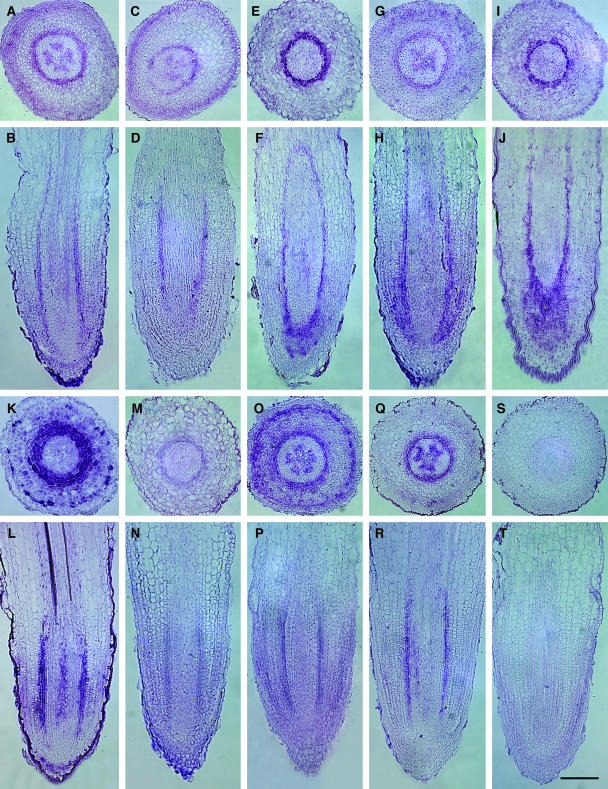
In situ hybridization using digoxigenin (DIG)-labeled antisense RNA probes showed the accumulation of all nine protoberberine alkaloid gene transcripts in one or more cell layers at the interface between the developing stele and ground tissues near the root apical meristem (Figure 3). All gene transcripts were localized to the immature endodermis (i.e., lacking a Casparian strip) and the pericycle ∼100 to 300 μm from the root apex. Most gene transcripts were associated with the innermost cell layers of the developing cortex adjacent to the endodermis. SOMT and 4′OMT gene transcripts were also abundant in outer cortical cell layers proximal to the exodermis (Figures 3K and 3O). TYDC, NCS, CNMT, SOMT, and CYP719A gene transcripts were associated with spokes of developing xylem in the same apical region of the root (Figures 3A, 3C, 3G, 3O, and 3Q). The inherent curvature of young roots produced longitudinal sections tangential to the stele in isolated regions of some samples, which led to the apparent distribution of gene transcripts beyond cell layers peripheral to the pericycle (Figures 3F and 3J). Protoberberine alkaloid biosynthetic gene transcripts were not detected in mature endodermal and cortical cells or in the pericycle of root sections undergoing secondary growth.
Figure 3.
Protoberberine Alkaloid Biosynthetic Gene Transcripts Are Localized to the Immature Endodermis and Surrounding Tissues in T. flavum Roots.
(A) to (R) In situ hybridization using DIG-labeled antisense probes for TYDC ([A] and [B]), NCS ([C] and [D]), 6OMT ([E] and [F]), CNMT ([G] and [H]), CYP80B ([I] and [J]), 4′OMT ([K] and [L]), BBE ([M] and [N]), SOMT ([O] and [P]), and CYP719A ([Q] and [R]) performed on cross ([A], [C], [E], [G], [I], [K], [M], [O], and [Q]) and longitudinal ([B], [D], [F], [H], [J], [L], [N], [P], and [R]) root sections.
(S) and (T) In situ hybridization using a DIG-labeled sense probe for CYP719A performed on cross (S) and longitudinal (T) root sections. Bar = 50 μm for (A) to (T).
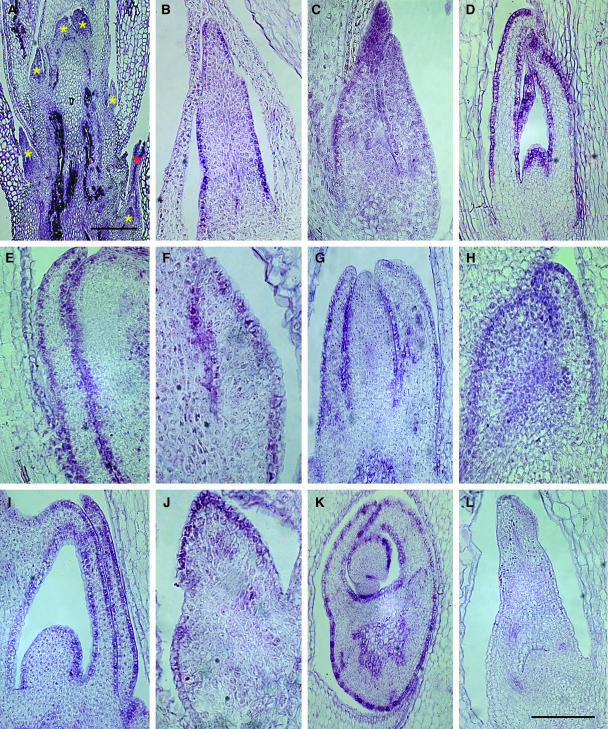
The rhizomes of T. flavum are composed of several compact internodes, from which leaf primordia and axillary buds are produced (Figure 4A). In situ hybridization using DIG-labeled antisense RNA probes showed the accumulation of all nine protoberberine alkaloid gene transcripts in the protoderm of leaf primordia (Figures 4B to 4J). Hybridization signals were associated with protoderm cells extending around the entire circumference of the leaf primordia from the base to the apex (Figure 4K). Protoberberine alkaloid biosynthetic gene transcripts were not detected in any other tissues, including the cortex and the pith, at any stage of rhizome development.
Figure 4.
Protoberberine Alkaloid Biosynthetic Gene Transcripts Are Localized to the Protoderm of Leaf Primordia in T. flavum Rhizomes.
(A) Rhizome longitudinal section stained with toluidine blue O.
(B) to (K) In situ hybridization using DIG-labeled antisense probes for TYDC (B), NCS (C), 6OMT ([D] and [K]), CNMT (E), CYP80B (F), 4′OMT (G), BBE (H), SOMT (I), and CYP719A (J) performed on longitudinal ([B] to [J]) and cross (K) rhizome sections.
(L) In situ hybridization using a DIG-labeled sense probe for CYP719A performed on a longitudinal rhizome section.
Yellow and red asterisks in (A) show the location of leaf primordia and axillary buds, respectively. Bar = 250 μm in (A) and 50 μm in (L). Bar in (L) also applies to (B) to (K).
Hybridization signals were not detected in any tissue when root (Figures 3S and 3T) or rhizome (Figure 4L) sections were exposed to sense RNA probes for CYP719A or to sense RNA probes corresponding to any other protoberberine alkaloid biosynthetic gene (data not shown). Sense probes were used at a fivefold higher concentration than that used for antisense RNA probes to confirm the specificity of hybridization between the antisense RNA probes and endogenous transcripts found in specific root and rhizome tissues.
Cell Type–Specific Localization of Protoberberine Alkaloids in Roots and Rhizomes
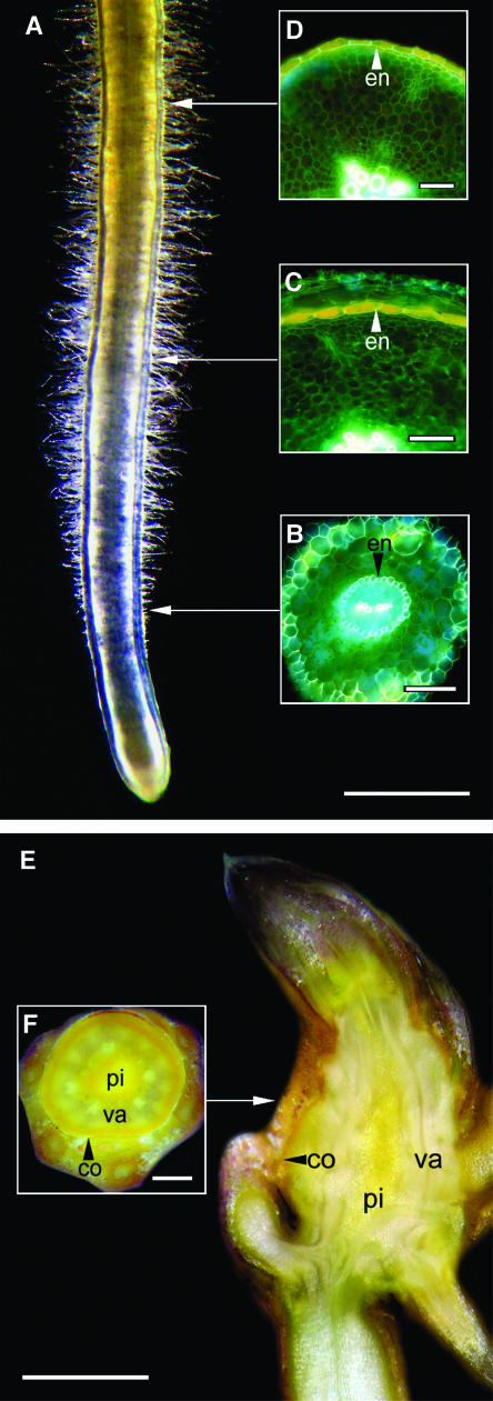
Protoberberine alkaloids can be readily visualized in fresh plant tissues because of the bright yellow color of berberine and related compounds. Protoberberine alkaloids were apparent along the length of T. flavum roots but were absent from segments extending ∼4 mm basipetal to the apical meristem (Figure 5A). The yellow color gradually became visible in root segments displaying maximum root hair extension. Quaternary protoberberine alkaloids, such as berberine, also exhibit strong yellow fluorescence under UV illumination at certain wavelengths. The fluorescence characteristics of T. flavum roots at various stages of development were examined (Figures 5D to 5F). In root sections near the apical meristem, an epidermis surrounded about five cortical cell layers, which enclosed a central stele (Figure 5B). The autofluorescence of the cell walls indicated the presence of an exodermis and an endodermis within the cortex (Figure 5B). The lignified secondary wall of the xylem elements also fluoresced strongly. In young roots lacking secondary growth, a yellow fluorescence corresponding to protoberberine alkaloids was absent (Figure 5B). When the root reached ∼4 mm in diameter, secondary growth was identified by the periclinal division of pericycle cells internal to the endodermis (Figure 5C). By contrast, the endodermis showed anticlinal division and expansion at the onset of secondary growth, and an abundance of protoberberine alkaloids appeared in the endodermal cells (Figure 5C). The endodermis continued to undergo extensive anticlinal cell division and expansion and ultimately displaced all external tissues in mature roots (Figure 5D), which expanded to ∼6 mm in diameter at the base of the stem. Protoberberine alkaloid accumulation was confined to the outermost region of mature roots, including the endodermis (Figure 5D) and three to four layers of adjacent pericycle cells (data not shown).
Figure 5.
Protoberberine Alkaloids Accumulate in the Mature Endodermis of Roots, and in the Pith and Cortex of Rhizomes, in T. flavum.
(A) to (D) Accumulation of protoberberine alkaloids in a fresh whole root (A) shown by light microscopy and in 0.5-mm cross sections of fresh roots at various stages of development ([B] to [D]) revealed by epifluorescence microscopy.
(E) and (F) Accumulation of protoberberine alkaloids in fresh longitudinal sections (E) and cross sections (F) of a rhizome shown by light microscopy.
ap, apical meristem; co, cortex; en, endodermis; ep, epidermis; ex, exodermis; pe, petiole; pi, pith; ro, root; va, vascular tissue. Bars = 500 μm in (A), 40 μm in (B) to (D), and 500 μm in (E) and (F).
The distribution of protoberberine alkaloids in fresh hand-cut sections of rhizomes was also determined (Figures 5E and 5F). In rhizomes, protoberberine alkaloids were abundant throughout the pith and the cortex but were absent from vascular tissues, including the secondary phloem and xylem. Similarly, protoberberine alkaloids were detected in the rib parenchyma of older petioles (Figure 5F).
DISCUSSION
We have shown that the cell type–specific localization of protoberberine alkaloid biosynthetic gene transcripts and product accumulation are temporally and spatially separated in T. flavum roots and rhizomes, respectively. A model depicting these relationships is shown in Figure 6. In roots, gene transcripts for nine consecutive biosynthetic enzymes (essentially the entire pathway) were localized to immature endodermis, pericycle, and, in some cases, adjacent cortical cells proximal to the apical meristem (Figure 3). In rhizomes, all gene transcripts were restricted to the protoderm of leaf primordia (Figure 4). By contrast, protoberberine alkaloid accumulation was restricted to the pith and cortex in rhizomes and to mature endodermis after the initiation of secondary growth in roots (Figure 5) but was also found in the pericycle once the endodermis was sloughed off (Samanani et al., 2002). Protoberberine alkaloid biosynthetic enzymes are almost certainly localized to the same cell types as their corresponding gene transcripts. However, immunolocalization studies would provide insight into the stability of these enzymes in the endodermis and pericycle of roots and in the protoderm of leaf primordia. In particular, the temporal distinction between the localization of gene transcripts and alkaloid product accumulation in the root endodermis implies that all biosynthetic enzymes remain active well after mRNA levels are no longer detectable. Nevertheless, our results clearly show that the biosynthesis and accumulation of protoberberine alkaloids in T. flavum are unique with respect to the cell type–specific localization of alkaloid metabolism in other plants (Facchini, 2001), including the evolutionarily related benzylisoquinoline alkaloid pathways in P. somniferum (Bird et al., 2003).
Figure 6.
The Cell Type–Specific Localization of Protoberberine Alkaloid Gene Transcripts and Product Accumulation Are Temporally and Spatially Distinct in T. flavum Roots and Rhizomes, Respectively.
(A) Model showing the relationship between the sites of protoberberine alkaloid biosynthetic gene expression (blue) and alkaloid accumulation (yellow) in a root.
(B) Model showing the relationship between the sites of protoberberine alkaloid biosynthetic gene expression (blue) and alkaloid accumulation (yellow) in a rhizome.
ap, apical meristem; co, cortex; en, endodermis; pi, pith; va, vascular tissue.
Berberine is the predominant yellow fluorescent compound in T. flavum, although other protoberberine alkaloids also accumulate (Velcheva et al., 1992). In contrast with phytoalexins, which are produced in response to pathogen challenge, berberine is a constitutive secondary metabolite with potent antimicrobial properties and, thus, is often referred to as a phytoanticipin (Luijendijk et al., 1996). Berberine inhibits DNA and protein synthesis (Ghosh et al., 1985) and confers protection against gram-positive and gram-negative bacteria and other microorganisms (Schmeller et al., 1997; Isawa et al., 1998). The abundant accumulation of berberine in T. flavum roots and rhizomes, which are surrounded by a multitude of soil-borne pathogens, is in agreement with the purported biological function of protoberberine alkaloids as phytoanticipins. The berberine content of roots and rhizomes increases as each organ grows (Samanani et al., 2002), suggesting that protoberberine alkaloid biosynthesis occurs throughout plant development.
The rhizome and older petioles of T. flavum are conspicuously yellow due to the abundance of protoberberine alkaloids throughout the pith, cortex, and rib parenchyma (Figures 5E and 5F). By contrast, protoberberine alkaloids were confined to endodermal cells in T. flavum roots (Figures 5A to 5D), except in the oldest parts of the root where three to four pericycle cell layers also accumulate alkaloids (Samanani et al., 2002). The initial pale blue autofluorescence of endodermal cells in the young root was due to the presence of phenolics in the cell wall (Figure 5B). Upon the initiation of secondary growth, protoberberine alkaloids were detected in the endodermis (Figures 5C and 5D). In T. flavum, the endodermis remained intact despite a rapid expansion of the stele (Figure 5C) and emerged as the outer cell layer of the mature root (Figure 5D). The accumulation of berberine in the endodermis and, subsequently, in the pericycle ensures the presence of a protective layer of peripheral cells containing potent antimicrobial alkaloids (Samanani et al., 2002).
The tissue-specific biosynthesis and accumulation of protoberberine alkaloids and the unusual development of the endodermis might have evolved as a mechanism to protect roots from soil-borne pathogens and perhaps other harmful organisms. Other antimicrobial secondary metabolites exhibit an abundant accumulation in peripheral root tissues. Saponins have been localized to the epidermal cells of oat (Avena sativa) roots as an initial barrier to fungal infection (Osbourn et al., 1994; Osbourn, 1996). Flavonoids and flavonoid biosynthetic enzymes also accumulate in the epidermis and adjacent cortical cells of Arabidopsis thaliana roots (Saslowsky and Winkel-Shirley, 2001). The tissue-specific association of protoberberine alkaloids with the unusual endodermis and pericycle of T. flavum roots suggests that these cells also act as an initial barrier to potential pathogens.
Endodermal and pericycle root tissues have been implicated in the biosynthesis and accumulation of several types of alkaloids. Putrescine N-methyltransferase and hyoscyamine 6β-hydroxylase catalyze the first and last steps in the biosynthesis of the tropane alkaloid scopolamine and are exclusively localized to the pericycle (Hashimoto et al., 1991; Suzuki et al., 1999). Putrescine N-methyltransferase also catalyzes the first step in nicotine biosynthesis and has been localized to endodermis, outer cortex, and xylem tissues in Nicotiana sylvestris (Shoji et al., 2000, 2002). However, tropinone reductase I, an intermediate enzyme in tropane alkaloid metabolism, resides in the endodermis and nearby cortical cells (Nakajima and Hashimoto, 1999); thus, tropane alkaloid pathway intermediates must be transported between cell types. Intercellular translocation of monoterpenoid indole alkaloid pathway intermediates has also been suggested to occur between internal phloem, epidermis, laticifers, and idioblasts in the leaves of C. roseus (St-Pierre et al., 1999; Irmler et al., 2000; Burlat et al., 2004). Moreover, tropane alkaloids and nicotine are transported from roots to shoots for storage (Facchini, 2001). By contrast, protoberberine alkaloid biosynthesis and accumulation in T. flavum roots appear to occur in the same cell, although we cannot rule out the possibility that products are translocated between endodermis and pericycle or transported to other organs. The formation and storage of acridone alkaloids were also associated with endodermis in Ruta graveolens (Junghanns et al., 1998). Localization of alkaloid biosynthetic enzymes to endodermis and pericycle provides efficient access to amino acid precursors from the phloem and to vascular conduits for the systemic transport of metabolic products.
The occurrence of benzylisoquinoline alkaloids in basal angiosperm families suggests an ancient evolutionary origin for this group of secondary metabolites (Facchini et al., 2004). A common phylogeny is supported by the extensive sequence homology among enzymes from different plant families operating at corresponding points in the benzylisoquinoline alkaloid pathway (Table 1). For example, molecular clones encoding BBE, which is involved in the formation of protoberberine and benzophenanthridine alkaloids, have been isolated from members of Papaveraceae, Berberidaceae, and Ranunculaceae and exhibit extensive sequence similarity. Despite the extensive similarity between biosynthetic enzymes, benzylisoquinoline alkaloid metabolism involves vascular cell types in P. somniferum (Bird et al., 2003) and does not involve endodermis, pericycle, protoderm, cortex, or pith tissues as in T. flavum. The emergence of a complex benzylisoquinoline alkaloid pathway in an ancestor common to the basal angiosperms is in contrast with the proposed independent recruitment of pyrrolizidine alkaloid biosynthetic enzymes in at least four different angiosperm lineages (Reimann et al., 2004). Two scenarios have emerged concerning the relationship between the cell type–specific localization and evolutionary origin of plant alkaloids. In the case of pyrrolizidine alkaloids, the differential localization of a key pathway enzyme has been interpreted to support the independent origin of the biosynthetic pathway in various lineages (Anke et al., 2004). By contrast, the differential cell type–specific localization of benzylisoquinoline alkaloid gene transcripts in T. flavum and P. somniferum implicates the migration of established pathways between cell types as a key feature of phytochemical evolution. Thus, it is possible that benzylisoquinoline alkaloid pathways have become established in cell types other than those identified in T. flavum and P. somniferum.
In T. flavum rhizomes, biosynthetic gene transcripts were specifically localized to the protoderm of leaf primordia (Figure 4), which was in sharp contrast with the widespread accumulation of protoberberine alkaloids throughout the cortex and pith (Figures 5E and 5F). The spatial separation of product formation and storage implicates the intercellular transport of protoberberine alkaloids from the protoderm to cortical and pith cells. A molecular clone for a multidrug-resistance protein (MDR)–type ATP binding cassette (ABC) transporter (Cjmdr1), identified as a putative berberine transporter, has been isolated (Yazaki et al., 2001) and characterized (Shitan et al., 2003) from C. japonica. RNA gel blot and in situ hybridization analyses showed the abundant and preferential expression of Cjmdr1 in the rhizome (Yazaki et al., 2001) and suggested the localization of gene transcripts to xylem tissues (Shitan et al., 2003). In C. japonica, berberine accumulates mainly in the rhizome, whereas alkaloid biosynthetic enzymes have been associated with roots (Fujiwara et al., 1993). CjMDR1 was localized to the plasma membrane and suggested to function as an influx pump in the transport of berberine from roots to rhizomes by unloading protoberberine alkaloids from the xylem (Shitan et al., 2003).
The involvement of an ABC transporter in the secretion of berberine into the medium by cultured C. japonica and T. minus cells has also been demonstrated (Sakai et al., 2002; Terasaka et al., 2003). However, cultured T. flavum cells do not secrete berberine into the culture medium, but instead sequester protoberberine alkaloids to the vacuole (N. Samanani and P. Facchini, unpublished results). Vacuolar accumulation of protoberberine alkaloids was also observed in endodermal, pericycle, pith, and cortical cells of T. flavum plants (Samanani et al., 2002). Screening the same T. flavum cell culture cDNA library that contained a multitude of molecular clones encoding protoberberine alkaloid biosynthetic enzymes with Cjmdr1 yielded a low abundance (i.e., one Cjmdr1 cDNA among ∼300,000 plaque-forming units) partial clone encoding a protein with 94% amino acid identity over the final 143 C-terminal residues (N. Samanani and P. Facchini, unpublished results). Thus, the inability of T. flavum cells to secrete berberine into the culture medium might be due to the low level of a plasma membrane–associated ABC transporter. The sequestration of protoberberine alkaloids to the vacuole suggests a different mechanism of intracellular transport in T. flavum cells. The accumulation of protoberberine alkaloids in the endodermis and pericycle (Samanani et al., 2002) of T. flavum roots also implicates a different transport mechanism compared with C. japonica. It is not known whether berberine biosynthesis in C. japonica roots is associated with endodermis and pericycle, but the products mostly accumulate in the rhizome (Fujiwara et al., 1993). Modulations in the expression and substrate specificity of transporters, such as CjMDR1, might be a major force in the evolution of secondary metabolism in addition to the recruitment of new catalytic functions after gene duplication events (Pichersky and Gang, 2000).
METHODS
Plant Cultivation
Thalictrum flavum ssp glaucum seeds were surface sterilized with 20% (v v−1) sodium hypochlorite for 15 min, rinsed with sterile water, and incubated on phytoagar at 4°C for 14 d. The seeds were transferred to phytagar containing B5 salts and vitamins (Gamborg et al., 1968) supplemented with 100 mg L−1 myo-inositol, 1 g L−1 hydrolyzed casein, 20 g L−1 sucrose, and 5 mg/L of GA3 and incubated at 23°C. After germination, T. flavum plants were maintained in vitro on phytagar-containing B5 salts and vitamins under a photoperiod of 16 h light/8 h dark at 23°C.
RNA Isolation and Analysis
Total RNA was isolated using the method of Logemann et al. (1987), and poly(A)+ RNA was selected by oligo(dT) cellulose chromatography. For gel blot analysis, 15 μg of RNA was fractionated on a 1.0% (w v−1) agarose gel, containing 7.5% (w v−1) formaldehyde, before transfer to a nylon membrane. Blots were hybridized with a random primer 32P-labeled, full-length NCS probe. Hybridizations were performed at 65°C in 0.25 mM sodium phosphate buffer, pH 8.0, 7% (w v−1) SDS, 1% (w v−1) BSA, and 1 mM EDTA. Blots were washed at 65°C, twice with 2× SSC and 0.1% (w/v) SDS and twice with 0.2× SSC and 0.1% (w v−1) SDS (1× SSC is 0.15 M NaCl and 0.015 M sodium citrate, pH 7.0), and autoradiographed with an intensifier at −80°C.
Molecular Cloning of cDNAs Encoding Berberine Biosynthetic Enzymes
A unidirectional oligo(dT)-primed cDNA library constructed in λUni-ZAPII XR (Stratagene, La Jolla, CA) using poly(A)+ RNA isolated from T. flavum cell suspension cultures (Samanani et al., 2004) was screened with probes for eight genes encoding enzymes involved in the biosynthesis of berberine, or other benzylisoquinoline alkaloids, from related plant species. The T. flavum NCS cDNA was isolated as described previously (Samanani et al., 2004).
Tissue Fixation and Embedding
Plant materials were immersed in FAA [50% (v v−1) ethanol, 5% (v v−1) acetic acid, and 3.7% (v v−1) formaldehyde] and fixed overnight at 4°C. The tissues were dehydrated using an ethanol/tertiary butanol (t-butanol) series (4:1:5, 5:2:3, 5:3.5:1.5, 4.5:5.5:0, 2.5:7.5:0, and 0:1:0 ethanol:t-butanol:water) with a 2-h incubation in each solution except for the final step, which was overnight. Paraplast Plus (Oxford Labware, St. Louis, MO) was added to a paraffin infiltration series (1:1, 6.7:3.3, and 1:0 wax:t-butanol) with overnight incubations for each step. Embedded tissues were cut into 10-μm sections using an American Optical 620 microtome. Sections were placed onto aminopropyltriethoxysilane-coated slides and incubated overnight at 37°C to firmly adhere sections to the slides.
In Situ RNA Hybridization
The NCS cDNA in pBluescript SK+ was used to synthesize sense and antisense DIG-labeled RNA probes using T3 and T7 RNA polymerases, respectively. DIG-labeled probes were hydrolyzed at 60°C in 40 mM sodium carbonate buffer, pH 10, to produce 200 to 400 nucleotide fragments. The pH was neutralized using 10% (v v−1) acetic acid and the precipitated RNA resuspended in 50 μL of deionized water. Sections were deparaffinized with xylene and rehydrated using an ethanol series (1:0, 1:0, 9.5:0.5, 7:3, and 1:1 ethanol:water), incubated for 30 min in prehybridization buffer (100 mM Tris-HCl, pH 8.0, and 50 mM EDTA) containing 2 to 10 μg mL−1 proteinase K (Roche Diagnostics, Indianapolis, IN), and blocked in TBS (10 mM Tris-HCl, pH 7.5, and 150 mM NaCl) containing 2 mg mL−1 Gly. Sections were postfixed in 3.7% (v v−1) formaldehyde in PBS (100 mM sodium phosphate buffer, pH 7.2, and 140 mM NaCl), incubated in 100 mM triethanolamine buffer, pH 8.0, containing 0.5% (v v−1) acetic anhydride, and rinsed in TBS. Slides were inverted onto 100 μL of hybridization buffer (10 mM Tris-HCl, pH 6.8, 10 mM sodium phosphate buffer, pH 6.8, 40% [v v−1] deionized formamide, 10% [w v−1] dextran sulfate, 300 mM NaCl, 5 mM EDTA, 1 mg mL−1 yeast tRNA, 0.8 units mL−1 RNase inhibitor [Invitrogen, Carlsbad, CA], and 200 to 1000 ng mL−1 of DIG-RNA) spread over a cover slip. Slides were sealed in a Petri dish lined with filter paper soaked in 50% (v v−1) formamide, incubated overnight at 50°C, then immersed in 2× SSC at 37°C. Sections were incubated in 50 μg mL−1 RNase A (Roche Diagnostics) in 500 mM NaCl, 10 mM Tris-HCl, pH 7.5, and 1 mM EDTA for 30 min at 37°C, and washed in 2 liters of the following solutions for 1 h: 2× SSC and 1× SSC at room temperature and 0.1× SSC at 65°C. Slides were rinsed in TBST (10 mM Tris-HCl, pH 7.5, 150 mM NaCl, and 0.3% [v v−1] Triton X-100), blocked for 1 h in TBST containing 2% (w v−1) BSA, inverted onto cover slips carrying 100 μL of goat anti-DIG alkaline phosphatase (AP) conjugate (Roche Diagnostics) diluted 1:200 in TBST containing 1% (w v−1) BSA, incubated for 2 h in sealed Petri dishes lined with TBST-soaked filter paper, and rinsed in TBST buffer. Color development was performed in AP buffer containing 400 μM 5-bromo-4-chloro-3-indolyl phosphate and 428 μM nitroblue tetrazolium for 24 h.
Localization of Berberine in Fresh Tissues
Fresh hand sections of T. flavum roots, rizomes, and petioles were prepared as described previously (Samanani et al., 2002) and examined for the presence of protoberberine alkaloids. A Leica Aristoplan fluorescence microscope (Leica Microsystems, Wetzlar, Germany) was used to detect the autofluorescence of protoberberine alkaloids in root sections using a Leica A-UV filter combination (excitation, 340 to 380 nm; emission, 425 nm). Protoberberine alkaloids were visualized in root and rhizome sections under visible light using a Leica MZ125 stereomicroscope.
Light Microscopy
Results of in situ hybridization were viewed and recorded using a Leica DM RXA2 microscope and a Retiga EX digital camera mounted with a RGB color liquid crystal filter (QImaging, Burnaby, BC, Canada). Images were digitally captured and color corrected using Open Lab version 2.09 (Improvision, Coventry, UK).
Sequence data for T. flavum have been deposited with the EMBL/GenBank data libraries under the following accession numbers: TYDC, AF314150; NCS, AY376412; 6OMT, AY610507; CNMT, AY610508; CYP80B, AY610509; 4′OMT, AY610510; BBE, AY610511; SOMT, AY610512; CYP719A, AY610513; MDR1, AY780675.
Acknowledgments
P.J.F. holds the Canada Research Chair in Plant Biotechnology. S.-U.P. was the recipient of a postdoctoral fellowship from the Alberta Ingenuity Fund. This work was funded by a grant from the Natural Sciences and Engineering Research Council of Canada to P.J.F.
The author responsible for distribution of materials integral to the findings presented in this article in accordance with the policy described in the Instructions for Authors (www.plantcell.org) is: Peter J. Facchini (pfacchin@ucalgary.ca).
Article, publication date, and citation information can be found at www.plantcell.org/cgi/doi/10.1105/tpc.104.028654.
References
- Anke, S., Niemüller, D., Moll, S., Hänsch, R., and Ober, D. (2004). Polyphyletic origin of pyrrolizidine alkaloids within the Asteraceae: Evidence from differential tissue expression of homospermidine synthase. Plant Physiol. 136, 4037–4047. [DOI] [PMC free article] [PubMed] [Google Scholar]
- Bird, D.A., Franceschi, V.R., and Facchini, P.J. (2003). A tale of three cell types: Alkaloid biosynthesis is localized to sieve elements in opium poppy. Plant Cell 15, 2626–2635. [DOI] [PMC free article] [PubMed] [Google Scholar]
- Burlat, V., Oudin, A., Courtois, M., Rideau, M., and St-Pierre, B. (2004). Co-expression of three MEP pathway genes and geraniol10-hydroxylase in internal phloem parenchyma of Catharanthus roseus implicates multicellular translocation of intermediates during the biosynthesis of monoterpene indole alkaloids and isoprenoid-derived primary metabolites. Plant J. 38, 131–141. [DOI] [PubMed] [Google Scholar]
- Choi, K.B., Morishige, T., Shitan, N., Yazaki, K., and Sato, F. (2002). Molecular cloning and characterization of coclaurine N-methyltransferase from cultured cells of Coptis japonica. J. Biol. Chem. 277, 830–835. [DOI] [PubMed] [Google Scholar]
- Facchini, P.J. (2001). Alkaloid biosynthesis in plants: Biochemistry, cell biology, molecular regulation, and metabolic engineering applications. Annu. Rev. Plant Physiol. Plant Mol. Biol. 52, 29–66. [DOI] [PubMed] [Google Scholar]
- Facchini, P.J., Bird, D.A., and St-Pierre, B. (2004). Can Arabidopsis make complex alkaloids? Trends Plant Sci. 9, 116–122. [DOI] [PubMed] [Google Scholar]
- Facchini, P.J., and De Luca, V. (1994). Differential and tissue-specific expression of a gene family for tyrosine/dopa decarboxylase in opium poppy. J. Biol. Chem. 269, 26684–26690. [PubMed] [Google Scholar]
- Facchini, P.J., and De Luca, V. (1995). Phloem-specific expression of tyrosine/dopa decarboxylase genes and the biosynthesis of isoquinoline alkaloids in opium poppy. Plant Cell 7, 1811–1821. [DOI] [PMC free article] [PubMed] [Google Scholar]
- Facchini, P.J., Penzes, C., Johnson, A.G., and Bull, D. (1996). Molecular characterization of berberine bridge enzyme genes from opium poppy. Plant Physiol. 112, 1669–1677. [DOI] [PMC free article] [PubMed] [Google Scholar]
- Fujiwara, H., Takeshita, N., Terano, Y., Fitchen, J.H., Tsujita, T., Katagiri, Y., Sato, F., and Yamada, Y. (1993). Expression of (S)-scoulerine 9-O-methyltransferase in Coptis japonica plants. Phytochemistry 34, 949–954. [Google Scholar]
- Gamborg, O.L., Miller, R.O., and Ojima, K. (1968). Nutrient requirements of suspension cultures of soybean root cells. Exp. Cell Res. 50, 151–158. [DOI] [PubMed] [Google Scholar]
- Ghosh, A.K., Bhattacharyya, F.K., and Ghosh, D.K. (1985). Leishmania donovani: Amatigote inhibition and mode of action of berberine. Exp. Parasitol. 60, 404–413. [DOI] [PubMed] [Google Scholar]
- Hashimoto, T., Hayashi, A., Amano, Y., Kohno, J., Iwanari, H., Usuda, S., and Yamada, Y. (1991). Hyoscyamine 6β-hydroxylase, an enzyme involved in tropane alkaloid biosynthesis, is localized at the pericycle of the root. J. Biol. Chem. 266, 4648–4653. [PubMed] [Google Scholar]
- Ikezawa, N., Tanaka, M., Nagayoshi, M., Shinkyo, R., Sakaki, T., Inouye, K., and Sato, F. (2003). Molecular cloning and characterization of CYP719, a methylenedioxy bridge-forming enzyme that belongs to a novel P450 family, from cultured Coptis japonica cells. J. Biol. Chem. 278, 38557–38565. [DOI] [PubMed] [Google Scholar]
- Irmler, S., Schröder, G., St-Pierre, B., Crouch, N.P., Hotze, M., Schmidt, J., Strack, D., Matern, U., and Schröder, J. (2000). Indole alkaloid biosynthesis in Catharanthus roseus: New enzyme activities and identification of cytochrome P450 CYP72A1 as secologanin synthase. Plant J. 24, 797–804. [DOI] [PubMed] [Google Scholar]
- Isawa, K., Nanba, H., Lee, D.U., and Kang, S.I. (1998). Structure-activity relationships of protoberberines having antimicrobial activity. Planta Med. 64, 748–751. [DOI] [PubMed] [Google Scholar]
- Junghanns, K.T., Kneusel, R.E., Gröger, D., and Matern, U. (1998). Differential regulation and distribution of acridone synthase in Ruta graveolens. Phytochemistry 49, 403–411. [DOI] [PubMed] [Google Scholar]
- Logemann, J., Schell, J., and Willmitzer, L. (1987). Improved method for the isolation of RNA from plant tissues. Anal. Biochem. 163, 16–20. [DOI] [PubMed] [Google Scholar]
- Luijendijk, T.J.C., Vandermeijden, E., and Verpoorte, R. (1996). Involvement of strictosidine as a defensive chemical in Catharanthus roseus. J. Chem. Ecol. 22, 1355–1366. [DOI] [PubMed] [Google Scholar]
- McCaskill, D., Gershenzon, J., and Croteau, R. (1992). Morphology and monoterpene biosynthetic capabilities of secretory cell clusters isolated from glandular trichomes of peppermint (Mentha piperita L.). Planta 187, 445–454. [DOI] [PubMed] [Google Scholar]
- Moll, S., Anke, S., Kahmann, U., Hansch, R., Hartmann, T., and Ober, D. (2002). Cell-specific expression of homospermidine synthase, the entry enzyme of the pyrrolizidine alkaloid pathway in Senecio vernalis, in comparison with its ancestor, deoxyhypusine synthase. Plant Physiol. 130, 47–57. [DOI] [PMC free article] [PubMed] [Google Scholar]
- Morishige, T., Tsujita, T., Yamada, Y., and Sato, F. (2000). Molecular characterization of the S-adenosyl-L-methionine:3′-hydroxy-N-methylcoclaurine 4′-O-methyltransferase involved in isoquinoline alkaloid biosynthesis in Coptis japonica. J. Biol. Chem. 275, 23398–23405. [DOI] [PubMed] [Google Scholar]
- Nakajima, K., and Hashimoto, T. (1999). Two tropinone reductases, that catalyze opposite stereospecific reductions in tropane alkaloid biosynthesis, are localized in plant root with different cell-specific patterns. Plant Cell Physiol. 40, 1099–1107. [DOI] [PubMed] [Google Scholar]
- Osbourn, A.E. (1996). Saponins and plant defence—A soap story. Trends Plant Sci. 1, 4–9. [Google Scholar]
- Osbourn, A.E., Clarke, B.R., Lunness, P., Scott, P.R., and Daniels, M.J. (1994). An oat species lacking avenacin is susceptible to infection by Gaeumannomyces graminin var. tritici. Physiol. Mol. Plant Pathol. 45, 457–467. [Google Scholar]
- Pichersky, E., and Gang, D.R. (2000). Genetics and biochemistry of secondary metabolites in plants: An evolutionary perspective. Trends Plant Sci. 5, 439–445. [DOI] [PubMed] [Google Scholar]
- Reimann, A., Nurhayati, N., Backenkohler, A., and Ober, D. (2004). Repeated evolution of the pyrrolizidine alkaloid-mediated defense system in separate angiosperm lineages. Plant Cell 16, 2772–2784. [DOI] [PMC free article] [PubMed] [Google Scholar]
- Reinold, S., and Hahlbrock, K. (1997). In situ localization of phenylpropanoid biosynthetic mRNAs and proteins in parsley (Petroselinum crispum). Bot. Acta 110, 431–443. [Google Scholar]
- Sakai, K., Shitan, N., Sato, F., Ueda, K., and Yazaki, K. (2002). Characterization of berberine transport into Coptis japonica cells and the involvement of ABC protein. J. Exp. Bot. 53, 1879–1886. [DOI] [PubMed] [Google Scholar]
- Samanani, N., and Facchini, P.J. (2001). Isolation and partial characterization of norcoclaurine synthase, the first committed step in benzylisoquinoline alkaloid biosynthesis, from opium poppy. Planta 213, 898–906. [DOI] [PubMed] [Google Scholar]
- Samanani, N., and Facchini, P.J. (2002). Purification and characterization of norcoclaurine synthase. The first committed enzyme in benzylisoquinoline alkaloid biosynthesis in plants. J. Biol. Chem. 277, 33878–33883. [DOI] [PubMed] [Google Scholar]
- Samanani, N., Liscombe, D.K., and Facchini, P.J. (2004). Molecular cloning and characterization of norcoclaurine synthase, an enzyme catalyzing the first committed step in benzylisoquinoline alkaloid biosynthesis. Plant J. 40, 302–313. [DOI] [PubMed] [Google Scholar]
- Samanani, N., Yeung, E.C., and Facchini, P.J. (2002). Cell type-specific protoberberine alkaloid accumulation in Thalictrum flavum. J. Plant Physiol. 139, 1189–1196. [Google Scholar]
- Saslowsky, D., and Winkel-Shirley, B. (2001). Localization of flavonoid enzymes in Arabidopsis roots. Plant J. 27, 37–48. [DOI] [PubMed] [Google Scholar]
- Schmeller, T., Latz-Brüning, B., and Wink, M. (1997). Biochemical activities of berberine, palmitine and sanguinarine mediating chemical defense against microorganisms and herbivores. Phytochemistry 44, 257–266. [DOI] [PubMed] [Google Scholar]
- Shitan, N., Bazin, I., Dan, K., Obata, K., Kigawa, K., Ueda, K., Sato, F., Forestier, C., and Yazaki, K. (2003). Involvement of CjMDR1, a plant multidrug-resistance-type ATP-binding cassette protein, in alkaloid transport in Coptis japonica. Proc. Natl. Acad. Sci. USA 100, 751–756. [DOI] [PMC free article] [PubMed] [Google Scholar]
- Shoji, T., Winz, R., Iwase, T., Nakajima, K., Yamada, Y., and Hashimoto, T. (2002). Expression patterns of two tobacco isoflavone reductase-like genes and their possible roles in secondary metabolism in tobacco. Plant Mol. Biol. 50, 427–440. [DOI] [PubMed] [Google Scholar]
- Shoji, T., Yamada, Y., and Hashimoto, T. (2000). Jasmonate induction of putrescine N-methyltransferase genes in the root of Nicotiana sylvestris. Plant Cell Physiol. 41, 831–839. [DOI] [PubMed] [Google Scholar]
- St-Pierre, B., Vazquez-Flota, F.A., and De Luca, V. (1999). Multicellular compartmentation of Catharanthus roseus alkaloid biosynthesis predicts intercellular translocation of a pathway intermediate. Plant Cell 11, 887–900. [DOI] [PMC free article] [PubMed] [Google Scholar]
- Suzuki, K., Yamada, Y., and Hashimoto, T. (1999). Expression of Atropa belladonna putrescine N-methyltransferase gene in root pericycle. Plant Cell Physiol. 40, 289–297. [DOI] [PubMed] [Google Scholar]
- Takeshita, N., Fujiwara, H., Mimura, H., Fitchen, J.H., Yamada, Y., and Sato, F. (1995). Molecular cloning and characterization ofS-adenosyl-L-methionine:scoulerine-9-O-methyltransferase from cultured cells of Coptis japonica. Plant Cell Physiol. 36, 29–36. [PubMed] [Google Scholar]
- Terasaka, K., Sakai, K., Sato, F., Yamamoto, H., and Yazaki, K. (2003). Thalictrum minus cell cultures and ABC-like transporter. Phytochemistry 62, 483–489. [DOI] [PubMed] [Google Scholar]
- Velcheva, M., Dutschewska, H., and Samuelsson, G. (1992). The alkaloids of the roots of Thalictrum flavum L. Acta Pharm. Nord. 4, 57–58. [PubMed] [Google Scholar]
- Yazaki, K., Shitan, N., Takamatsu, H., Ueda, K., and Sato, F. (2001). A novel Coptis japonica multidrug-resistant protein preferentially expressed in the alkaloid-accumulating rhizome. J. Exp. Bot. 52, 877–879. [DOI] [PubMed] [Google Scholar]


