

Abstract
Objectives:
Major surgical approaches for volar plating of the distal radius include the standard flexor carpi radialis (FCR) approach, the extended FCR (eFCR) approach, and the extended FCR approach combined with radial-sided carpal tunnel release (eFCR+CTR). The purpose of this study was to determine which of these three surgical approaches offers the greatest exposure and visualization of the distal radius.
Methods:
Sequential dissections were performed on each of 30 fresh frozen below elbow cadaveric samples in order to simulate the three surgical approaches for distal radius volar plating, starting with the standard FCR approach, advancing to eFCR, and finishing with eFCR+CTR. Prior to the initial dissection of each cadaveric sample, radiographs were taken in order to calculate the total area of the distal radius. Then, following each sequential dissection, photographs were taken of each specimen and analyzed with an image measuring software in order to obtain the area of distal radius exposed. The percentage of total distal radius exposure was then calculated for each of the three surgical approaches.
Results:
The eFCR+CTR approach offered the greatest average distal radius exposure at 87% of total distal radius visualized. The eFCR approach provided the next greatest exposure with an average of 73% visualized, followed by the standard FCR approach with an average of 61% visualized.
Conclusion:
The extended FCR approach with radial-sided carpal tunnel release is both safe and efficacious for osteosynthesis of distal radius fractures in the setting of concomitant carpal tunnel syndrome. This study demonstrates that an additional advantage of this approach includes improved surgical exposure and visualization of the distal radius. This surgical approach is a valuable addition to any upper extremity surgeon’s armamentarium and should be considered when treating difficult distal radius fractures.
Key Words: Carpal tunnel syndromes, Distal radius fracture, Fracture osteosynthesis, Internal fracture fixation, Open fracture reductions
Introduction
Multiple surgical approaches have been described for volar plating of the distal radius including the standard flexor carpi radialis (FCR) approach, the extended FCR approach, and the extended FCR approach combined with radial-sided carpal tunnel release [Figure 1].1 Which approach affords the most exposure of the distal radius has yet to be determined. Reviewing our prior clinical experience treating complex distal radius fractures with concomitant carpal tunnel syndrome, we noticed that the addition of the radial-sided carpal tunnel release to the extended FCR approach appeared to have the added advantage of greater visualization of the distal radius fracture fragments intraoperatively. Greater visualization may lead to easier and more accurate fracture reduction. The purpose of this study is to determine which surgical approach affords the most exposure of the distal radius and to quantify how much of the distal radius is exposed with each approach. The three surgical approaches in question are: 1) the standard FCR approach, 2) the extended FCR approach, and 3) the extended FCR approach combined with a radial-sided carpal tunnel release.1-5 We hypothesize that performing an extended FCR approach combined with a radial-sided carpal tunnel release will yield better overall visualization of the distal radius compared to other well-described approaches.
Figure 1.

Diagram of the extended flexor carpi radialis approach with radial-sided carpal tunnel release (eFCR + CTR)
Materials and Methods
Sequential dissections were performed on 30 cadaveric specimens in this study. All cadaver specimens were fresh frozen below elbow preparations and ranged in age from 18 to 99 years of age. Wrist circumference of the specimens ranged from 13.5 centimeters (cm) to 21 cm. Given the varying sizes of wrists, area was reported as an overall percentage based on an initial radiograph. The cadavers were examined prior to the procedure in order to assess for deformities, surgery or fractures that could skew the results. None of the specimens demonstrated any visual or radiographic evidence of injury distal to their elbows nor was there any reported history of injury or previous surgery. An a priori power analysis was performed on the basis of three preliminary cadaveric dissections which concluded that 30 below the elbow cadaver arms would provide 80% power ( = 0.05) to detect a clinically meaningful, statistically significant difference between surgical approaches. This sample size provided similar power to a previous cadaver study evaluating exposure visualization.6 Pre-dissection radiographs were obtained on all cadaver specimens; of note, these radiographs included size calibration markers for later digital measurement reference. Each of the 30 cadaveric arms was then dissected sequentially starting with a standard FCR approach, advancing to an extended FCR approach and, finally, adding a radial-sided carpal tunnel release. The standard FCR approach, or volar approach to the wrist as originally described by Henry in 1973, was performed in this experiment by making an initial incision along the boundary between the FCR and the radial artery7—this incision extended distally from the wrist crease to seven centimeters proximally from the wrist crease in all cadavers [Figure 2]. Of note, the FCR sheath was then released along this same seven-centimeter span. This initial dissection was expanded to an extended FCR approach as described by Orbay et al. in 2001 by extending the distal limb of the incision over the thenar eminence8; in this experiment, this distal extension was standardized to three centimeters in length on all cadavers [Figure 3]. In this extended FCR approach, the FCR sheath was further released by the same three-centimeter span distally. Lastly, a radial-sided carpal tunnel release was added to the extended FCR approach as originally described by Weber and Sanders in 1997 by dividing both the superficial and deep leaves of the transverse carpal ligament (TCL) at the margin between the radially retracted FCR and the ulnarly retracted flexor pollicis longus (FPL) [Figures 1 and 4].9
Figure 2.
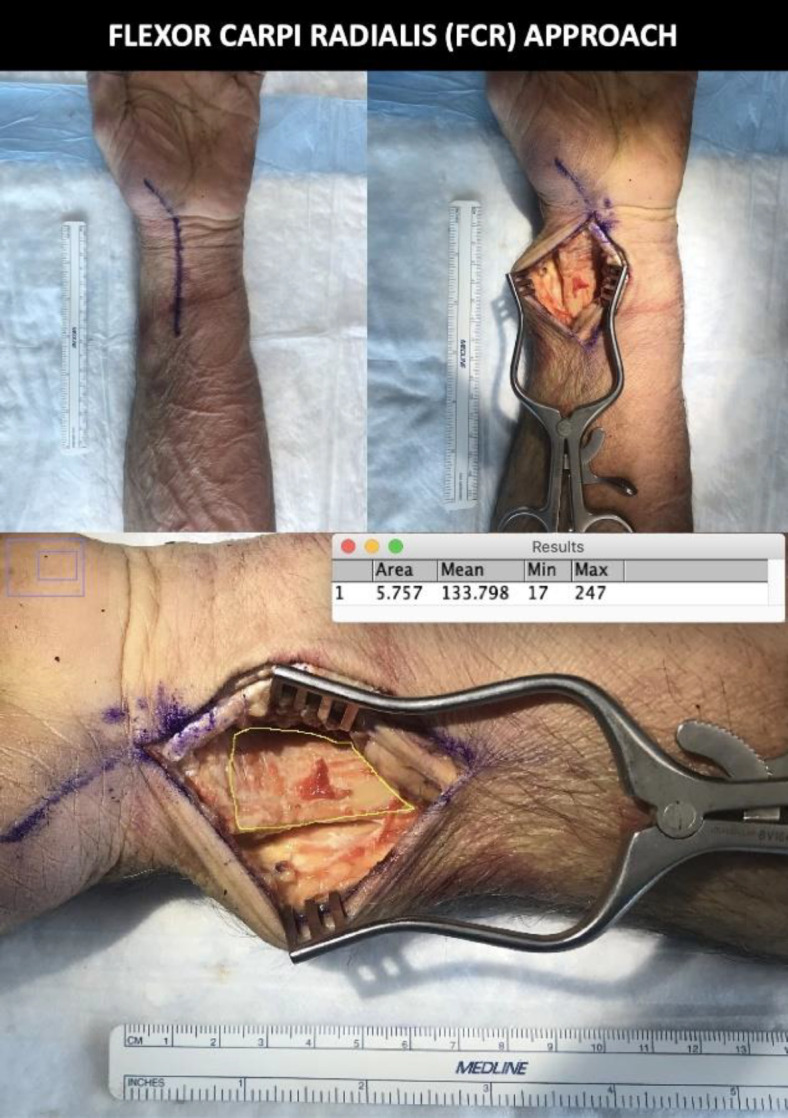
Example of flexor carpi radialis (FCR) approach with calculated area
Figure 3.

Example of extended flexor carpi radialis (eFCR) approach with calculated area
Figure 4.

Example of extended flexor carpi radialis approach with radial-sided carpal tunnel release (eFCR + CTR) and calculated exposure
Immediately after each sequential dissection of the cadaver specimen, a photo was taken of the exposure from the surgeon’s vantage point immediately adjacent to the surgeon dissector’s eye level using a smartphone camera. Similarly to the pre-dissection radiographs, included in each photo was a ruler positioned next to the cadaver sample in order to serve as a size calibration marker for later digital measurement reference. Of important note, the same standardized weitlaner retractor and the same centralized position was used in all cases. Additionally, the retractor was opened maximally to ensure uniformity throughout the dissection and data collection process.
After the pre-dissection radiograph, the three sequential dissections, and the three accompanying smartphone camera photos were performed for each of the 30 cadavers, attention was turned to the digital processing of the data in order to determine the area of distal radius exposure afforded by each surgical approach. This was accomplished by utilizing the ImageJ software program (National Institutes of Health, Bethesda, MD, USA). The ImageJ program has been previously validated and allows for quantification of the area of a two-dimensional space using a calibrated digital image.10 The first step in the digital processing of this data entailed determining the “total distal radius area” by manually outlining this corresponding area on each cadaver’s pre-dissection radiograph in ImageJ [Figure 5]. Of note, the proximal boundary of this outlined area (the line transecting the radial diaphysis) was standardized utilizing a calibrated set scale: First, the camera photo from each cadaver’s initial dissection (standard FCR approach) was used as reference, and the distance from the central aspect of the distal radius articular surface to the most proximal portion of visualized radial shaft was measured in ImageJ. Then, this same distance was manually traced, after recalibration, over the pre-dissection radiograph in ImageJ proximally from the central articular surface of the distal radius in order to set the proximal boundary of the eventual “total distal radius” outline [Figure 6]. Once this outline was traced over each pre-dissection radiograph, the ImageJ program then allowed for automatic calculation of the total area bound by each distal radius outline [Figure 5]. In a similar fashion, the outline of distal radius visible in each of the three surgical exposures was then manually traced in ImageJ for each of the three corresponding dissection images, allowing for automatic calculation of the area of distal radius visualization obtained with each exposure. Figures 2-4 illustrate examples of each dissection and its corresponding image area calculation using the ImageJ software in units of square centimeters [Figures 2-4].
Figure 5.

Radiograph of pre-dissected cadaver arm to obtain total area of distal radius measured with ImageJ software (National Institutes of Health, Bethesda, MD, USA)
Figure 6.

Illustrated example of the calibrated set scale system employed to standardize the proximal boundary of the “total distal radius area” manually traced by the experimenter in the ImageJ software program (National Institutes of Health, Bethesda, MD, USA) for each cadaver’s pre-dissection radiograph. Abbreviations used: “FCR” = flexor carpi radialis
The area of the exposure was reported as a percentage of the total distal radius area as measured from the pre-dissection radiographs ((area of surgical distal radius exposure/radiographic total distal radius area) x100). Visual exposures from the three distinct surgical approaches were compared using a one-way analysis of variance with =0.05.
Of note in this study, all radiographic and cadaver measurements and calculations in ImageJ as well as all dissections were performed by the same, single surgeon/observer.
Results
All cadaveric specimens were obtained from Science Care (Phoenix, AZ) and included 16 males and 14 females. Of note, this distribution of male versus female cadaveric specimens was based solely on cadaver availability at the time of this study rather than an intentional distribution of male versus female specimens.
The standard FCR approach demonstrated an average percentage of total distal radius exposed of 61% (range of 45%-76% exposure). The extended FCR approach demonstrated an average of 73% of distal radius exposed (range of 58%-93% exposure). Finally, the extended FCR approach with radial-sided carpal tunnel release demonstrated an average of 87% of distal radius exposed (range of 67%-100% exposure). One-way analysis of variance demonstrated that there was a significant difference in average percentage of distal radius exposed for each surgical approach compared to the other (P<0.05) [Table 1].
Table 1.
Distal Radius Exposure Attained by Surgical Approach—Standard FCR vs. Extended FCR vs. Extended FCR with Radial-Sided CTR
| Measure/Surgical Approach | Average Value— All Specimens (N=30) |
|---|---|
| Baseline Radiograph: Total Surface Area of Distal Radius (cm2) | 9.24 cm2 |
| Standard FCR Approach: Surface Area of Distal Radius Visualized (cm2) | 5.63 cm2 |
| Standard FCR Approach: Percentage of Total Distal Radius Exposed (%) | *61% |
| Extended FCR Approach: Surface Area of Distal Radius Visualized (cm2) | 6.65 cm2 |
| Extended FCR Approach: Percentage of Total Distal Radius Exposed (%) | *73% |
| Extended FCR Approach w/Radial-Sided CTR: Surface Area of Distal Radius Visualized (cm2) | 8.01 cm2 |
| Extended FCR Approach w/Radial-Sided CTR: Percentage of Total Distal Radius Exposed (%) | *87% |
FCR = flexor carpi radialis. CTR = carpal tunnel release. *Indicates statistically significant difference in percentage of total distal radius exposed when compared to the other two surgical approaches (P<0.05)
Discussion
The traditional volar Henry approach for distal radius fracture fixation does not provide enough surgical exposure for concomitant carpal tunnel release through the same incision.11,12 Alternative surgical exposures to decompress the median nerve during volar plate osteosynthesis include a second separate incision directly over the carpal tunnel or the volar Henry approach with extension across the wrist crease over the carpal tunnel on the ulnar side of palmaris longus, which places the palmar cutaneous branch at risk.13 Alternatively, the volar ulnar approach between the digital flexor tendons and the flexor carpi ulnaris does allow for concomitant distal radius fracture fixation as well as concomitant carpal tunnel release. However, Lattmann et al.14 recommended abandoning this approach for distal radius fracture fixation due to increased frequency of median nerve symptoms due to traction neuropraxia after surgery that were often permanent. Tannan et al.5 demonstrated that the extended FCR approach with radial-sided carpal tunnel release was both efficacious and safe with regards to the treatment of distal radius fractures and concomitant carpal tunnel release. The authors of this study did not report any neuropraxias of the median nerve or injury to the recurrent motor branch. All patients had improvement in functional outcome scores and sensibility by 6 weeks postoperatively.5
The results of our study supported our hypothesis by demonstrating that the extended FCR approach combined with a radial-sided carpal tunnel release provides wider surgical exposure with greater surface area of the distal radius visualized. Greater visualization of the distal radius can facilitate fracture reduction. Improved visibility may be helpful when working alone, without relying on an assist for retraction [Figure 7]. The extended FCR approach with a radial-sided carpal tunnel release has the additional advantage of avoidance of median nerve neuropraxia and temporary or permanent median nerve dysfunction that have been reported with the volar-ulnar approach.
Figure 7.

Example of flexor carpi radialis approach with radial-sided carpal tunnel release (eFCR + CTR) for isolated volar Barton’s fracture dislocation with nascent (3-week-old) malunion
In the setting of concomitant median nerve contusion or acute carpal tunnel syndrome, the extended FCR approach with radial-sided carpal tunnel release may be used to treat challenging distal radius fractures and release the carpal tunnel successfully and safely without the need for a separate incision in the palm of the hand.
The findings of this study should be considered along with its limitations. All measurements were performed by a single surgeon. There may be minor differences in how the area exposure dimensions of the distal radius are manually drawn for each sample and dissection, which affects how the overall area is calculated. We attempted to offset this by measuring each area three different times and reporting the average of each measurement. In addition, areas are reported as percentages instead of an actual unit of measurement. This was performed given that each cadaver arm differs in size. We aimed to account for this variation by reporting the surgically exposed areas as percentages of the distal radius area as seen on radiographs. This reporting, however, adds another manual measurement and thus additional potential for variation in the data. Lastly, subtle variations in retractor placement or force of retraction can alter the exposed area. We standardized our retractors throughout the dissections to include one centrally placed self-retaining weitlaner retractor opened maximally to ensure uniformity throughout the dissection and data collection process.
Conclusion
In conclusion, the extended FCR approach with radial-sided carpal tunnel release is both safe and efficacious for osteosynthesis of distal radius fractures in the setting of concomitant carpal tunnel syndrome. This study demonstrates that an additional advantage of this approach includes improved surgical exposure with regards to the distal radius. This surgical approach is thus a valuable addition to any upper extremity surgeon’s armamentarium and should be considered when treating difficult distal radius fractures, particularly when an assist is not available for manual retraction.
Acknowledgment
Not applicable
Contribution of authors:
IM and ET researched literature and conceived the study. IM was responsible for protocol development, acquisition of cadaveric samples, performance of the surgical procedures and measurements, data analysis, and writing the first draft of the manuscript. All authors reviewed and edited the manuscript and approved the final version of the manuscript.
Conflict of interest:
IM and RS have received funding from Acumed [Hillsboro, Oregon, USA].
Funding:
This work was supported by Acumed [Hillsboro, Oregon, USA].
References
- 1.Wijfells MM, Orbay JL, Indriago I, Ring D. The extended flexor carpi radialis approach for partially healed malaligned fractures of the distal radius. Injury. 2012;43(7):1204–8. doi: 10.1016/j.injury.2012.04.002. [DOI] [PubMed] [Google Scholar]
- 2.Dennison DG. Median nerve injuries associated with distal radius fractures. Tech Orthop. 2006;21(1):48–53. [Google Scholar]
- 3.Gwathmey FW, Brunton LM, Pensy RA, Chhabra AB. Volar plate osteosynthesis of distal radius fractures with concurrent prophylactic carpal tunnel release using a hybrid flexor carpi radialis approach. J Hand Surg. 2010;35(7):1082–1088. doi: 10.1016/j.jhsa.2010.03.043. [DOI] [PubMed] [Google Scholar]
- 4.Pensy RA, Brunton LM, Parks BG, Higgins JP, Chhabra AB. Single-incision extensile volar approach to the distal radius and concurrent carpal tunnel release: cadaveric study. J Hand Surg. 2010;35(2):217–222. doi: 10.1016/j.jhsa.2009.11.011. [DOI] [PubMed] [Google Scholar]
- 5.Tannan SC, Pappou IP, Gwathmey FW, Freilich AM, Chhabra AB. The extended flexor carpi radialis approach for concurrent carpal tunnel release and volar plate osteosynthesis for distal radius fracture. J Hand Surg. 2015;40(10):2026–2031. doi: 10.1016/j.jhsa.2015.07.001. [DOI] [PubMed] [Google Scholar]
- 6.Jockel CR, Zlotolow DA, Butler RB, Becker EH. Extensile surgical exposures of the radius: a comparative anatomic study. J Hand Surg. 2013;38(4):745–752. doi: 10.1016/j.jhsa.2012.12.029. [DOI] [PubMed] [Google Scholar]
- 7.Henry AK. Extensile exposures. 2st ed . Edinburgh: Churchill Livingston; 1973. [Google Scholar]
- 8.Orbay JL, Badia A, Indriago IR, et al. The extended flexor carpi radialis approach: a new perspective for the distal radius fracture. Tech Hand up Extrem Surg. 2001;5(4):204–211. doi: 10.1097/00130911-200112000-00004. [DOI] [PubMed] [Google Scholar]
- 9.Weber RA, Sanders WE. Flexor carpi radialis approach for carpal tunnel release. J Hand Surg. 1997;22(1):120–6. doi: 10.1016/S0363-5023(05)80191-1. [DOI] [PubMed] [Google Scholar]
- 10.Lichstein PM, Kleimeyer JP, Githens M, et al. Does the Watson-Jones or Modified Smith-Petersen approach provide superior exposure for femoral neck fracture fixation? Clin Orthop. 2018;476(7):1468–1476. doi: 10.1097/01.blo.0000533627.07650.bb. [DOI] [PMC free article] [PubMed] [Google Scholar]
- 11.Tordjman D, Hinds RM, Ayalon O, Yang SS, Capo JT. Volar-ulnar approach for fixation of the volar lunate facet fragment in distal radius fractures: a technical tip. J Hand Surg. 2016;41(12):e491–e500. doi: 10.1016/j.jhsa.2016.09.007. [DOI] [PubMed] [Google Scholar]
- 12.Zemirline A, Taleb C, Naito K, Vernet P, Liverneaux P, Lebailly F. Distal radius fracture fixation with a volar locking plate and endoscopic carpal tunnel release using a single 15 mm approach: feasbility study. Hand Surg and Rehab. 2018;37(4):231–237. doi: 10.1016/j.hansur.2018.03.006. [DOI] [PubMed] [Google Scholar]
- 13.Conti Mica MA, Bindra R, Moran SL. Anatomic considerations when performing the modified Henry approach for exposure of distal radius fractures. J Orthop. 2017;14(1):104–107. doi: 10.1016/j.jor.2016.10.015. [DOI] [PMC free article] [PubMed] [Google Scholar]
- 14.Lattmann T, Dietrich M, Meier C, Kilgus M, Platz A. Comparison of 2 surgical approaches for volar locking plate osteosynthesis of the distal radius. J Hand Surg. 2008;33(7):1135–1143. doi: 10.1016/j.jhsa.2008.03.016. [DOI] [PubMed] [Google Scholar]