Abstract
Background
Stress hyperglycemia ratio (SHR), associated with adverse outcomes in patients with ST-segment elevation myocardial infarction (STEMI), has several definitions. This study aims to assess the prognostic value of SHR, derived from hemoglobin A1c (HbA1c) or glycated albumin (GA), to mortality.
Methods
The study comprised 1,643 STEMI patients who underwent percutaneous coronary intervention (PCI) in two centers. SHR1 was calculated using fasting blood glucose (FBG)/GA, while SHR2 was calculated using the formula FBG/(1.59*HbA1c-2.59). The primary endpoints were in-hospital death and all-cause mortality, with a median follow-up duration of 1.56 years.
Results
Higher SHR1 and SHR2 values are associated with increased risks of in-hospital death and all-cause mortality. Each standard deviation increase in SHR1 corresponded to a 39% and 22% escalation in in-hospital death and all-cause mortality, respectively. The respective increases for SHR2 were 51% and 26%. Further examinations validated these relationships as linear. Additionally, the areas under the curve (AUC) for in-hospital death were not significantly different between SHR1 and SHR2 (p > 0.05). Incorporating SHR1 or SHR2 into the base model significantly improved the discrimination and risk reclassification for in-hospital and all-cause mortality. A subgroup analysis revealed that the effects of SHR1 and SHR2 were more pronounced in patients with hypercholesteremia.
Conclusion
SHR1 and SHR2 have emerged as robust and independent prognostic markers for STEMI patients undergoing PCI. The SHR calculation based on either HbA1c or GA can provide additional predictive value for mortality beyond traditional risk factors, helping to identify high-risk STEMI patients.
Supplementary Information
The online version contains supplementary material available at 10.1186/s12933-023-02061-6.
Keywords: Stress-induced hyperglycemia, ST-segment elevation myocardial infarction, Glycated albumin, Glycated hemoglobin A1c, Prognosis
Introduction
Cardiovascular disease, is a leading cause of morbidity and mortality globally and frequently presents clinically as acute myocardial infarction (AMI) for the first time [1, 2]. Despite a relative decrease, ST-segment elevation myocardial infarction (STEMI) remains approximately 40% of all AMI presentations [3]. The STEMI mortality rate has been reduced due to advancements in percutaneous coronary intervention (PCI) and pharmaceutical therapies. However, this decrease has reached a plateau, and the mortality rate remains high [4].
Stress hyperglycemia ratio (SHR), a novel indicator of stress-induced hyperglycemia status, pertains to the temporary spike in blood glucose levels in critically ill patients [5–7]. Previous studies have suggested that SHR is linked to a poor prognosis in acute coronary syndrome (ACS) patients [8–10]. However, these studies predominantly concentrated on unstable angina and non-STEMI [8] or simply evaluated the association between SHR and short-term mortality [9, 10]. Furthermore, the literature reveals varying definitions of SHR, dictated by different average glucose level formulas. Hemoglobin A1c (HbA1c) is typically used to calculate this average. A recent study has revealed that glycated albumin (GA), a marker reflecting 2–3 weeks glucose levels, may provide a more accurate calculation [11, 12]. The association between diverse SHR definitions based on HbA1c or GA and the mortality risk associated with STEMI patients undergoing PCI remains unexplored, thus necessitating further investigation.
Therefore, our study’s objective is to assess the prognostic value of different definitions of SHR, calculated on either HbA1c or GA, regarding in-hospital death and long-term mortality in STEMI patients who underwent PCI.
Methods
Study design and population
This retrospective cohort study was conducted in two tertiary academic hospitals: The Second Affiliated Hospital of Zhejiang University School of Medicine and the First People's Hospital of Yulin. A total of 1,643 patients who underwent emergency PCI for STEMI between January 1, 2016, and December 31, 2021, were included in the study. The diagnosis criteria of STEMI was established based on definitive clinical signs of myocardial ischemia and novel ischemic alterations on the electrocardiogram, specifically novel ST-segment elevation in two sequential leads (V2-V3 leads ≥ 2 mm in males aged ≥ 40 years, ≥ 2.5 mm in males aged < 40 years, and ≥ 1.5 mm in females irrespective of age; other leads: ≥ 1 mm). Patients were excluded if they: (1) lacked essential laboratory data, such as fasting blood glucose (FBG), GA, and HbA1c; (2) presented with cardiogenic shock; (3) had a life expectancy of less than 9 months; (4) were pregnant or lactating. Coronary angiography and PCI aligned with pertinent guidelines [2, 13]. All patients were administered 300 mg of aspirin, 180 mg of ticagrelor (or 300–600 mg of clopidogrel), and 100 IU/kg of heparin. The PCI was performed via radial or femoral artery access deploying a 6 or 7-Fr catheter, adhering to the standard technique employed by experienced cardiologists. The utilization of glycoprotein IIb/IIIa antagonists was determined by the operator, contingent on the patient's specific clinical state. The study was approved by the local committee. The requirement for written consent was waived because the study was retrospective and non-intrusive.
Data collection
In this study, demographic and clinical data including ischemic time, age, sex, hypertension, diabetes, hypercholesteremia, atherosclerotic cardiovascular disease (ASCVD), and smoking status, were collected from all participating patients. The diabetes was identified based on historical medical records, documented use of oral hypoglycemic agents or insulin, or an HbA1c level that surpassed 6.5%. Hypercholesterolemia was defined as total cholesterol ≥ 200 mg/dL, high-density lipoprotein (HDL) < 40 mg/dL, low-density lipoprotein (LDL) ≥ 130 mg/dL, or documented use of cholesterol-lowering drugs. Additionally, the gathered clinical data incorporated laboratory parameters, angiographic characteristics, and the treatment regimens implemented at admission and upon discharge. The presence of stenosis exceeding 70% in a minimum of two distinct coronary arteries served as the definition for multivessel coronary disease.
Fasting venous blood samples were obtained within 24 h upon patients’ admission and swiftly transported to the laboratory. GA was expressed as a percentage of total serum albumin to denote GA levels, while HbA1c was a proportion of glycated hemoglobin to total serum hemoglobin. Two measures, namely, SHR1 = FBG (mmol/L)/GA (%) [12] and SHR2 = (FBG (mmol/L))/ (1.59 × HbA1c (%)—2.59) [8], were employed to delineate stress hyperglycemia. The choice of utilizing fasting glucose as the numerator, rather than admission blood glucose (ABG), was anchored in its enhanced prognostic significance in patients with acute cardiovascular disease [14, 15], relative insensitivity to food or other sugar infusions, and limited inter-individual variability [17, 18]. Furthermore, patients were stratified into four distinct subgroups based on the SHR1 quartiles (Q1 ≤ 0.359, Q2 0.359–0.410, Q3 0.410–0.477, and Q4 > 0.477) or SHR2 (Q1 ≤ 0.746, Q2 0.746–0.843, Q3 0.843–0.979, and Q4 > 0.979).
Follow-up and endpoint
The primary endpoints were defined as in-hospital death and all-cause mortality in STEMI patients treated with PCI. The designated cut-off for long-term follow-up was set for October 1, 2022, ensuring a minimum of 9 months of follow-up. The follow-up data acquisition was facilitated by specially trained independent staff, who collated clinical outcomes via telephonic communication, outpatient consultations, or medical record evaluation. Thus, the data obtained were subject to an adjudication conducted by independent medical experts intentionally kept uninformed regarding the specific study details.
Statistical analysis
A multivariable logistic regression was employed, and odds ratio (OR) and 95% confidence interval (95% CI) were calculated to evaluate the relationship between SHR1, SHR2, and in-hospital mortality. In contrast, a multivariable Cox regression was utilized to assess the association between SHR1, SHR2, and all-cause mortality, and hazard ratio (HR) and 95% CI were calculated. Three multivariable adjustment models were constructed. Model 1 was adjusted for age and sex, while Model 2 was adjusted for ischemic time, smoking status, hypertension, diabetes, hypercholesterolemia, ASCVD, and estimated glomerular filtration rate (eGFR). Model 3 was adjusted for the culprit vessel and multi-vessel disease. SHR1 and SHR2 were analyzed as continuous and categorical variables (quartiles). The Q1 group was designated as the reference group in the quartile-based analysis. Tests for linear trends were also conducted by including the median value of each category of SHR1 and SHR2 as continuous variables in these models. Restricted cubic splines were employed to comprehensively describe the dose–response curves between SHR1, SHR2, and the outcome. The likelihood ratio test was used to assess the nonlinearity of the risk curves. Subgroup analyses were performed including age, sex, smoking status, hypertension, diabetes, and hypercholesterolemia. The product term p-value was used to evaluate potential interactions. Sensitivity analyses and re-performed logistic and Cox regression analyses were conducted after excluding individuals previously diagnosed with ASCVD.
Furthermore, the receiver operating characteristic (ROC) curve analysis was conducted to evaluate the predictive ability of SHR1 and SHR2, with the respective areas under the curve (AUC) determined. The improvement in model performance, discrimination, and risk stratification after adding SHR1 and SHR2 to the baseline model was quantified using Harrell's C-statistic and C-index, integrated discrimination improvement (IDI), and net reclassification improvement (NRI). A two-sided p < 0.05 was recognized as statistical significance.
Results
Baseline characteristics
The present study enrolled a cohort of 1,643 STEMI patients who underwent PCI treatment. The mean ischemic duration was 6.13 h (interquartile range: 4.00–10.78 h), and the patient’s average age was 62.46 ± 12.58 years. The cohort included 1,323 male patients, constituting 80.52% of the total sample. The mean SHR1 and SHR2 values were 0.44 ± 0.14 and 0.89 ± 0.26, respectively. Table 1 and Additional file 2: Table S1 present demographic data stratified into quartiles based on the SHR1 and SHR2 levels. The higher SHR1 quartile (Q4) had a higher proportion of female patients than the lower SHR1 quartile (Q1). This study has an increased prevalence of associated comorbidities, including hypertension, diabetes, and hyperlipidemia. Additionally, Q4 had marked elevation in the TG and LDL levels and a higher incidence of patients with TIMI flow grade 0 and reduced LVEF, than in the Q1 group.
Table 1.
Baseline demographic and clinical data
| Total | SHR1 | ||||
|---|---|---|---|---|---|
| Q1 (n = 411) | Q2 (n = 409) | Q3 (n = 412) | Q4 (n = 411) | ||
| Ischemia time, hours | 6.13 (4.00, 10.78) | 7.00 (4.23, 11.00) | 6.57 (4.00, 11.00) | 6.00 (4.00, 10.04) | 6.00 (3.84, 10.12) |
| Age, years | 62.46 ± 12.58 | 65.14 ± 12.39 | 61.59 ± 12.71 | 60.64 ± 12.59 | 62.46 ± 12.24 |
| Sex, male, n (%) | 1323 (80.52) | 325 (79.08) | 335 (81.91) | 349 (84.71) | 314 (76.40) |
| Smoking history, n (%) | |||||
| Current | 787 (47.90) | 179 (43.55) | 209 (51.10) | 222 (53.88) | 177 (43.07) |
| Past | 128 (7.79) | 31 (7.54) | 27 (6.60) | 33 (8.01) | 37 (9.00) |
| Medical history, n (%) | |||||
| Hypertension | 928 (56.48) | 228 (55.47) | 211 (51.59) | 233 (56.55) | 256 (62.29) |
| Diabetes | 556 (33.84) | 130 (31.63) | 106 (25.92) | 115 (27.91) | 205 (49.88) |
| Hypercholesteremia | 386 (23.49) | 72 (17.52) | 88 (21.52) | 111 (26.94) | 115 (27.98) |
| ASCVD | 231 (14.06) | 71 (17.27) | 52 (12.71) | 47 (11.41) | 61 (14.84) |
| Laboratory examinations | |||||
| eGFR, ml/min/1.73m2 | 95.68 ± 18.14 | 92.99 ± 18.02 | 97.26 ± 17.44 | 96.89 ± 16.04 | 95.58 ± 20.53 |
| FBG, mmol/L | 7.01 ± 3.05 | 5.34 ± 1.51 | 5.97 ± 1.50 | 6.76 ± 1.92 | 9.94 ± 4.06 |
| HbA1c, % | 6.58 ± 1.54 | 6.56 ± 1.54 | 6.30 ± 1.22 | 6.43 ± 1.38 | 7.04 ± 1.85 |
| GA, % | 16.10 ± 4.43 | 17.08 ± 4.77 | 15.52 ± 3.78 | 15.37 ± 4.35 | 16.42 ± 4.55 |
| Triglycerides, μmol/L | 1.38 (1.02, 2.04) | 1.27 (0.95, 1.76) | 1.40 (1.06, 2.01) | 1.42 (1.01, 2.07) | 1.52 (1.06, 2.25) |
| HDL, μmol/L | 1.05 ± 0.25 | 1.06 ± 0.26 | 1.05 ± 0.27 | 1.04 ± 0.23 | 1.05 ± 0.26 |
| LDL, μmol/L | 2.76 ± 0.95 | 2.57 ± 0.92 | 2.82 ± 0.92 | 2.80 ± 0.84 | 2.85 ± 1.08 |
| LVEF, % | 51.45 ± 16.14 | 52.69 ± 16.83 | 52.95 ± 15.36 | 51.65 ± 16.36 | 48.48 ± 15.61 |
| Procedural information | |||||
| Culprit vessel, n (%) | |||||
| LM | 18 (1.10) | 4 (0.97) | 1 (0.24) | 2 (0.49) | 11 (2.68) |
| LAD | 885 (53.86) | 223 (54.26) | 209 (51.10) | 222 (53.88) | 231 (56.20) |
| LCX | 177 (10.77) | 47 (11.44) | 58 (14.18) | 38 (9.22) | 34 (8.27) |
| RCA | 563 (34.27) | 137 (33.33) | 141 (34.47) | 150 (36.41) | 135 (32.85) |
| Multivessel disease, n (%) | |||||
| Two-vessel | 407 (24.77) | 114 (27.74) | 92 (22.49) | 102 (24.76) | 99 (24.09) |
| Three-vessel | 263 (16.01) | 60 (14.60) | 66 (16.14) | 63 (15.29) | 74 (18.00) |
| TIMI flow, n (%) | |||||
| 0 | 665 (40.47) | 123 (29.93) | 154 (37.65) | 193 (46.84) | 195 (47.45) |
| 1 | 43 (2.62) | 9 (2.19) | 12 (2.93) | 11 (2.67) | 11 (2.68) |
| 2 | 86 (5.23) | 25 (6.08) | 18 (4.40) | 22 (5.34) | 21 (5.11) |
| 3 | 849 (51.67) | 254 (61.80) | 225 (55.01) | 186 (45.15) | 184 (44.77) |
| Thrombectomy, n (%) | 774 (47.17) | 136 (33.17) | 182 (44.61) | 230 (55.83) | 226 (54.99) |
| Number of stents | 1.47 ± 0.80 | 1.54 ± 0.92 | 1.47 ± 0.79 | 1.52 ± 0.80 | 1.37 ± 0.67 |
| Diameter of stents | 3.00 (2.75, 3.50) | 3.00 (2.50, 3.50) | 3.00 (2.75, 3.50) | 3.00 (2.75, 3.50) | 3.00 (2.75, 3.50) |
| Length of stents | 31.50 (24.00, 46.00) | 32.00 (23.00, 45.25) | 32.00 (23.00, 48.00) | 32.00 (24.00, 50.00) | 30.00 (24.00, 40.00) |
| Medication, n (%) | |||||
| Statins | 1598 (97.26) | 406 (98.78) | 403 (98.53) | 404 (98.06) | 385 (93.67) |
| ACEI or ARB | 1302 (79.25) | 318 (77.37) | 338 (82.64) | 330 (80.10) | 316 (76.89) |
| β-Blocker | 1506 (91.66) | 375 (91.24) | 382 (93.40) | 377 (91.50) | 372 (90.51) |
Data are means ± SD, median (interquartile range), or n (%)
ACEI Angiotensin converting enzyme inhibitors, ARB Angiotensin II receptor blockers, FBS fasting blood sugar, GA glycated albumin, HbA1c Glycosylated Hemoglobin, Type A1c, HDL-C High density lipoprotein cholesterol, LAD Left anterior descending artery, LCX Left circumflex artery, LDL-C Low density lipoprotein cholesterol, LM left main artery, LVEF Left ventricular ejection fraction, RCA Right coronary artery, SHR stress hyperglycemia ratio, TIMI Thrombolysis in myocardial infarction
Association between SHR1, SHR2 and in-hospital death
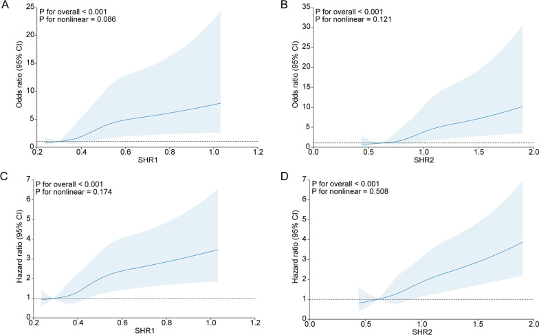
After adjusting variables, including ischemic time, age, sex, hypertension, diabetes, hypercholesteremia, ASCVD, smoking status, eGFR, culprit vessels, and multivessel disease, a notable correlation between both SHR1 and SHR2 and in-hospital death in STEMI patients was identified. Table 2 illustrates that OR for Q2, Q3, and Q4 was 1.35 (0.49, 3.76), 2.31 (0.91, 5.86), and 4.26 (1.85, 9.83), respectively, denoting a significant trend (p for trend < 0.001) compared to the Q1 group (the reference group). Similarly, Table 2 displays a significant trend of 0.70 (0.25, 2.01), 0.76 (0.28, 2.06), and 3.44 (1.63, 7.26) for Q2, Q3, and Q4, respectively (p for trend < 0.001), compared to OR. Furthermore, Fig. 1 highlights a linear relationship between SHR1, SHR2, and in-hospital mortality after adjusting the confounders previously mentioned (p for nonlinearity > 0.05). A rise in one standard deviation in SHR1 corresponded to a 39% increase in in-hospital mortality risk. A similar result was observed with SHR2, where a rise in one standard deviation indicated a 51% increase in mortality risk.
Table 2.
Multivariable Logistic and Cox regression analyses for mortality according to SHR1
| SHR1 | Per SD increment in SHR1 | |||||
|---|---|---|---|---|---|---|
| ≤ 0.359 | 0.359–0.410 | 0.410–0.477 | > 0.477 | Ptrend | ||
| In-hospital death | ||||||
| Model 1 | Reference | 1.18 (0.43, 3.18) | 2.04 (0.83, 5.01) | 4.83 (2.18, 10.72) | < 0.001 | 1.52 (1.30, 1.77) |
| Model 2 | Reference | 1.43 (0.52, 3.95) | 2.50 (0.98, 6.33) | 4.88 (2.13, 11.15) | < 0.001 | 1.43 (1.21, 1.68) |
| Model 3 | Reference | 1.35 (0.49, 3.76) | 2.31 (0.91, 5.86) | 4.26 (1.85, 9.83) | < 0.001 | 1.39 (1.17, 1.66) |
| All-cause mortality | ||||||
| Model 1 | Reference | 0.83 (0.48, 1.43) | 1.19 (0.71, 1.97) | 2.34 (1.51, 3.64) | < 0.001 | 1.28 (1.18, 1.38) |
| Model 2 | Reference | 0.89 (0.51, 1.55) | 1.38 (0.82, 2.33) | 2.23 (1.42, 3.51) | < 0.001 | 1.24 (1.13, 1.35) |
| Model 3 | Reference | 0.93 (0.53, 1.61) | 1.40 (0.83, 2.35) | 2.14 (1.36, 3.37) | < 0.001 | 1.22 (1.10, 1.34) |
Model 1: adjusted for age, sex
Model 2: further adjusted for ischemia time, hypertension, hypercholesterolemia, diabetes, ASCVD, smoking status, eGFR
Model 3: further adjusted for culprit vessel, multivessel lesion
Fig. 1.
Association of SHR1 and SHR2 with poor prognosis. A SHR1 and in-hospital death; B SHR2 and in-hospital death; C SHR1 and all-cause mortality; D SHR2 and all-cause mortality
Association between SHR1, SHR2 and all-cause mortality
A total of 143 patients died during a follow-up period of 2,566 person-years (with a median follow-up of 1.56 years). The Kaplan–Meier curve was shown in Additional file 1: Figure S1. Following adjusting for previously mentioned confounders, a distinct correlation was revealed between SHR1, SHR2, and all-cause mortality in STEMI patients. Table 2 depicts that HR for Q2, Q3, and Q4 groups were 0.93 (0.53, 1.61), 1.40 (0.83, 2.35), and 2.14 (1.36, 3.37), respectively, indicating a statistically significant trend (p for trend < 0.001) compared to the SHR1 Q1 group. Similarly, Table 3 reveals an escalation in the risk of all-cause mortality in proportion to SHR2, with HR for Q2, Q3, and Q4 of 0.87 (0.50, 1.52), 1.17 (0.71, 1.93), and 1.89 (1.21, 2.93), respectively (p for trend < 0.001). The RCS demonstrated a linear association between SHR1, SHR2, and all-cause mortality in patients (p for nonlinearity > 0.05). The risk of all-cause mortality augmented by 22% and 26%, with each unit increase in standard deviation in SHR1 and SHR2, respectively.
Table 3.
Multivariable Logistic and Cox regression analyses for mortality according to SHR2
| SHR2 | Per SD increment in SHR2 | |||||
|---|---|---|---|---|---|---|
| ≤ 0.746 | 0.746–0.843 | 0.843–0.979 | > 0.979 | Ptrend | ||
| In-hospital death | ||||||
| Model 1 | Reference | 0.62 (0.22, 1.72) | 0.74 (0.28, 1.96) | 3.95 (1.93, 8.08) | < 0.001 | 1.63 (1.38, 1.92) |
| Model 2 | Reference | 0.73 (0.26, 2.07) | 0.81 (0.30, 2.19) | 3.98 (1.90, 8.33) | < 0.001 | 1.53 (1.29, 1.81) |
| Model 3 | Reference | 0.70 (0.25, 2.01) | 0.76 (0.28, 2.06) | 3.44 (1.63, 7.26) | < 0.001 | 1.51 (1.26, 1.81) |
| All-cause mortality | ||||||
| Model 1 | Reference | 0.73 (0.42, 1.27) | 1.09 (0.66, 1.79) | 2.11 (1.37, 3.25) | < 0.001 | 1.33 (1.22, 1.44) |
| Model 2 | Reference | 0.88 (0.50, 1.54) | 1.23 (0.75, 2.03) | 2.04 (1.32, 3.17) | < 0.001 | 1.29 (1.18, 1.41) |
| Model 3 | Reference | 0.87 (0.50, 1.52) | 1.17 (0.71, 1.93) | 1.89 (1.21, 2.93) | < 0.001 | 1.26 (1.14, 1.40) |
Model 1: adjusted for age, sex
Model 2: further adjusted for ischemia time, hypertension, hypercholesterolemia, diabetes, ASCVD, smoking status, eGFR
Model 3: further adjusted for culprit vessel, multivessel lesion
Predictive value of SHR1 and SHR2 for mortality
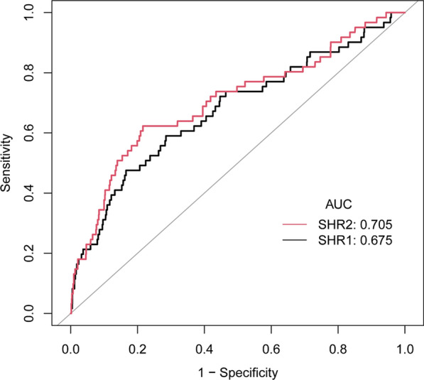
ROC curve analysis (Fig. 2) demonstrated that SHR1 and SHR2 had a moderate predictive capacity for in-hospital mortality in STEMI patients who had undergone PCI (AUC for SHR1: 0.675, 95% CI 0.598–0.752; AUC for SHR2: 0.705, 95% CI 0.629–0.782). The AUC for SHR1 and SHR2 did not differ significantly (p > 0.05). Furthermore, Table 4 presents that the C-statistic for the base model is 0.783 (95% CI 0.723—0.843). Notably, adding SHR1 to the model significantly enhanced its predictive power, raising the C-statistic to 0.808 (0.747–0.868; p < 0.05). Similarly, incorporating SHR2 into the model led to a significant improvement, elevating the C-statistic to 0.819 (0.761–0.877), thereby indicating an advancement compared to the base model (p < 0.05).
Fig. 2.

ROC curves of SHR1 and SHR2 to predict the in-hospital death in the overall study population. The AUC of SHR1 was 0.675, 95% CI 0.598–0.752; the AUC of SHR2 was 0.705, 95% CI 0.629–0.782 (p = 0.06)
Table 4.
C-statistics for discrimination ability of different measures of stress hyperglycemia for in-hospital death
| C-Statistic (95% CI) | P value | |
|---|---|---|
| Established risk factors | 0.783 (0.723, 0.843) | reference |
| Established risk factors + SHR1 | 0.808 (0.747, 0.868) | 0.03 |
| Established risk factors + SHR2 | 0.819 (0.761, 0.877) | 0.01 |
Established risk factors included age, sex, ischemia time, hypertension, hypercholesterolemia, diabetes, ASCVD, smoking status, eGFR, culprit vessel, multivessel lesion
Introducing SHR1 and SHR2 into the base model notably improved the prediction potential for all-cause mortality. Table 5 indicates that the inclusion of SHR1 raised the C-index from 0.782 (95% CI 0.740–0.825) to 0.793 (95% CI 0.750—0.836), and further inclusion of SHR2 increased it to 0.798 (95% CI 0.756–0.840). After adding SHR1 and SHR2 to the base model, the IDI reported a significant advancement of 1.70% (95% CI 0.20–4.30%, p = 0.007) and 2.10% (95% CI 0.40—5.20%, p < 0.001), respectively. The NRI affirmed significance for SHR2 (21.00%, 95% CI 4.50–32.80%, p = 0.02), but not for SHR1 (11.60%, 95% CI −2.20 to 23.60%, p = 0.13).
Table 5.
Improvement in discrimination and risk reclassification for all-cause mortality after the addition of SHR1 or SHR2
| Model | C-index (95%CI) | IDI (%) (95%CI) | P-value | NRI (%) (95%CI) | P-value |
|---|---|---|---|---|---|
| Established risk factors | 0.782 (0.740, 0.825) | Ref | Ref | Ref | Ref |
| Established risk factors + SHR1 | 0.793 (0.750, 0.836) | 1.70 (0.20, 4.30) | 0.007 | 11.60 (−2.20, 23.60) | 0.13 |
| Established risk factors + SHR2 | 0.798 (0.756, 0.840) | 2.10 (0.40, 5.20) | < 0.001 | 21.00 (4.50, 32.80) | 0.02 |
Established risk factors include age, sex, ischemia time, hypertension, hypercholesterolemia, diabetes, ASCVD, smoking status, eGFR, culprit vessel, multivessel lesion
Subgroup and sensitivity analyses
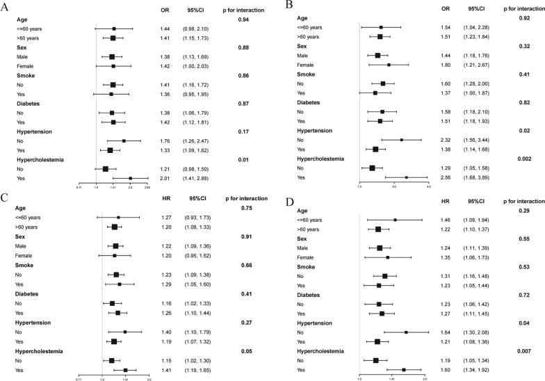
We conducted subgroup analysis to explore the relationship between SHR1, SHR2, and mortality within diverse subgroups and ascertain the robustness of our results (Fig. 3). We identified a direct correlation between SHR1 and SHR2 and in-hospital mortality, as well as all-cause mortality, upon adjusting confounding variables. Our analysis revealed that the impact of SHR1 and SHR2 was markedly augmented in the patients with hypercholesterolemia (p for interaction ≤ 0.05). Additionally, we excluded patients with a prior diagnosis of ASCVD, and the results were sustained and consistent (Additional file 3: Table S2 and Additional file 4: Table S3). We made additional adjustments to the LVEF and TIMI flow, and the results remained steady and coherent (Additional file 5: Table S4 and Additional file 6: Table S5).
Fig. 3.
Subgroup analyses of SHR1 and SHR2 in different populations. A In-hospital death according to SHR1; B In-hospital death according to SHR2; C All-cause mortality according to SHR1; D All-cause mortality according to SHR2
Discussion
This cohort study identified a robust correlation between mortality in STEMI patients who received PCI treatment and the SHR, calculated using GA or HbA1c. This association remained independent of traditional risk factors, such as age, sex, smoking habits, hypertension, hyperlipidemia, diabetes, and renal impairment. Our findings underscore the role of SHR as a risk factor for the unfavorable prognosis in STEMI patients. Our analysis revealed a linear relationship, indicating that incorporating SHR1 or SHR2, besides traditional risk factors, improved the prediction of in-hospital and all-cause mortality in STEMI patients. Additionally, our study included various subgroup and sensitivity analyses; the outcomes were stable. We discovered that the effects of SHR1 and SHR2 were enhanced in patients with hypercholesteremia. To our knowledge, this is the first study to compare the predictive value between different calculations of SHR among STEMI patients treated with PCI.
Mechanism and detrimental effects of stress hyperglycemia
Stress-induced hyperglycemia has been activated by the hypothalamic–pituitary–adrenal axis, elevating cortisol and adrenaline secretion and exacerbating the hyperglycemia state [16]. Furthermore, this condition has been associated with the upregulation of pro-inflammatory cytokines, including interleukin-1, interleukin-6, and tumor necrosis factor-α. These cytokines may impair insulin secretion and augment insulin resistance [17–19]. These inflammatory mediators have been implicated in the worsening of atherosclerosis [20, 21]. Additionally, stress-induced hyperglycemia may contribute to increased thrombogenic activity, culminating in a hypercoagulable state [22, 23]. Previous studies exhibited that higher SHR was associated with a larger thrombus burden and lower TIMI flow grade during angiography [24, 25].
Our study's conclusions partially align with those of certain previous investigations. Previous studies have recognized stress-induced hyperglycemia as an adverse prognostic risk factor in ACS patients [8, 9, 11, 26, 27]. This study determined that the SHR, derived from either HbA1c or GA, was associated with in-hospital mortality and long-term adverse prognosis in STEMI patients. The possibility of a non-linear relationship between SHR and adverse prognosis has been debated in prior studies. Yang et al. enrolled 5,562 ACS patients, 410 of whom had STEMI, and identified a U-shaped correlation between SHR, based on HbA1c, and adverse prognosis [8]. Similarly, Wang et al. identified a U-shaped relationship between SHR, as determined by GA, and adverse prognosis in 5,190 ACS patients, including 1,320 STEMI cases [11]. Similar results were found in patients with heart failure and diabetes [28]. Conversely, Fang et al.'s found no U-shaped relationship between SHR and in-hospital death among 8,196 patients (3,001 with ACS and the remainder with stable angina [9]. Our analysis discovered a linear association between SHR, calculated from HbA1c or GA, and both in-hospital and all-cause mortality. This discrepancy could be attributed to earlier studies using ABG levels for SHR calculation, which various factors may influence. Another plausible explanation is the predominance of unstable angina patients in the cohorts of previous investigations, whereas our study solely encompassed STEMI patients.
Different definitions of stress hyperglycemia
Several indices are employed to assess the extent of stress-induced hyperglycemia, including ABG, FBG, and SHR. However, ABG and FBG are deemed insufficient to accurately represent stress-induced hyperglycemia because baseline blood glucose levels influence them. In contrast, the SHR, calculated by dividing the current blood glucose level by the average blood glucose level, may more precisely reflect stress-induced hyperglycemia states. Nevertheless, a consensus on the optimal method to determine average blood glucose levels is lacking. However, earlier studies frequently utilized HbA1c to estimate past blood glucose levels. Recent investigations have suggested that GA may provide a superior measure due to its independence from renal insufficiency and anemia [29, 30]. To our knowledge, our study is the first to evaluate the impact of different SHR calculations on prognostic prediction. Our study outcomes reveal that the prognostic accuracy of SHR1 for in-hospital death was comparable to SHR2 via a comparative evaluation of ROC curves. Additionally, the discrimination capabilities of SHR1 and SHR2 for in-hospital death and all-cause mortality were significantly superior to the base model, including traditional risk factors.
Previous studies have debated the influence of diabetes status on the relationship between SHR and mortality. Wang et al. demonstrated that SHR exhibited no substantial connection with all-cause mortality and cardiovascular death among ACS patients in a non-diabetic population [11]. Conversely, Cui et al. found a significant association between SHR and in-hospital death among myocardial infarction individuals, regardless of their diabetes status [9]. Our study discovered that SHR, determined by either HbA1c or GA, was associated with in-hospital mortality and all-cause mortality in diabetic and non-diabetic cohorts. Furthermore, we noted a significant enhancement in the association between SHR and mortality in hypercholesterolemia patients, indicating a potential interactive effect between hypercholesterolemia and SHR. Previous studies have suggested that insulin resistance may precipitate lipid irregularities, identifiable by reduced HDL levels [31, 32]. Inversely, HDL can influence glucose metabolism, with a correlation between higher HDL and lower blood glucose levels [33–36]. The stress-induced hyperglycemia and lipid abnormalities may provoke endothelial dysfunction, consequently elevating the risk of cardiovascular events.
Predictive value of SHR
Currently, the most used prognostic model for STEMI patients is the TIMI risk score [37, 38]. However, the TIMI risk score primarily focuses on cardiovascular-related risk factors and does not incorporate important metabolic factors into the scoring system. Previous studies have demonstrated that the predictive ability of the TIMI risk score is generally limited [39, 40]. Some studies have suggested that adding SHR to the risk score may be useful for early risk stratification [10, 41, 42]. This study found that SHR remained an independent risk factor for in-hospital death and all-cause mortality in STEMI patients even after adjusting for traditional risk factors. Moreover, FBG, HbA1c, and GA are easily obtained in clinic settings at low cost. This study discovered that integrating the SHR and the traditional risk factors improved the predictive ability of short- and long-term adverse outcomes in STEMI patients. This study suggests that the SHR should be included as an independent risk factor in constructing new prognostic models for STEMI patients.
Many guidelines exist to regulate blood glucose in critically ill patients, signifying the persisting variations in stress hyperglycemia management [43–45]. Despite the efficacy of numerous medications in glucose control, the advantages of hypoglycemic intervention in stress hyperglycemia patients remain debatable [46–52]. However, these studies primarily concentrated on AMI patients with diabetes, not on stress-induced hyperglycemia. Future research is necessitated to establish whether hypoglycemic therapy could enhance the prognosis in AMI patients with stress-induced hyperglycemia.
Strengths and limitations
Our study’s primary strength is to compare different SHR calculation methods and their association with the prognosis of STMEI patients. To our knowledge, our study is the first to compare the predicted value of different formulas of SHR on in-hospital death and all-cause mortality. Moreover, our study analyzed short- and long-term outcomes, yielding consistent results. Additionally, we performed subgroup and sensitivity analyses to support the robustness of our findings further. However, several limitations should be acknowledged. First, the study was executed across two tertiary academic hospitals and included a comparatively limited sample size, consisting entirely of Asian patients, necessitating a prudent result interpretation. Second, the inherent design of the cohort study precludes inference of a causal relationship within our investigation; verification of these outcomes requires additional prospective studies. Lastly, the lack of data regarding the hypoglycemic therapy application during the monitoring period inhibits our ability to gauge its impact on patients with STEMI.
Conclusion
Our study highlighted association between SHR, calculated from either GA or HbA1c and in-hospital death and all-cause mortality in STEMI patients who received PCI. The discriminating ability of SHR derived from GA was similar to that derived from HbA1c. In summary, SHR based on either GA or HbA1c proved an effective stratification marker for risk assessment in STEMI patients.
Supplementary Information
Additional file 1: Figure S1. Kaplan–Meier curve for SHR1 (A) and SHR2 (B).
Additional file 2: Table S1. Baseline demographic and clinical data
Additional file 3: Table S2. Regression analyses for mortality according to SHR1 after excluding participants with ASCVD
Additional file 4: Table S3. Regression analyses for mortality according to SHR2 after excluding participants with ASCVD
Additional file 5: Table S4. Multivariable Logistic and Cox regression analyses for mortality according to SHR1
Additional file 6: Table S5. Multivariable Logistic and Cox regression analyses for mortality according to SHR2
Acknowledgements
We would like to extend our earnest appreciation to everyone who has significantly contributed to the successful accomplishment of this endeavor.
Abbreviations
- ABG
Admission blood glucose
- ACS
Acute coronary syndrome
- AMI
Acute myocardial infarction
- ASCVD
Atherosclerotic cardiovascular disease
- AUC
Areas under the curve
- eGFR
Estimated glomerular filtration rate
- FBG
Fasting blood glucose
- GA
Glycated albumin
- HbA1c
Hemoglobin A1c
- HDL
High-density lipoprotein
- IDI
Integrated discrimination improvement
- LDL
Low-density lipoprotein
- NRI
Net reclassification improvement
- PCI
Percutaneous coronary intervention
- ROC
Receiver operating characteristic
- SHR
Stress hyperglycemia ratio
- STEMI
ST-segment elevation myocardial infarction
Author contributions
QCW, JJ, and ZDW were responsible for study conceptualization and design. WL, QCW, RRG, ML, ZLL, LJS, HHC, QYG, KDR, and YBL expertly managed the data gathering task. WL and YWC shouldered the responsibility of conducting the statistical analysis. WL prepared the primary draft of the manuscript. All authors participated in a rigorous manuscript review, ensuring the focus was on key intellectual content before approving the article’s final version.
Funding
No.
Availability of data and materials
A reasonable request to the corresponding author will provide access to the data utilized in this study.
Declarations
Ethics approval and consent to participate
In adherence to the principles outlined in the Helsinki Declaration, our investigation obtained the necessary authorization from the ethics review boards of the Second Affiliated Hospital of Zhejiang University and the First People's Hospital of Yulin.
Consent for publication
Not applicable.
Competing interests
There are no competing interests to declare by the authors.
Footnotes
Publisher's Note
Springer Nature remains neutral with regard to jurisdictional claims in published maps and institutional affiliations.
Contributor Information
Zhengdong Wang, Email: 18078700090@163.com.
Jun Jiang, Email: jiang-jun@zju.edu.cn.
Qucheng Wei, Email: wqc2060@zju.edu.cn.
References
- 1.GBD 2017 Causes of Death Collaborators Global, regional, and national age-sex-specific mortality for 282 causes of death in 195 countries and territories, 1980–2017: a systematic analysis for the Global Burden of Disease Study 2017. Lancet. 2018;392:1736–88. doi: 10.1016/S0140-6736(18)32203-7. [DOI] [PMC free article] [PubMed] [Google Scholar]
- 2.Byrne RA, Rossello X, Coughlan JJ, Barbato E, Berry C, Chieffo A, et al. 2023 ESC Guidelines for the management of acute coronary syndromes. Eur Heart J. 2023 doi: 10.1093/eurheartj/ehad191. [DOI] [PubMed] [Google Scholar]
- 3.Tern PJW, Ho AKH, Sultana R, Ahn Y, Almahmeed W, Brieger D, et al. Comparative overview of ST-elevation myocardial infarction epidemiology, demographics, management, and outcomes in five Asia-Pacific countries: a meta-analysis. Eur Heart J Qual Care Clin Outcomes. 2021;7:6–17. doi: 10.1093/ehjqcco/qcaa057. [DOI] [PubMed] [Google Scholar]
- 4.Vogel B, Claessen BE, Arnold SV, Chan D, Cohen DJ, Giannitsis E, et al. ST-segment elevation myocardial infarction. Nat Rev Dis Primers. 2019;5:39. doi: 10.1038/s41572-019-0090-3. [DOI] [PubMed] [Google Scholar]
- 5.Lee TF, Drake SM, Roberts GW, Bersten A, Stranks SN, Heilbronn LK, et al. Relative hyperglycemia is an independent determinant of in-hospital mortality in patients with critical illness. Crit Care Med. 2020;48:e115–e122. doi: 10.1097/CCM.0000000000004133. [DOI] [PubMed] [Google Scholar]
- 6.Peng Z, Song J, Li L, Guo C, Yang J, Kong W, et al. Association between stress hyperglycemia and outcomes in patients with acute ischemic stroke due to large vessel occlusion. CNS Neurosci Ther. 2023;29:2162–2170. doi: 10.1111/cns.14163. [DOI] [PMC free article] [PubMed] [Google Scholar]
- 7.Mone P, Lombardi A, Salemme L, Cioppa A, Popusoi G, Varzideh F, et al. Stress hyperglycemia drives the risk of hospitalization for chest pain in patients with ischemia and nonobstructive coronary arteries (INOCA) Diabetes Care. 2023;46:450–454. doi: 10.2337/dc22-0783. [DOI] [PMC free article] [PubMed] [Google Scholar]
- 8.Yang J, Zheng Y, Li C, Gao J, Meng X, Zhang K, et al. The Impact of the stress hyperglycemia ratio on short-term and long-term poor prognosis in patients with acute coronary syndrome: insight from a large cohort study in Asia. Diabetes Care. 2022;45:947–956. doi: 10.2337/dc21-1526. [DOI] [PubMed] [Google Scholar]
- 9.Cui K, Fu R, Yang J, Xu H, Yin D, Song W, et al. The impact of fasting stress hyperglycemia ratio, fasting plasma glucose and hemoglobin A1c on in-hospital mortality in patients with and without diabetes: findings from the China acute myocardial infarction registry. Cardiovasc Diabetol. 2023;22:165. doi: 10.1186/s12933-023-01868-7. [DOI] [PMC free article] [PubMed] [Google Scholar]
- 10.Xu W, Yang Y-M, Zhu J, Wu S, Wang J, Zhang H, et al. Predictive value of the stress hyperglycemia ratio in patients with acute ST-segment elevation myocardial infarction: insights from a multi-center observational study. Cardiovasc Diabetol. 2022;21:48. doi: 10.1186/s12933-022-01479-8. [DOI] [PMC free article] [PubMed] [Google Scholar]
- 11.Wang M, Su W, Cao N, Chen H, Li H. Prognostic implication of stress hyperglycemia in patients with acute coronary syndrome undergoing percutaneous coronary intervention. Cardiovasc Diabetol. 2023;22:63. doi: 10.1186/s12933-023-01790-y. [DOI] [PMC free article] [PubMed] [Google Scholar]
- 12.Pan Y, Cai X, Jing J, Meng X, Li H, Wang Y, et al. Stress hyperglycemia and prognosis of minor ischemic stroke and transient ischemic attack: the CHANCE study (clopidogrel in high-risk patients with acute nondisabling cerebrovascular events) Stroke. 2017;48:3006–3011. doi: 10.1161/STROKEAHA.117.019081. [DOI] [PubMed] [Google Scholar]
- 13.Neumann F-J, Sousa-Uva M, Ahlsson A, Alfonso F, Banning AP, Benedetto U, et al. 2018 ESC/EACTS guidelines on myocardial revascularization. Eur Heart J. 2019;40:87–165. doi: 10.1093/eurheartj/ehy394. [DOI] [PubMed] [Google Scholar]
- 14.Qin Y, Yan G, Qiao Y, Wang D, Luo E, Hou J, et al. Predictive value of random blood glucose versus fasting blood glucose on in-hospital adverse events in patients with ST-segment elevation acute myocardial infarction. BMC Cardiovasc Disord. 2020;20:95. doi: 10.1186/s12872-020-01394-4. [DOI] [PMC free article] [PubMed] [Google Scholar]
- 15.Upur H, Li J-L, Zou X-G, Hu Y-Y, Yang H-Y, Abudoureyimu A, et al. Short and long-term prognosis of admission hyperglycemia in patients with and without diabetes after acute myocardial infarction: a retrospective cohort study. Cardiovasc Diabetol. 2022;21:114. doi: 10.1186/s12933-022-01550-4. [DOI] [PMC free article] [PubMed] [Google Scholar]
- 16.Marik PE, Zaloga GP. Adrenal insufficiency in the critically ill: a new look at an old problem. Chest. 2002;122:1784–1796. doi: 10.1378/chest.122.5.1784. [DOI] [PubMed] [Google Scholar]
- 17.Mizock BA. Alterations in fuel metabolism in critical illness: hyperglycaemia. Best Pract Res Clin Endocrinol Metab. 2001;15:533–551. doi: 10.1053/beem.2001.0168. [DOI] [PubMed] [Google Scholar]
- 18.Heesen M, Bloemeke B, Heussen N, Kunz D. Can the interleukin-6 response to endotoxin be predicted? Studies of the influence of a promoter polymorphism of the interleukin-6 gene, gender, the density of the endotoxin receptor CD14, and inflammatory cytokines. Crit Care Med. 2002;30:664–669. doi: 10.1097/00003246-200203000-00028. [DOI] [PubMed] [Google Scholar]
- 19.Ruan H, Miles PDG, Ladd CM, Ross K, Golub TR, Olefsky JM, et al. Profiling gene transcription in vivo reveals adipose tissue as an immediate target of tumor necrosis factor-alpha: implications for insulin resistance. Diabetes. 2002;51:3176–3188. doi: 10.2337/diabetes.51.11.3176. [DOI] [PubMed] [Google Scholar]
- 20.de Rekeneire N, Peila R, Ding J, Colbert LH, Visser M, Shorr RI, et al. Diabetes, hyperglycemia, and inflammation in older individuals: the health, aging and body composition study. Diabetes Care. 2006;29:1902–1908. doi: 10.2337/dc05-2327. [DOI] [PubMed] [Google Scholar]
- 21.Stentz FB, Umpierrez GE, Cuervo R, Kitabchi AE. Proinflammatory cytokines, markers of cardiovascular risks, oxidative stress, and lipid peroxidation in patients with hyperglycemic crises. Diabetes. 2004;53:2079–2086. doi: 10.2337/diabetes.53.8.2079. [DOI] [PubMed] [Google Scholar]
- 22.Nieuwdorp M, van Haeften TW, Gouverneur MCLG, Mooij HL, van Lieshout MHP, Levi M, et al. Loss of endothelial glycocalyx during acute hyperglycemia coincides with endothelial dysfunction and coagulation activation in vivo. Diabetes. 2006;55:480–486. doi: 10.2337/diabetes.55.02.06.db05-1103. [DOI] [PubMed] [Google Scholar]
- 23.Stegenga ME, van der Crabben SN, Blümer RME, Levi M, Meijers JCM, Serlie MJ, et al. Hyperglycemia enhances coagulation and reduces neutrophil degranulation, whereas hyperinsulinemia inhibits fibrinolysis during human endotoxemia. Blood. 2008;112:82–89. doi: 10.1182/blood-2007-11-121723. [DOI] [PMC free article] [PubMed] [Google Scholar]
- 24.Chu J, Tang J, Lai Y, Gao Y, Ye Z, Guan C, et al. Association of stress hyperglycemia ratio with intracoronary thrombus burden in diabetic patients with ST-segment elevation myocardial infarction. J Thorac Dis. 2020;12:6598–6608. doi: 10.21037/jtd-20-2111. [DOI] [PMC free article] [PubMed] [Google Scholar]
- 25.Stalikas N, Papazoglou AS, Karagiannidis E, Panteris E, Moysidis D, Daios S, et al. Association of stress induced hyperglycemia with angiographic findings and clinical outcomes in patients with ST-elevation myocardial infarction. Cardiovasc Diabetol. 2022;21:140. doi: 10.1186/s12933-022-01578-6. [DOI] [PMC free article] [PubMed] [Google Scholar]
- 26.Abdu FA, Galip J, Qi P, Zhang W, Mohammed A-Q, Liu L, et al. Association of stress hyperglycemia ratio and poor long-term prognosis in patients with myocardial infarction with non-obstructive coronary arteries. Cardiovasc Diabetol. 2023;22:11. doi: 10.1186/s12933-023-01742-6. [DOI] [PMC free article] [PubMed] [Google Scholar]
- 27.Lin Z, Liang X, Zhang Y, Dai Y, Zeng L, Chen W, et al. Positive association between stress hyperglycemia ratio and pulmonary infection in patients with ST-segment elevation myocardial infarction undergoing percutaneous coronary intervention. Cardiovasc Diabetol. 2023;22:76. doi: 10.1186/s12933-023-01799-3. [DOI] [PMC free article] [PubMed] [Google Scholar]
- 28.Zhou Y, Liu L, Huang H, Li N, He J, Yao H, et al. ’Stress hyperglycemia ratio and in-hospital prognosis in non-surgical patients with heart failure and type 2 diabetes. Cardiovasc Diabetol. 2022;21:290. doi: 10.1186/s12933-022-01728-w. [DOI] [PMC free article] [PubMed] [Google Scholar]
- 29.English E, Idris I, Smith G, Dhatariya K, Kilpatrick ES, John WG. The effect of anaemia and abnormalities of erythrocyte indices on HbA1c analysis: a systematic review. Diabetologia. 2015;58:1409–1421. doi: 10.1007/s00125-015-3599-3. [DOI] [PubMed] [Google Scholar]
- 30.Zelnick LR, Batacchi ZO, Ahmad I, Dighe A, Little RR, Trence DL, et al. Continuous glucose monitoring and use of alternative markers to assess glycemia in chronic kidney disease. Diabetes Care. 2020;43:2379–2387. doi: 10.2337/dc20-0915. [DOI] [PMC free article] [PubMed] [Google Scholar]
- 31.Ormazabal V, Nair S, Elfeky O, Aguayo C, Salomon C, Zuñiga FA. Association between insulin resistance and the development of cardiovascular disease. Cardiovasc Diabetol. 2018;17:122. doi: 10.1186/s12933-018-0762-4. [DOI] [PMC free article] [PubMed] [Google Scholar]
- 32.Goldberg IJ. Clinical review 124: diabetic dyslipidemia: causes and consequences. J Clin Endocrinol Metab. 2001;86:965–971. doi: 10.1210/jcem.86.3.7304. [DOI] [PubMed] [Google Scholar]
- 33.Barter PJ, Rye K-A, Tardif J-C, Waters DD, Boekholdt SM, Breazna A, et al. Effect of torcetrapib on glucose, insulin, and hemoglobin A1c in subjects in the investigation of lipid level management to understand its impact in atherosclerotic events (ILLUMINATE) trial. Circulation. 2011;124:555–562. doi: 10.1161/CIRCULATIONAHA.111.018259. [DOI] [PubMed] [Google Scholar]
- 34.Drew BG, Rye K-A, Duffy SJ, Barter P, Kingwell BA. The emerging role of HDL in glucose metabolism. Nat Rev Endocrinol. 2012;8:237–245. doi: 10.1038/nrendo.2011.235. [DOI] [PubMed] [Google Scholar]
- 35.Drew BG, Duffy SJ, Formosa MF, Natoli AK, Henstridge DC, Penfold SA, et al. High-density lipoprotein modulates glucose metabolism in patients with type 2 diabetes mellitus. Circulation. 2009;119:2103–2111. doi: 10.1161/CIRCULATIONAHA.108.843219. [DOI] [PubMed] [Google Scholar]
- 36.Lehti M, Donelan E, Abplanalp W, Al-Massadi O, Habegger KM, Weber J, et al. High-density lipoprotein maintains skeletal muscle function by modulating cellular respiration in mice. Circulation. 2013;128:2364–2371. doi: 10.1161/CIRCULATIONAHA.113.001551. [DOI] [PMC free article] [PubMed] [Google Scholar]
- 37.Morrow DA, Antman EM, Parsons L, de Lemos JA, Cannon CP, Giugliano RP, et al. Application of the TIMI risk score for ST-elevation MI in the National Registry of Myocardial Infarction 3. JAMA. 2001;286:1356–1359. doi: 10.1001/jama.286.11.1356. [DOI] [PubMed] [Google Scholar]
- 38.Morrow DA, Antman EM, Charlesworth A, Cairns R, Murphy SA, de Lemos JA, et al. TIMI risk score for ST-elevation myocardial infarction: a convenient, bedside, clinical score for risk assessment at presentation: an intravenous nPA for treatment of infarcting myocardium early II trial substudy. Circulation. 2000;102:2031–2037. doi: 10.1161/01.CIR.102.17.2031. [DOI] [PubMed] [Google Scholar]
- 39.Khan SQ, Quinn P, Davies JE, Ng LL. N-terminal pro-B-type natriuretic peptide is better than TIMI risk score at predicting death after acute myocardial infarction. Heart. 2008;94:40–43. doi: 10.1136/hrt.2006.108985. [DOI] [PubMed] [Google Scholar]
- 40.Yanqiao L, Shen L, Yutong M, Linghong S, Ben H. Comparison of GRACE and TIMI risk scores in the prediction of in-hospital and long-term outcomes among East Asian non-ST-elevation myocardial infarction patients. BMC Cardiovasc Disord. 2022;22:4. doi: 10.1186/s12872-021-02311-z. [DOI] [PMC free article] [PubMed] [Google Scholar]
- 41.Chen Q, Su H, Yu X, Chen Y, Ding X, Xiong B, et al. The stress hyperglycemia ratio improves the predictive ability of the GRACE score for in-hospital mortality in patients with acute myocardial infarction. Hellenic J Cardiol. 2023;70:36–45. doi: 10.1016/j.hjc.2022.12.012. [DOI] [PubMed] [Google Scholar]
- 42.Luo J, Xu S, Li H, Li Z, Gong M, Qin X, et al. Prognostic impact of stress hyperglycemia ratio in acute myocardial infarction patients with and without diabetes mellitus. Nutr Metab Cardiovasc Dis. 2022;32:2356–2366. doi: 10.1016/j.numecd.2022.07.004. [DOI] [PubMed] [Google Scholar]
- 43.American Diabetes Association Executive summary: standards of medical care in diabetes–2014. Diabetes Care. 2014;37(Suppl 1):S5–13. doi: 10.2337/dc14-S005. [DOI] [PubMed] [Google Scholar]
- 44.COIITSS Study Investigators. Annane D, Cariou A, Maxime V, Azoulay E, D’honneur G, et al. Corticosteroid treatment and intensive insulin therapy for septic shock in adults: a randomized controlled trial. JAMA. 2010;303:341–8. doi: 10.1001/jama.2010.2. [DOI] [PubMed] [Google Scholar]
- 45.American Diabetes Association Professional Practice Committee 9. Pharmacologic approaches to glycemic treatment: standards of medical care in diabetes-2022. Diabetes Care. 2022;45:S125–43. doi: 10.2337/dc22-S009. [DOI] [PubMed] [Google Scholar]
- 46.Marfella R, Sardu C, D’Onofrio N, Fumagalli C, Scisciola L, Sasso FC, et al. SGLT-2 inhibitors and in-stent restenosis-related events after acute myocardial infarction: an observational study in patients with type 2 diabetes. BMC Med. 2023;21:71. doi: 10.1186/s12916-023-02781-2. [DOI] [PMC free article] [PubMed] [Google Scholar]
- 47.Ritsinger V, Malmberg K, Mårtensson A, Rydén L, Wedel H, Norhammar A. Intensified insulin-based glycaemic control after myocardial infarction: mortality during 20 year follow-up of the randomised Diabetes Mellitus Insulin Glucose Infusion in Acute Myocardial Infarction (DIGAMI 1) trial. Lancet Diabetes Endocrinol. 2014;2:627–633. doi: 10.1016/S2213-8587(14)70088-9. [DOI] [PubMed] [Google Scholar]
- 48.Mellbin LG, Malmberg K, Norhammar A, Wedel H, Rydén L, DIGAMI 2 Investigators Prognostic implications of glucose-lowering treatment in patients with acute myocardial infarction and diabetes: experiences from an extended follow-up of the Diabetes Mellitus Insulin-Glucose Infusion in Acute Myocardial Infarction (DIGAMI) 2 Study. Diabetologia. 2011;54:1308–17. doi: 10.1007/s00125-011-2084-x. [DOI] [PubMed] [Google Scholar]
- 49.Malmberg K, Rydén L, Wedel H, Birkeland K, Bootsma A, Dickstein K, et al. Intense metabolic control by means of insulin in patients with diabetes mellitus and acute myocardial infarction (DIGAMI 2): effects on mortality and morbidity. Eur Heart J. 2005;26:650–661. doi: 10.1093/eurheartj/ehi199. [DOI] [PubMed] [Google Scholar]
- 50.Lexis CPH, van der Horst ICC, Lipsic E, Wieringa WG, de Boer RA, van den Heuvel AFM, et al. Effect of metformin on left ventricular function after acute myocardial infarction in patients without diabetes: the GIPS-III randomized clinical trial. JAMA. 2014;311:1526–1535. doi: 10.1001/jama.2014.3315. [DOI] [PubMed] [Google Scholar]
- 51.Paolisso P, Bergamaschi L, Gragnano F, Gallinoro E, Cesaro A, Sardu C, et al. Outcomes in diabetic patients treated with SGLT2-Inhibitors with acute myocardial infarction undergoing PCI: the SGLT2-I AMI PROTECT Registry. Pharmacol Res. 2023;187:106597. doi: 10.1016/j.phrs.2022.106597. [DOI] [PMC free article] [PubMed] [Google Scholar]
- 52.Dandona P, Chaudhuri A, Ghanim H. Acute myocardial infarction, hyperglycemia, and insulin. J Am Coll Cardiol. 2009;53:1437–1439. doi: 10.1016/j.jacc.2009.01.040. [DOI] [PubMed] [Google Scholar]
Associated Data
This section collects any data citations, data availability statements, or supplementary materials included in this article.
Supplementary Materials
Additional file 1: Figure S1. Kaplan–Meier curve for SHR1 (A) and SHR2 (B).
Additional file 2: Table S1. Baseline demographic and clinical data
Additional file 3: Table S2. Regression analyses for mortality according to SHR1 after excluding participants with ASCVD
Additional file 4: Table S3. Regression analyses for mortality according to SHR2 after excluding participants with ASCVD
Additional file 5: Table S4. Multivariable Logistic and Cox regression analyses for mortality according to SHR1
Additional file 6: Table S5. Multivariable Logistic and Cox regression analyses for mortality according to SHR2
Data Availability Statement
A reasonable request to the corresponding author will provide access to the data utilized in this study.


