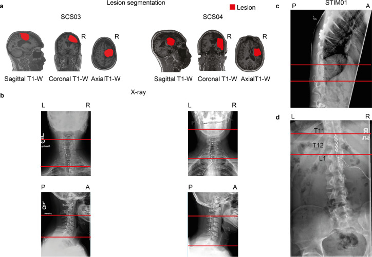
Extended Data Fig. 6 |. Lesion characterization and SCS implantation in humans with paralysis.
a, Sagittal, coronal, and axial T1-weighted magnetic-resonance imaging (MRI) 2D projections for SCS03 and SCS04 using a 3T Prisma system (Siemens) using a 64-channel head and neck coil (T1-weighted structural image was captured using a magnetization-prepared rapid gradient echo sequence: repetition time = 2,300 ms; echo time = 2.9 ms; field of view = 256 × 256 mm2; 192 slices, slice thickness = 1.0 mm, in-plane resolution = 1.0 × 1.0 mm). The segmented lesion is shown in red for both participants and performed as described in6. R indicates the Right hemisphere. b, X-rays for SCS03 and SCS04 showing the position of the spinal leads. The red lines mark the same anatomical location across the X-rays to facilitate interpretation. Minimal displacement occurred after initial implantation. c-d, X-rays for STIM01 showing the position of the spinal leads and the lesion. The red lines mark the same anatomical location across the X-rays to facilitate interpretation. Minimal displacement occurred after initial implantation.

