Abstract
The conformational ensembles of G protein-coupled receptors (GPCRs) include inactive and active states. Spectroscopy techniques, including NMR, show that agonists, antagonists and other ligands shift the ensemble toward specific states depending on the pharmacological efficacy of the ligand. How receptors recognize ligands and the kinetic mechanism underlying this population shift is poorly understood. Here, we investigate the kinetic mechanism of neurotensin recognition by neurotensin receptor 1 (NTS1) using 19F-NMR, hydrogen-deuterium exchange mass spectrometry and stopped-flow fluorescence spectroscopy. Our results indicate slow-exchanging conformational heterogeneity on the extracellular surface of ligand-bound NTS1. Numerical analysis of the kinetic data of neurotensin binding to NTS1 shows that ligand recognition follows an induced-fit mechanism, in which conformational changes occur after neurotensin binding. This approach is applicable to other GPCRs to provide insight into the kinetic regulation of ligand recognition by GPCRs.
Subject terms: Solution-state NMR, Biophysical chemistry, G protein-coupled receptors
GPCRs include inactive and active states. 19F-NMR and stopped-flow fluorescence kinetic assays reveal that neurotensin activates the prototypical peptide-binding GPCR, neurotensin receptor 1, through an induced-fit mechanism, where ligand binding precedes receptor conformational changes.
Introduction
G protein-coupled receptors (GPCRs) are the largest superfamily of membrane proteins, which transduce signals from a wide range of stimuli across the plasma membrane to activate intracellular effector proteins, most importantly G proteins, GPCR kinases, and the β-arrestin family of proteins1. Ligand binding to the extracellular, so-called orthosteric, ligand-binding pocket of GPCRs induces global conformational alterations in the seven transmembrane helix domain (TMD), including the prominent outward movement of helix 6 (TM6) and inward movement of TM7 in the cytosolic region2. These conformational changes open the intracellular crevice for the binding and subsequent activation of G proteins3. Activation of G protein is followed by desensitization of the receptor largely via GPCR receptor kinase (GRK) phosphorylation-induced β-arrestin-mediated receptor internalization4.
GPCRs usually exhibit a basal level of activity in the apo-state that is modulated in an efficacy-dependent manner upon ligand binding to the extracellular orthosteric binding site, where agonists fully activate the receptor and inverse agonists suppress the basal activity. The functional output of the receptor can be further modulated by binding of allosteric ligands to secondary binding pockets or by biased ligands that selectively activate one of the signalling pathways over the others5,6. Central concepts for understanding such pharmacological behaviour in GPCR signalling are (1) a pre-existing equilibrium of fully inactive to fully active conformations in the conformational landscape of apo-state receptor and (2) a population shift towards the active conformation upon agonist binding7 that has been supported by NMR, EPR, single molecule studies as well as MD simulations8–13. For example, recent NMR studies on Adenosine 2A (A2A) receptor indicate sampling of active state conformations of the receptor that are further populated in the presence of ligand and heterotrimeric G proteins8. The population-shift model addresses the equilibrium aspects of GPCR signalling and allostery, irrespective of the kinetic pathways underlying the coupling of population shift and ligand binding. In the case of two pre-dominant conformations, such as an inactive receptor conformation R1 and an active conformation R2, there are two pathways along which a population shift from R1 to R2 during ligand binding can occur14 (Supplementary Fig. 1): an induced-fit pathway15, along which ligand binding to R1 and formation of encounter complexes precede the conformational change to R2 and induce the population shift; and a conformation-selection pathway16 in which the conformational change from R1 to R2 precedes ligand binding. Despite the pharmaceutical importance of GPCRs, the kinetic mechanism underlying population shifts in GPCRs is poorly understood. Insight into this mechanism may aid tailoring of selective designer molecules with desired pharmacological output against GPCRs.
Neurotensin receptor 1 (NTS1) is a class A GPCR that is primarily expressed in the central nervous system and gastrointestinal tract17 and activated by the endogenous 13-residue linear peptide neurotensin, pELYENKPRRPYIL18. The last six residues of NT (NT8-13) have been demonstrated to be the primary epitope of the peptide for high affinity receptor binding and activation and have been used as a scaffold for development of NTS1 targeting drug candidates18. NTS1 regulates neurological processes including dopamine transmission and GABAergic system modulation19 and is considered as a promising target for treatment of addiction and schizophrenia20. Crystal structures of NTS1 in complex with different ligands showed efficacy-dependent modification of the volume of the ligand-binding pocket, where agonists contract the binding pocket and inverse agonists expand its volume21. The conformational changes of the binding pocket of NTS1 have been further investigated by NMR22. However, how receptor conformational changes and dynamics are kinetically linked to ligand recognition remains unclear.
In this study, we combined 19F NMR experiments, hydrogen–deuterium exchange mass spectrometry (HDX-MS) and stopped-flow fluorescence kinetics to address the mechanism underlying NT recognition and activation of NTS1. Our ligand-observed 19F-NMR experiments, on fluorinated full agonist analogues of NT, and receptor-observed experiments, on NTS1 labelled with fluorinated unnatural amino acids, revealed formation of NTS1 conformers upon ligand binding that are in slow conformational exchange. HDX-MS demonstrated that this conformational heterogeneity arises from the interaction between the N-terminal region of the receptor and extracellular loop 2 (ECL2). Further kinetic analysis of binding of NT to NTS1 using stopped-flow fluorescence proposes an induced-fit mechanism of binding underlying NT recognition by NTS1.
Results
Development and characterization of fluorinated NT analogues for structural studies
NMR is well-suited to obtain atomic resolution insight into the dynamics of biomolecular systems. However, common isotope labelling schemes, including 13C and 15N, are cumbersome to study the dynamics of large systems such as GPCRs. Recently, 19F-labelled aromatics have re-gained popularity as NMR probes to study conformational dynamics of GPCRs due to the sensitivity of 19F-aromatics as chemical microenvironmental sensors, resulting in high resolution NMR spectra23. For example, fluorinated ligands have been used to unravel the conformational heterogeneity in the orthosteric pocket of the neurokinin receptor24. Moreover, the large gyromagnetic ratio of 19F enables working with low concentrations of the receptor. In this context we were inspired to develop 19F-NT analogues, by substituting Tyr11 in NT, to investigate the mechanisms of ligand recognition by the receptor25–28. We substituted Tyr11 with para-trifluoromethyl-phenylalanine (tfmF) to produce a sensitive fluorinated analogue of NT (Y11tfmF-NT) (Supplementary Fig. 2) with minimal modification. This substitution reduced the affinity of the peptide for wt-rNTS1 by 100-fold (Supplementary Fig. 3a), which may be due to the steric bulk of the CF3 group and its low tendency to form hydrogen bonds29. Perturbing hydrogen bonding of Tyr11 by phenylalanine substitution shows similar effects, but less pronounced than tfmF probably due to the smaller size of phenylalanine compared to tfmF30. However, the presence of the CF3 moiety leaves the affinity of Y11tfmF-NT for the engineered NTS1 variant, enNTS131, unaffected (Supplementary Fig. 3b), suggesting that the introduced mutations during thermostabilization of the receptor (Supplementary Fig. 4) may account for this gain of affinity. Nonetheless, these probes are equally efficacious in BRET assays measuring G protein (Supplementary Fig. 3c) and β-arrestin (Supplementary Fig. 3d) recruitment and can fully activate wt-rNTS1 providing us with a promising tool to investigate the conformational dynamics of ligand binding to the extracellular region of enNTS1.
The conformational dynamics of ECL2 revealed by 19F-labelled neurotensin
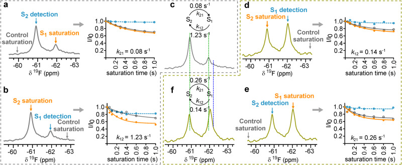
The synthesized Y11tfmF-NT gives a sharp NMR signal (Supplementary Fig. 5a), that is not perturbed in the presence of DDM (Supplementary Fig. 5b), indicating negligible interaction of the fluorinated peptide with DDM micelles. However, upon complexation of Y11tfmF-NT with enNTS1, two distinct resonances were observed in the 19F NMR spectrum, a highly populated state of ~75%, and an additional upfield state populated at ~25% (Supplementary Fig. 5c). Both signals are lost upon addition of unlabelled NT validating that both signals correspond to the peptide bound in the orthosteric pocket (Supplementary Fig. 5d); we name them S1 and S2, respectively (Supplementary Fig. 5e). Such signal splitting can be observed in the truncated version of 19F-NT (Supplementary Fig 5f), Y11tfmF-NT8-13, indicating minimal effect from the first seven residues of NT to the conformation of NT in the bound state. Mutation of Pro10 to alanine, P10A-Y11tfmF-NT8-13, also preserves the conformational heterogeneity (Supplementary Fig. 5g) confirming that the heterogeneity is not due to cis/trans isomerization of Pro10 in Y11tfmF-NT8-13. The signal splitting in the presence of receptor proposes a slow exchange conformational equilibrium in the complex of enNTS1 with Y11tfmF-NT that can be measured using saturation transfer difference (STD) experiments32. 19F STD experiments on Y11tfmF-NT8-13 bound to enNTS1 show that the exchange rate constants from S1 to S2, k12, and from S2 to S1, k21, are 1.23 s−1 and 0.08 s−1, respectively (Fig. 1a–c).
Fig. 1. 19F NMR of Y11tfmF-NT8-13 shows slow-exchanging conformational heterogeneity in complex with enNTS1 and enNTS1-R213.
19F STD experiments on a, b the complex of Y11tfmF-NT8-13 and enNTS1 and d, e Y11tfmF-NT8-13 and enNTS1-R213. In each experiment the intensity of the detected peak was measured by on-resonance irradiation (orange line, solid symbols) and equidistant off-resonance irradiation (grey line, open symbols) from the detected peak as control. The on- and off-resonance spectra were subtracted to obtain the saturation transfer exchange plots (dashed light blue line, filled symbols). In (c, f), the exchange rates extracted from the saturation plots are summarized above the spectra of Y11tfmF-NT8-13 in complex with enNTS1 (grey) (c) and enNTS1-R213 (olive) (f). The chemical shift of S1 and S2 are highlighted with green dotted lines. The dark blue line designates the chemical shift of free peptide. In all experiments, a train of 50 ms gaussian shaped pulses with a field-strength of 50 Hz was used for on and off-resonance saturation. Source data for this figure are provided as a Source data file.
Such slow time scale dynamics has been linked to the ligand-independent and large amplitude conformational changes of the extracellular regions of other GPCRs, for example the neurokinin 1 receptor24. To support that the source of the conformational dynamics of the receptor is due to fluctuations in the extracellular surface of NTS1, several receptor mutants were made. Two criteria were considered in designing receptor mutants: (1) the mutation should not cause major loss of interactions between the peptide and the receptor based on the crystal structures and (2) the mutation should have minimal effect on the topology of the orthosteric binding pocket. In enNTS1 position 213 is mutated from wild-type arginine to leucine during directed evolution thermostabilization (Supplementary Fig. 4)31. Position 213 is not considered as a critical point of direct interaction with bound NT and comparison of the published structures with (PDB: 4XEE)33 and without (PDB: 4BUO)34 mutation in position 213 show the topology of the binding pocket is the same. It proposes position 213 as an appropriate candidate for mutation to address the source of conformational heterogeneity. The back mutation of Leu213 to arginine in enNTS1 (enNTS1-R213), resulted in significant stabilization of the S1 conformer, increasing its population from ~25% in enNTS1 to ~60% in enNTS1-R213 (Fig. 1f). However, mutation from a hydrophobic residue, Leu213 in enNTS1, to a positively charged residue did not cause any significant chemical shift perturbations, suggesting that the chemical environment around the probe and the topology of the binding pocket of enNTS1 and enNTS1-R213 are similar (Fig. 1f). As expected, the dynamics between the S1 and S2 conformers are modified, whereby k12 is 0.14 s−1 and k21 is 0.26 s−1, reflecting a change to the dynamics of ECL2 (Fig. 1d, e).
G protein binding to GPCRs extends the residence time of ligands in the orthosteric binding pocket via allosterically inducing conformational changes in the extracellular surface of the receptor including receptor lidding35. To monitor the effect of G protein binding on the conformational dynamics of the complex of enNTS1 and Y11tfmF-NT8-13 we titrated enNTS1-R213 bound to Y11tfmF-NT8-13 with chimeric Gαiq. This chimeric construct binds enNTS1 with an affinity of 1 µM and has been shown to induce small intensity or chemical shift changes to the 13CεH3 resonances of Met-204, which is near the orthosteric binding pocket36. However, titration of Y11tfmF-NT8-13 bound to enNTS1-R213 with Gαiq did not induce any further changes to the conformational dynamics of the complex (Supplementary Fig. 6) which may suggest that NT sufficiently stabilizes the active conformational state within the orthosteric region or that as the enNTS1 variant used in this study is thermostabilized and only partially active in cells (Supplementary Fig. 3d, e), Gαiq cannot completely stabilize a fully active state.
Neurotensin fine-tunes the capping event in enNTS1
The ECL2 of many GPCRs forms a lid over the ligand-bound orthosteric binding pocket of the receptor via interaction with the N-terminal region or ECL3 of the receptor, which can increase the affinity of the ligand by slowing down its dissociation rate35,37. Although such a formation has not been confirmed experimentally for NTS1, all structures published to date on the complex of NT with NTS1 show close contacts between the N-terminal region and the ECL2 of NTS1 (PDB: 4XEE, 6YVR, 4BUO)21,33,34. However, the structure of these parts of NTS1 in the apo-state or when complexed with small molecule ligands, including full agonist SRI-9829, partial agonist RTI-3a, and the two inverse agonists SR48692 and SR142948A, cannot be resolved, suggesting flexibility of ECL2 and the N-terminal region and that interaction with NT promotes the closed conformation21. We hypothesized that the observed conformational dynamics in the complex of fluorinated peptides and the NTS1 variants originates from contact between the receptor N-terminal region and the tip of ECL2 that consequently shrinks the orthosteric binding pocket of the receptor21. According to the crystal structures of NTS1 in complex with NT8-13, Pro51 is the first point of contact between the receptor N-terminal region and ECL2, which we designate here as the NECL complex. Regions N-terminal to Pro51 appear of least importance in ligand binding, and are typically truncated in bacterially expressed receptors to minimize heterogeneity through proteolysis38. To investigate the interaction of the N-terminal region with ECL2, and how this is influenced by peptide binding, we introduced 19F labels at Gly50, (Supplementary Fig. 4) close enough to the NECL association point to report on conformational changes in the region and far enough from the NT binding site to minimize the impact of introduced label on ligand binding.
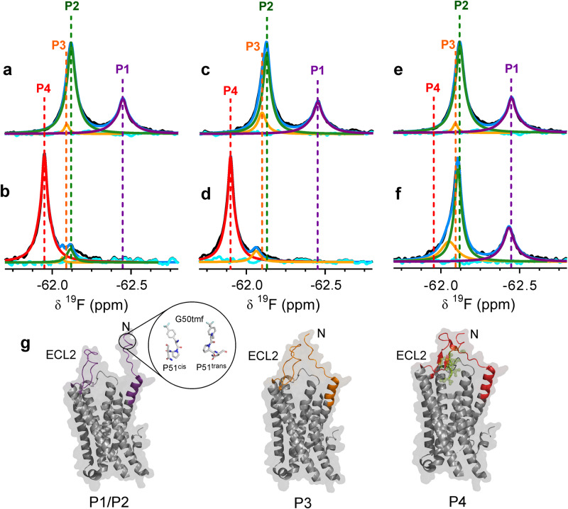
We introduced tfmF at Gly50 of enNTS1, by using the previously engineered tRNA and tRNA synthetase pair for incorporation of tfmF in Escherichia coli39. This system allowed us to produce highly purified, site-specifically labelled receptors for NMR studies (Supplementary Fig. 7). The spectrum of the apo-G50tfmF-enNTS1-R213 showed the presence of two major populations, labelled P1 and P2 (Fig. 2a). Close inspection of P2 showed the presence of downfield asymmetry, whereby deconvolution of P2 suggests an additional population, P3. Mutation of Pro51 in enNTS1-R213 to Ala (G50tfmF-enNTS1-R213-P51A) eliminated the P1 signal but did not change the asymmetry of the peak for P2 and P3, suggesting P1 arises from the cis isomer of Pro51 and P2 and P3 belong to the trans isomer (Supplementary Fig. 8). Titration of NT8-13 into the protein solution promoted sampling of a new substate downfield of P3, labelled P4, to ~90% coupled with line narrowing of the signal (Fig. 2b). The small chemical shift difference between P3 and P4 prevented measurement of the kinetics of exchange between these states by 19F STD experiments.
Fig. 2. The N-terminal regions of enNTS1-R213 and enNTS1 show different conformational dynamics in response to binding agonist and inverse agonist.
19F NMR spectra of a apo G50tfmF-enNTS1-R213 and b in complex with saturating concentrations of NT8-13. Complexation with NT8-13 promotes formation of P4 (red) and reduces the population of P3 (orange). Despite being saturated, ligand cannot remove P2 (green). P1 (purple), which is due to cis/trans isomerization of Pro51 (Supplementary Fig. 7) disappears. The blue and cyan line in each spectrum indicate the sum and residuals of the deconvoluted spectrum, respectively. 19F NMR spectra of c apo G50tfmF-enNTS1 (residue 213 is Leu) and d in complex with saturating concentrations of NT8-13. Colour scheme is the same as described in (a, b). P3 is more populated in apo G50tfmF-enNTS1 compared to G50tfmF-enNTS1-R213. Upon binding to NT8-13, P1 disappears and P4 formation is promoted, which is slightly shifted downfield compared to P4 of G50tfmF-enNTS1-R213 (b). 19F NMR spectra of e apo G50tfmF-enNTS1-R213 and f in complex with inverse agonist SR142948A. Colour scheme is the same as described in (a, b). P3 is increased in population in the complex with inverse agonist, but the signal P4 is not observed compared to spectra in the presence of peptide agonist. P1 is also observed, suggesting that the N-terminal region retains flexibility. The chemical shift and population of each state is summarized in Supplementary Table 1. g Structural models of enNTS1 where P1/P2 show significant flexibility of the N-terminal region. P4 shows the N-terminal region and ECL2 form a closed conformation upon binding of NT (green). An intermediate state, P3, is formed by loose interaction of ECL2 with the N-terminal region of the receptor. P3 is populated in both apo state and ligand-bound state of the receptor.
If the NECL is formed upon ligand binding and P4 reports this conformational state, repeating the experiments with G50tfmF-enNTS1 (where residue 213 is Leu) should affect the thermodynamics of the system. In 19F spectra of G50tfmF-enNTS1 the population of P3 increased from ~4% to ~14% (Fig. 2c and Supplementary Table 1). Also absent in the apo state of G50tfmF-enNTS1, the new substate P4, is again populated to ~90% upon binding of NT8-13. P4, however, is shifted further downfield compared to the P4 signal in the complex of NT8-13 with G50tfmF-enNTS1-R213 (Fig. 2b, d and Supplementary Table 1), suggesting a slight difference in conformation of the NECL in the two receptors. Another difference between G50tfmF-enNTS1-R213 and G50tfmF-enNTS1 is that under saturating concentrations of NT8-13 (10-fold excess), P2 is present in the ligand bound state of G50tfmF-enNTS1-R213 and is absent in G50tfmF-enNTS1 (Fig. 2b, d). Addition of the inverse agonist SR142948A leaves the 19F signals for G50tfmF-enNTS1-R213 largely unaffected, and hence is similar to the apo state (Fig. 2e, f). Most notably, P1 persists indicating that the cis/trans isomerism of Pro51 is not affected and there is no evidence of formation of the substate P4. The signal for P2/3 appears more asymmetric and deconvolution shows an increase in P3 (Fig. 2e, f and Supplementary Table 1). These results are largely consistent with the unresolved NECL in crystal structures of the receptor in the apo state and in complex with small molecule ligands, but NECL is strongly populated in the presence of bound peptide agonists21. Similar to enNTS1-R213 bound to Y11tfmF-NT8-13 (Supplementary Fig. 6), the conformational dynamics of G50tfmF-enNTS1-R213 does not appear affected by titration with the chimera Gαiq under our experimental conditions (Supplementary Fig. 9).
HDX-MS shows different dynamics in the extracellular surface of enNTS1 when bound to agonist and inverse agonist
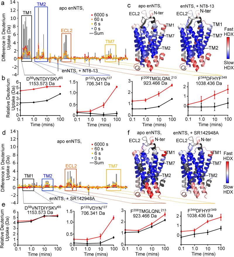
Hydrogen–deuterium exchange mass spectrometry (HDX-MS) is a powerful emerging technique to investigate the dynamics of proteins, inferred from deuterium uptake, that has been successfully applied to the characterization of membrane proteins40,41. The deuterium uptake rates during HDX experiments correlate with the structural fluctuations of hydrogen-bond opening and closing. Differences between protein states, such as apo- and ligand-bound, directly report on hydrogen bond stability in the ligand binding site or of the conformational dynamics of the protein states. To support our 19F NMR experiments of the receptor and the assignment of the fluorine signals to different conformational states of the receptor, the conformational dynamics of enNTS1 in apo state and in complex with the agonist NT8-13 or the inverse agonist SR142948A were investigated by HDX-MS (Fig. 3). Over the time course of the experiment (0–100 min) there is limited or no hydrogen–deuterium exchange for the helical regions of the TMD embedded within the DDM micelles. The helices of the TMD also showed lower proteolytic cleavage. Nonetheless, the HDX-MS experiments revealed important insight into the effect of ligand binding on the conformational dynamics of the extracellular region of enNTS1 and how ligand binding promotes these changes. Inspection of the available crystal structures shows that the backbone NH of the first 14 residues (G50 to S63) do not engage in hydrogen bonds upon ligand binding. As expected, peptides from this region are insensitive to exchange in both the apo and ligand bound state (Fig. 3). However, peptides that include the first residues of TM1 (D56VNTDIYSKV65) show marked differences upon complexation with NT8-13 but not SR142948A (Fig. 3b, e), indicating that agonist, and not inverse agonist, binding increases the stability of this helix, which may be due to reduced dynamics of the N-terminal region, consistent with the downfield shift and lower water-accessibility of the resonance assigned to P4 in the 19F spectra of G50tfmF-enNTS1 (Fig. 2). Moreover, the absence of such protection in the complex of SR142948A with enNTS1 (Fig. 3e), indicates the peptide agonism-dependency of TM1 dynamics. The extracellular ends of TM2 (P122VDYN127) and TM7 (F344DFHYF349) (Fig. 3e) show an increase in protection from deuterium exchange for SR142948A bound receptor. SR142948A might impose such effects by direct interaction with the pocket formed between TM6 and TM7, as reported in crystal structures (PDB: 6Z4Q)21. Peptides from ECL2 show an increase in protection in either ligand bound state (Fig. 3a, d), although protection appears slightly higher for agonist NT8-13 bound than inverse agonist SR142948A bound (Fig. 3b, e). For the shortest observed peptide of enNTS1 from this region (F206TMGLQNL213), the NH of Thr207 forms a hydrogen bond at the tip of TM4, while Gly209 and Gln211 of this peptide form hydrogen bonds within ECL2 itself. The remaining residues of this peptide are not expected to form hydrogen bonds. Consistent with the crystal structures, the adamantyl group of SR142948A forms van der Waals interactions with the side chain of Tyr146, Pro227 and Met208 in the ECL2 as well as Leu234 and Ile238 in TM5 which likely reduce the movement of ECL2.
Fig. 3. Agonist but not inverse agonist reduces the conformational dynamics of enNTS1.
a Difference in deuterium uptake plots of enNTS1 in apo-state and bound to NT8-13 at the time points 0 s (blue); 6 s (yellow); 60 s (orange); 6000 s (red); sum of differences (grey). b Relative deuterium uptake plots of apo state (red) and NT8-13 bound (black) for regions that show significant differences in deuterium uptake including the extracellular regions of TM1, D56VNTDIYSKV65; TM2, P122VDVYN127; TM7, F344DFHYF349; and ECL2, F206TMGLQNL213. Data shown as intensity-weighted mean values ± standard deviation (n = 4). c Heatmap generated from DynamX 3.0 at 100 min overlaid on the structure of NTS1 (PDB: 4XEE) (blue, least exchange, to red, maximum exchange). Peptides not observed by MS are coloured grey. d Difference in deuterium uptake plots of enNTS1 in the apo-state and bound to inverse agonist SR142948A at time points 0 s (blue); 6 s (yellow); 60 s (orange); 6000 s (red); sum of differences (grey). e Relative deuterium uptake plots of apo state (red) and SR142948A-bound (black) show SR142948A has less impact on deuterium uptake than NT8-13 in the extracellular region of TM1 and ECL2 of enNTS1. Data shown as intensity-weighted mean values ± standard deviation (n = 3). f Heatmap generated from HDX data (blue, least exchange, to red, maximum exchange) in apo state and in the presence of SR142948A on the structure of NTS1 (PDB: 4XEE). Source data for this figure are provided as a Source data file.
Kinetic analysis of binding pathway
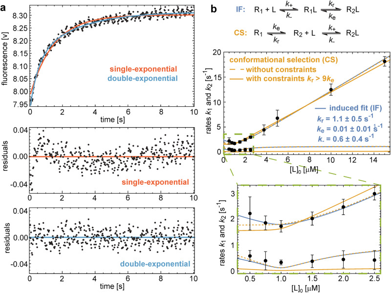
The appearance of the P4 resonance upon complex formation with NT8-13 and the conformational heterogeneity of the receptor-bound 19F-labelled peptide raises the question of whether ligand binding is through an induced-fit or conformational-selection mechanism. To address this question, we kinetically analysed the binding of NT to enNTS1 using stopped-flow fluorescence. We previously showed that upon binding of NT to enNTS1 the intrinsic fluorescence of Trp residues in the receptor increases in a dose-dependent manner, probably due to interaction of side chains of NT with Trp339 in ECL342. Equal volumes of NT and enNTS1 were rapidly mixed together using a fixed concentration of 2 µM of the receptor and varying concentrations of the peptide, and fluorescence change was tracked over time (Fig. 4a). Fitting the kinetic traces revealed a double-exponential trace with two characteristic rates k1 and k2 that depend on the total concentration [L]0 of the peptide ligand. The observed rates k1 and k2, as functions of [L]0, were jointly fitted using the equations described previously43 and summarized in ‘Methods’. Fitting the conformational-selection model without constraints on rate parameters leads to an implausibly large probability of the active conformation R2 in the unbound state, and fits in which this probability is constrained to less than 10% poorly match the data (Fig. 4b). In the induced-fit model, in contrast, the observed rates k1 and k2 can be well fitted with plausible conformational excitation and relaxation rate constants ke = 0.01 ± 0.01 s−1 and kr = 1.1 ± 0.5 s−1 and with a dissociation rate constant k- = 0.6 ± 0.4 s−1 of the bound excited state R1L. Moreover, the conformational exchange rate constants ke and kr obtained from the stopped-flow data in the induced-fit model are in good agreement with the exchange rate constants 0.08 s−1 and 1.23 s−1 measured in the 19F STD experiments of Y11tfmF-NT8-13 bound to enNTS1 within numerical accuracies (Fig. 1). This agreement of conformational exchange rates obtained from distinct experiments is a rather strong confirmation of induced-fit as the binding mechanism and indicates that the states R1L and R2L of the induced-fit model might correspond to the states S1 and S2 of the NMR experiments. A comparably strong kinetic proof of a binding mechanism based on the agreement of conformational exchange rates deduced from NMR and stopped-flow experiments has been previously reported for the rhodopsin kinase/recoverin system44.
Fig. 4. Kinetic analysis of binding of NT to enNTS1 shows an induced-fit mechanism.
a Single-exponential (red) and double-exponential (blue) fits of an exemplary stopped-flow time trace at 1 µM NT. Fit residuals and goodness of fits assessed with the Akaike Information Criterion (AIC) indicate double-exponential relaxation with two characteristic rates k1 and k2. AIC values −2280.7 and −2454.5 of single- and double-exponential fits, respectively, indicate a significantly better fit for double exponentials. b Error-weighted fits of the conformational-selection (CS) (orange lines) and induced-fit (IF) models (blue lines) to the rates k1 and k2 obtained from stopped-flow experiments at different total NT concentrations [L]0 (data points with errors). Data points represent mean values of rates from fits of 12 stopped-flow time traces at 0.5 µM NT; 6 traces at 0.75, 2 and 3.75 µM; 5 traces at 1 and 2.5 µM; 4 traces at 1.5, 10 and 15 µM; 3 traces at 5 µM. Error bars correspond to 95% confidence intervals estimated as 1.96 times standard errors (‘Methods’). For each model, data points are jointly fitted to the model equations for the rates k1 and k2 (‘Methods’) with the function Nonlinear Model Fit of Mathematica 1371 using the rate constants ke, kr, and k- of the CS and IF reaction schemes as fit parameters. The rate constant k+ is replaced by the experimentally determined KD value of 6 nM (Supplementary Fig. 11). For the CS model, an unconstrained fit (orange dashed lines) with AIC value 15.0 leads to the fit values ke = 1.2 ± 0.5 s−1, kr = 0.6 ± 0.4 s−1, and k- = 0.005 ± 0.002 s−1 and, thus, to an unrealistically large probability of the active state (R2) prior to ligand binding. For more realistic unbound-state probabilities of R2 smaller than 10% imposed by the constraint kr > 9 ke, the fit of the CS model (full lines) is poor with an AIC value of 33.8 that is significantly larger than the AIC value of 15.1 for the fit of the IF model. Source data for this figure are provided as a Source data file.
Discussion
NMR, EPR and single-molecule fluorescence experiments show the co-existence of several conformational states, ranging from inactive conformer to fully active conformer, in the conformational ensemble of apo-state GPCRs9,45,46. Ligands with different efficacies modify the population and dynamics of these conformers in the conformational landscape of GPCRs in favour of specific states. For example, in two-dimensional 1H,13C NMR experiments on the inverse agonist-bound β2AR, the 13CεH3 resonance of Met82 splits into two peaks that shift in an efficacy-dependent manner and collapse into one resonance in the presence of full agonist47. Similar observations have been made by NMR experiments on similar labelled residues in β1AR48, α1AAR49, ACKR350, MOR51, adenosine A2A52,53 and NTS122 receptors. Recently, the high sensitivity of 19F-NMR has enabled resolving lowly populated states as well as the measurement of transition rates between these conformations9,45,52,54–56.
Here, we monitored the conformational ensemble of thermostabilized variants of NTS1 by combining ligand-observed and receptor-observed 19F-NMR approaches. The fluorinated analogue of NT, Y11tfmF-NT8-13, samples two slow-exchanging conformational states when bound to enNTS1, likely arising from the conformational dynamics of the extracellular surface of enNTS1 that was further supported by receptor mutants at ECL2 (Fig. 1). Conformational change at the extracellular region of GPCRs upon ligand binding has been shown previously by crystallography21,57 as well as NMR studies22,24. The crystal structures of NTS1 in complex with NT (PDB: 6YVR) and small molecule antagonists (PDB: 6Z4Q) show contraction of the binding pocket in an efficacy-dependent manner21. Further, several structures show missing density for the N-terminal region and the ECL2 of the receptor in the apo state (PDB: 6Z66) and in complex with small non-peptide ligands (PDB: 6Z4Q, 6Z4S, 6Z8N, 6ZA8, 6ZIN)21, proposing conformational crosstalk between these regions that inspired our receptor-observed experiments. Labelling of the receptor in the N-terminal region at Gly50, G50tfmF-enNTS1, revealed such spatial proximity between the tip of ECL2 and the N-terminal region of the receptor that is further promoted by the agonist NT8-13 but not the inverse agonist SR142948A (Fig. 2). The lidding event of GPCRs is a well-established phenomenon for small molecule-bound GPCRs, where binding of ligand promotes folding of ECL2 of the receptor to cover the orthosteric binding pocket thus increasing the residence time of the ligand35,57. We believe our observation reports on similar phenomenon for enNTS1 where NT binding induces an interaction between ECL2 and the N-terminal region of the receptor and is responsible for the contraction of the binding pocket of NTS1 upon binding to NT8-1321, that is further confirmed by HDX-MS experiments on the complex of enNTS1 with agonist and inverse agonist ligands (Fig. 3).
Although NMR experiments delineate the conformational dynamics of GPCRs and how ligands modify the energy landscape of the receptor, the equilibrium nature of these experiments does not inform on how the population shift is kinetically regulated, which is critical to understand the mechanisms underlying ligand recognition and activation of GPCRs, especially that the output of GPCR function is proposed to be temporally and spatially regulated58,59. To address this issue, we successfully applied a label-free stopped-flow fluorescence approach to kinetically study the mechanisms of ligand recognition by enNTS1. Numerical analysis and mathematical modelling of binding data indicated an induced-fit mechanism underlies ligand recognition by enNTS1 including a relatively short-lived intermediate (R1L) state that transitions to the final active ligand-bound state (R2L) (Fig. 4). The physiological importance of such an intermediate state might formulate the kinetic bias that has been reported for other GPCRs, such as the dopamine receptor60. Of note, the transition rates calculated from our stopped-flow and 19F NMR data match the rates of Gs dissociation from β2AR61, proposing potential functional relevance of our observed intermediate states.
Whether NT binds to the orthosteric binding pocket of the intermediate state or occupies different conformations in distinct pockets remains to be fully answered. However, using transferred-NOE experiments we recently showed that NT forms an encounter complex with enNTS1 outside of the orthosteric binding pocket and possibly by engaging the N-terminal region and ECL242. Intermediate-state binding pockets have been attributed to a sequential activation mechanism of the free fatty acid receptor 2, FFA2, by the previously considered allosteric modulator, 4-CMTB62. 4-CMTB binds to the orthosteric binding pocket of FFA2, where it primarily initiates downstream orthosteric signalling followed by diffusion into an allosteric pocket to allosterically modulate receptor activity62. The transition of NTS1 from an encounter complex state to the final pose may play a similar role by temporally regulating biased signalling in NTS1. Indeed, impedance-based measurements of NTS1-expressing cell responses to NT and NT8-13 shows significant differences in the response kinetics, albeit at high concentration, where NT produces a more sustained signal compared to NT8-1363. Moreover, temporary residence of NT in an intermediate state might also indicate potential secondary binding pockets to be targeted for development of allosteric as well as bitopic ligands that simultaneously bind orthosteric and allosteric sites64–67. However, the conformation of NT in the intermediate state remains unresolved.
In this study, we have demonstrated that the binding of the linear peptide NT to NTS1 proceeds via an induced-fit rather than a conformational-selection mechanism. It is possible that peptide-binding GPCRs use induced-fit mechanisms, and the small molecule-binding GPCRs, such as the adrenoreceptors, use conformational-selection mechanisms. However, the slower association rate of ligands to the nanobody-activated or nucleotide free G protein-coupled GPCRs proposes that small molecule ligands may also favour to induce receptor activation through binding to the inactive state of the receptor35. The approach we applied here is adaptable to other ligand-GPCR pairs, such as small molecule-binding GPCRs, conditional on the observable change in the intrinsic fluorescence emission upon mixing of ligand with the receptor and providing the availability of NMR probes to investigate the conformational dynamics of the receptor. However, ligands can be conjugated to extrinsic fluorophores for stopped-flow fluorescence assays, in such cases appropriate controls should be conducted to avoid artefacts generated from non-specific interaction of the fluorophore with the membrane environment or receptor surface.
Methods
Peptide synthesis and purification
Peptides were synthesized using Fmoc-solid phase peptide synthesis method on a Fmoc-Leu-Wang resin in a 0.025 mmole scale. The peptides were deprotected and cleaved off the resin by using a mixture of 95% TFA, 2.5% water and 2.5% TIPS as a radical scavenging agent. The peptides were further purified using reverse-phase HPLC on a C18 column using a gradient buffer A (0.1% TFA in water) and buffer B (0.1% TFA in acetonitrile). A gradient of 20–30% of buffer B over 20 min was used for purification of these peptides. The peptides were characterized by LC-MS spectrometry on an Exactive Plus Orbitrap mass spectrometer, and the purity was checked (>95%) by analytical HPLC (Supplementary Fig. 2).
Expression and purification of enNTS1 and enNTS1-R213
The thermostabilized rat neurotensin receptor 1 variant enNTS1-(M208) has been described previously and will be referred to as enNTS1 in this study (Supplementary Fig. 4)31. Leu213 of this variant was back-mutated to Arg213 to provide a second receptor model, enNTS1-R213. The plasmids for these receptors were transformed into Escherichia coli C43 (DE3) cells and the receptors, N-terminally fused to maltose-binding protein (MBP) and C-terminally with green fluorescent protein (GFP), were expressed in autoinduction media ZYP5052 media supplemented with 1% glycerol, 0.3% lactose and 0.05% glucose68. The media was inoculated with 1% overnight preculture and incubated at 37 °C until an OD600 of ~1 was reached, then the culture was maintained at 20 °C for another 20–24 h.
All constructs of enNTS1 were purified as follows31. The frozen bacterial cell pellet was resuspended in ice-cold lysis buffer (50 mM HEPES pH 7.8, 500 mM NaCl, 20% glycerol, 10 mM imidazole, 4 mM MgSO4, 50 mg lysozyme, 10 mg DNase and one cOmplete protease inhibitor tablet). Two mL of buffer was added per gram of wet weight cells. The cells were stirred in a cold room at 5 °C to achieve complete cell resuspension followed by 8 min sonication on ice with cycles of 10 s on/20 s off. A solution mixture of 4% DDM, 2.4% CHAPS and 0.6% CHS was added to the cell lysate to reach final concentration of 1% DDM, 0.6% CHAPS and 0.12% CHS and lysis was continued for 2 h at 5 °C. The cell lysate was clarified by centrifugation and the supernatant was incubated for 1 h at 5 °C with TALON resin pre-equilibrated with base buffer (25 mM HEPES, 200 mM NaCl, 10% glycerol, 0.05% DDM, 10 mM imidazole). The resin was washed with 100 mL of base buffer containing 500 mM NaCl, 5 mM MgSO4 and 1 mM ATP. The protein was eluted with base buffer containing 200 mM imidazole. Imidazole was removed by buffer exchange and the fusion tags were cleaved off by addition of 1 mM TCEP, 100 mM Na2SO4 and 1 µM 3C-presicion protease to the protein solution and overnight incubation at 5 °C. The cleaved protein was incubated for 1 h with TALON resin pre-equilibrated with cleavage buffer supplemented with 5 mM imidazole and the protein was collected in flow-through. The receptor was concentrated and chromatographed over a Superdex 200 increase 10/300 GL size exclusion chromatography (SEC) column equilibrated in 50 mM phosphate buffer, 100 mM NaCl and 0.05% DDM pH 7.4. The purified protein fractions were pooled together and were either used freshly or snap frozen in liquid nitrogen and stored at −80 °C until required.
Expression and purification of chimeric Gαiq
The chimeric Gαiq was expressed and purified as follows36. 4 L of 2YT media, supplemented with 0.2% glucose and 100 µg/mL ampicillin, was inoculated with 1% overnight preculture of E. coli BL21 (DE3) cells harbouring the plasmid for expression of chimeric Gαiq N-terminally fused to maltose-binding protein (MBP). The cultures were grown at 37 °C to an OD600 of ~0.7. The culture was induced by 1 mM IPTG and expression continued for 16 h at 25 °C. The harvested cells were resuspended in 40 mM HEPES pH 7.5, 100 mM NaCl, 10 mM imidazole, 10% v/v glycerol, 5 mM MgSO4, 50 μM GDP, 10 mg of DNase I, 25 mg lysozyme, and 100 μM DTT by stirring in a cold room at 5 °C to achieve a homogenous suspension. The cells were then lysed by sonication on ice for 16 cycles (10 s on/ 20 s off) and the lysate was clarified by centrifugation at 30,000 × g. The supernatant was incubated with Ni-NTA resin pre-equilibrated with 25 mM HEPES, 100 mM NaCl, 10 mM imidazole pH 7.5 for 1 h at 5 °C. The resin was washed with 25 mM HEPES pH 7.5, 500 mM NaCl, 30 mM imidazole, 10% v/v glycerol, 1 mM MgSO4, 50 μM GDP in a gravity flow column and the protein was eluted with 25 mM HEPES, 100 mM NaCl, 300 mM imidazole, 10% v/v glycerol, 1 mM MgSO4, 50 μM GDP, pH 7.5. The imidazole was removed by buffer exchange and the solution was adjusted to 100 mM Na2SO4, 1 mM TCEP and 2 µM home-made 3C-Precision protease followed by overnight incubation at 5 °C. The cleaved protein solution was adjusted to 10 mM imidazole and incubated with Ni-NTA resin pre-equilibrated with 25 mM HEPES, 100 mM NaCl, 10% glycerol pH 7.5. After 1 h incubation at 5 °C, the flow-through was collected as Gαiq, concentrated and then chromatographed over a HiLoad 16/600 Superdex 75 pg column equilibrated in 50 mM potassium phosphate pH 7.4, 100 mM NaCl, 10% v/v glycerol, 1 mM MgSO4, 1 μM GDP, 100 μM TCEP. The Gαiq peak fractions were pooled, concentrated, and used either freshly or snap frozen and stored at −80 °C until further use.
Cell surface competition binding assay
100,000 HEK293F cells (Invitrogen) stably expressing receptor and the cytosolic mCherry fluorophore were suspended in complete phenol red free DMEM (10% FBS, 1% L-Glutamine, 1% penicillin-streptomycin) and were added to the relevant ligand concentrations in the presence of 5 nM Cy5-NT8-13 for enNTS1 and wt-rNTS1 or 2 nM FAM-NT8-13 for rNTS1-R213L. Cells were then gently agitated on an orbital shaker in the dark for 1 h at room temperature for ligand binding to reach a state of equilibrium. The mean fluorescence intensity of bound FAM-NT8-13 and Cy5-NT8-13 was measured with a CytoflexS (Beckman-Coulter) flow cytometer using the 525/40 and 660/20 filter, respectively. Data points gated for single cells that were mCherry positive as a marker of stable expression. Data was then analysed using the One site – Fit Ki function on GraphPad Prism (version: 9.3.1, Graphpad Software, San Diego, CA). The data for enNTS1 and wt-NTS1 are mean of four replicates (n = 4) and the assays with rNTS1-R213L are performed in triplicates (n = 3).
Cell-based assays
500,000 HEK293T cells were seeded per well in a 6-well plate in complete DMEM (10% FBS, 1% L-Glutamine, 1% penicillin-streptomycin). Cells were transfected the following day with a solution of Opti-mem and lipofectamine 2000 at a quantity of 1 µL/1 µg of DNA. For β-arrestin-2 recruitment assay cells were co-transfected with 250 ng of either pcDNA 3.1 zeo-HA-FLAG-wt-rNTS1-nLuc or pcDNA 3.1 zeo-HA-FLAG-rNTS1-R213L-nLuc and with 600 ng of pcDNA human β-arrestin-2-Venus. In G protein BRET assays, cells were co-transfected with 750 ng of pCSC-rNTS1-IRES-mCherry, pCSC-rNTS1-R213L-IRES-mCherry or pCSC-enNTS1-IRES-mCherry and with 250 ng of Gαi (for enNTS1) or Gq (for rNTS1 and rNTS1-R213L) BRET biosensor. Cells in each 6-well plate were then washed and harvested with DPBS the following day, then resuspended in 3 mL of complete phenol-red free DMEM (10% FBS, 1% L-Glutamine, 1% penicillin-streptomycin, 25 mM HEPES). Cells were then plated into a white opaque 96-well plate at 80 µL/well. The following day, the media of the cells were changed to 90 µL complete phenol-red free DMEM with 1/500 dilution of Promega Nano-Glo furimazine per well. The plate was then incubated at 37 °C for 10 min in a PHERAstar® FSX microplate reader (BMG LABTECH). Dual luminescence of 450/535 nm was then measured for 3 min before manually adding 10 µL of 10x ligand concentration or vehicle to respective wells before completing the time course measurement. The BRET ratio was calculated as Venus emission to nLuc emission. The following data analysis was performed using GraphPad Prism (version: 9.3.1, GraphPad). The BRET ratio response over time was first normalised to vehicle-treated controls. Concentration–response curves were calculated by first integrating the area under the BRET ratio over time plot using the Area Under the Curve function and then plotted against ligand concentration. Concentration–response curves were then fitted using three-parameter log(agonist) vs response function to calculate Log(EC50). All activity assays are performed in triplicate (n = 3).
Site specific labelling of enNTS1 and enNTS1-R213 with p-trifluoromethyl-phenylalanine
G50 on enNTS1 or enNTS1-R213, was mutated to the TAG amber stop codon and the plasmid was co-transformed into E. coli C43 (DE3) cells along with the plasmid containing the genes encoding the engineered tRNA and aminoacyl synthetase pair for incorporation of p-trifluoromethyl-phenylalanine (p-tfmF)39. The transformed cells were spread on LB-agar plates supplemented with 100 µg/mL ampicillin and 12.5 µg/mL tetracycline. A single colony was inoculated into an overnight preculture. Expression was conducted in media comprising 50 mM Na2HPO4, 50 mM KH2PO4, 25 mM (NH4)2SO4, 2 mM MgSO4, 1% glycerol, 0.5% Casamino acid (or a mixture of amino acids39) and 0.1% glucose. The media was supplemented with 100 µg/mL ampicillin, 12.5 µg/mL tetracycline, a mixture of trace metals68, 50 mL of 5% Asp solution pH 7.5 per litre of culture and 20 mL of 0.4% Leu solution per litre culture. The culture was started at 37 °C for 1–2 h, then adjusted to 1 mM p-tfmF and the culture continued until an OD600 of ~0.6, when protein expression was induced by 0.3 mM IPTG. Expression was continued at 20 °C for another 20–24 h. The protein was purified using the same procedure as for unlabelled enNTS1.
NMR spectroscopy
The peptide-receptor complex was formed by addition of a 10-fold excess of peptide to receptor. The solution was incubated on ice for one hour, followed by buffer exchange to remove free peptide in 50 mM potassium phosphate pH 7.4, 100 mM NaCl, 0.05% DDM, 50 µM EDTA, 0.02% NaN3. 100 μM of freshly formed complexes were mixed with 10 μM free receptor to ensure excess receptor. The samples were adjusted to 10% D2O, 500 μM DSS and 20 μM TFA as lock nuclei, 1H and 19F references, respectively.
For NMR experiments in the presence of Gαiq, enNTS1-R213 was incubated on ice with 5-fold Y11tfmF-NT8-13 over receptor for 30 min and then 5-fold Gαiq over receptor with incubation for another 30 min. Then the buffer was exchanged to G protein buffer, 50 mM potassium phosphate, 100 mM NaCl, 1 mM MgSO4, 100 µM TCEP and 1 µM GDP, pH 7.4, to remove the free peptide. The sample was concentrated and treated with 0.25 IU apyrase for 60 min at room temperature prior to data collection. 10% D2O, 20 µM TFA, 0.05% NaN3 and 500 µM SDS were added to the complex prior to NMR experiments.
30 µM of G50tfmF-enNTS1-R213 was buffer exchanged to G protein buffer and NMR spectra were collected in the presence and absence of 200 µM NT8-13. Another aliquot of the same batch of purified G50tfmF-enNTS1-R213 was buffer exchanged to G protein buffer and incubated on ice with 150 µM Gαiq on ice for 30 min. The sample was then treated with 0.25 IU apyrase followed by incubation for 60 min at ambient temperature prior to data collection. The NMR spectra were then collected in the absence and presence of 200 µM NT8-13. 10% D2O, 20 µM TFA, 0.05% NaN3 and 500 µM SDS were added to the complex prior to NMR experiments.
All NMR experiments were performed on a Bruker 700 MHz Avance IIIHD spectrometer running Topspin 3.2.7 and equipped with a 1H/13C/15N triple-resonance TCI cryoprobe, where the proton channel was tuned to 19F, ~658 MHz. The 1D spectra were collected in a pulse-acquire manner at 298 K, with the carrier frequency set at −65 ppm. 512 to 2048 scans were collected per spectrum with 16 K of data points and a spectral width of 49 ppm. 2680 scans were collected for the sample of Y11tfmF-NT8-13 in complex with enNTS1 in the presence of Gαiq. The spectrum on the complex of 19F-labelled peptides and 19F-labelled receptor were processed in Topspin vs3.4 using, respectively, 50 Hz and 10 Hz line broadening and zero filling prior to Fourier transformation. Spectral deconvolution was performed in MNova (version: 10.0.2-15465, Mestrelab Research).
19F saturation transfer difference (STD) experiments were acquired using the STDDiff pulse sequence from the Bruker library. On-resonance spectra (49 ppm spectral width, 16 K data points and 1.1 s relaxation delay) were collected with 0.05–1 s train of 50 ms gaussian shaped pulses of 50 Hz field strength. These were interleaved with equivalent off-resonance spectra, where the offset was set equidistant to the monitored and saturated peaks. The intensities of the monitored peak in the presence of on-resonance saturation were normalized against the spectra collected in the absence of saturation and subtracted from the intensities in the presence of the equidistant off-resonance spectra to account for off-resonance effects. The obtained intensities plotted against the saturation times were fitted to the Bloch-McConnell equation using GraphPad Prism (version: 9.3.1, GraphPad):
| 1 |
where IS(0) and IS(t) are intensities of spin S in the absence and presence of the saturating pulse; kSI and RS represent the exchange rate constant from spin S to I and the longitudinal relaxation rate for spin S.
Hydrogen–deuterium exchange mass spectrometry
HDX labelling of enNTS1 with and without ligands, NT8-13 and SR142948A, was performed at 20 °C for periods of 0, 6, 60, 6000 s using a PAL Dual Head HDX Automation manager (Trajan/LEAP) controlled by the ChronosHDX software (Trajan). Purified protein (~25 µM) was incubated on ice with 10-fold excess ligands (~250 µM) before the hydrogen–deuterium exchange reaction to achieve more than ~99% binding site saturation. In the case of NT8-13, which has a high affinity and slow dissociation rate, we diluted out the unbound peptide using Amicon filters down to 1.1-fold over the receptor to minimize potential non-specific interactions. Subsequently, 3 µL of the protein sample was transferred to 57 µL of non-deuterated (50 mM potassium phosphate pH 7.4 containing 100 mM NaCl and 0.02% DDM in H2O) or deuterated (50 mM potassium phosphate buffer pD 7 containing 100 mM NaCl and 0.02% DDM in D2O) buffer and incubated for the respective time. Quenching was performed by adding 50 µL of the deuterated protein to 50 µL of quench buffer (50 mM potassium phosphate buffer, pH 2.3 containing 100 mM NaCl, 0.02% DDM, 200 mM TCEP and 2 M guanidine hydrochloride) at 0 °C. For online pepsin digestion, 80 µL of the quenched sample was passed over an immobilized 2.1 × 30 mm Enzymate BEH pepsin column (Waters) equilibrated in 0.1% formic acid in water (solution A) at 100 µL/min. Proteolyzed peptides were captured and desalted by a C18 trap column (VanGuard BEH; 1.7 μm; 2.1 × 5 mm; (Waters)) and eluted with acetonitrile containing 0.1% formic acid gradient (Solution B) (5% to 40% solution B over 7 min, 40% to 95% solution B over 1 min, 95% solution B for 2 min) at a flow rate of 40 μL/min using an ACQUITY UPLC BEH C18 analytical column (1.7 μm, 1 × 100 mm, (Waters) delivered by ACQUITY UPLC I-Class Binary Solvent Manager (Waters).
For mass spectrometry, a SYNAPT G2-Si mass spectrometer (Waters) was used. Instrument settings were: 3.0 KV capillary and 40 V sampling cone with source and desolvation temperature of 100 and 40 °C, respectively. The desolvation and cone gas flow was at 800 L/h and 100 L/h, respectively. High energy ramp trap collision energy was from 20 to 40 V. All mass spectra were acquired using a 0.4 s scan time with continuous lock mass (Leu-Enk, 556.2771 m/z) for mass accuracy correction. Data were acquired in MSE mode and peptides from non-deuterated samples were identified using Protein Lynx Global Server (PLGS) v3.0 (Waters). To ensure high peptide selection stringency, we applied additional filter constraints of 0.3 fragments per residue, minimum intensity of 2000, maximum MH+ error of 5 ppm, retention time RSD of 10% and peptide has to be identified in at least 70% of the MSE files. The deuterium uptake values were calculated for each peptide using DynamX 3.0 (Waters). No adjustment was made for deuterium back-exchange during analysis, and therefore all results are reported as relative deuterium exchange levels expressed in mass unit (Da). Deuterium exchange experiments were performed in triplicate for each of the timepoints.
A hybrid significance test69 consisting of a two-prong statistical test, implemented in Deuteros 2.070 was used to identify peptides that show a significant difference (confidence interval of 95%) in deuterium uptake for each pair of HDX-MS experiments (apo versus NT8-13 bound, apo versus SR142948A bound). We also carried out an extra step of curating the data to represent only the peptides that are present in two or more of all replicates (Supplementary Fig. 10).
Stopped-flow fluorescence spectroscopy
Solutions of different concentrations of NT (1 µM to 30 µM) and a fixed concentration of the receptor (2 µM) were rapidly mixed symmetrically in an Applied Photophysics SX20 stopped-flow spectrophotometer running Pro-Data SX version 2.5 and equipped with a fluorescence detector. Tryptophans in the receptor were excited at 295 nm (slit width 1 mm) and the emitted light with wavelengths longer than 320 nm were collected after passing through a 320 nm cut-off filter. The experiments were performed in 50 mM potassium phosphate, pH 6 containing 100 mM NaCl, 0.05% DDM and the temperature was adjusted to ~10 ± 0.1 °C during experiments using a thermal controller. The slits were adjusted to 0.3 mm. At NT concentrations between 0.75 and 3.75 µM after mixing, 4 to 6 stopped-flow time traces with lengths up to 20 s were recorded. A larger number (12) of such time traces were recorded at 0.5 µM NT. At NT concentrations of 5, 10, and 15 µM, after mixing, 3 to 4 shorter time traces up to 5 s were recorded.
Fits of stopped-flow time traces
The time traces for NT concentrations up to 3.75 µM were individually fitted with double-exponential fit functions. Mean values of the two fit rates k1 and k2 were obtained by averaging the values from individual fits for the first 10 s of the time traces. For the faster rate k1, the error was determined as the standard error of the individual fit values at a given NT concentration. For the slower rate k2, the error was estimated as the standard deviation of the average values obtained for fits of the time traces up to 5, 10, and 20 s, because the variations of k2 values obtained for these different time windows are larger than the variations between individual trajectories for a given time window. For the NT concentrations ~5 µM and larger, the slower rate k2 cannot be determined reliably from double-exponential fits. Therefore, single-exponential individual fits of the time traces up to 0.5 s were used to determine k1. Fits were conducted with the function Nonlinear Model Fit of Mathematica 1371 using the method Differential Evolution72.
Near-equilibrium relaxation of induced-fit binding
Solving the rate equations of the induced-fit binding model (see Fig. 4b) is complicated by the fact that the binding step is a second-order reaction, which leads to products of the time-dependent concentrations [R1] and [L] of unbound receptors and unbound ligands in the equations. In the standard pseudo-first-order approximation, the rate equations are simplified by assuming that the total ligand concentration [L]0 greatly exceeds the total receptor concentration [R]0, so that the amount of ligand consumed during binding is negligible compared to the total amount of ligand. The concentration of the unbound ligand then can be taken to be constant, and the rate equations only contain terms that are linear in the time-dependent concentration of the receptor, which makes them solvable. In this solution, the time-dependent evolution of the concentrations is a double-exponential relaxation into equilibrium73,74. A general solution of the rate equations that holds for all total receptor and ligand concentrations [R]0 and [L]0 can be achieved by expanding the rate equations around the equilibrium concentrations of the bound and unbound receptors and ligands43. This expansion leads to a generally valid linearization of the rate equations and captures the final, double-exponential relaxation into equilibrium. The two rates of this double-exponential relaxation process are
| 2 |
| 3 |
With
| 4 |
| 5 |
and with the overall dissociation constant
| 6 |
of the induced-fit binding model. This general result for the two rates of the final, double-exponential relaxation includes the result derived in the pseudo-first approximation as a special case in the limit of large total ligand concentrations [L]0.
Near-equilibrium relaxation of conformational-selection binding
As in the case of the induced-fit binding model, a general solution of the conformational-selection binding model (see Fig. 4b), which holds for all total receptor concentrations [R]0 and ligand concentrations [L]0, can be achieved by expansion of the rate equations around the equilibrium concentrations of the bound and unbound receptors and ligands43. In this general solution, the two rates of the final, double-exponential relaxation into equilibrium are
| 7 |
| 8 |
With
| 9 |
| 10 |
and δ as in Eq. (5), and with the overall dissociation constant
| 11 |
of the conformational-selection binding model.
Biolayer interferometry (BLI) experiments
The affinity of NT for enNTS1 was measured by BLI experiments on an Octet R8 instrument (Sartorius) equipped with streptavidin (SA) decorated biosensors (Octet® SA Biosensors). The biotinylated receptors at the C-terminus avi-tag were immobilized on the surface of a SA-coated sensor. The free SA sites on the sensor were further quenched by immersing the receptor-coated sensor in a solution of 10 µg/mL biocytin for 10 min. The sensor was washed with buffer and NT association was started by immersing the sensor tip into a 96-well plate containing 200 µL of NT solution, (2–48 nM) and the light interference was measured for 600 s. All experiments were performed in 96-well plates at 15 °C in 50 mM phosphate buffer pH 6, 100 mM NaCl and 0.05% DDM. The BLI response, between 200–250 s, to different concentrations of NT was then calculated by the Octet® BLI analysis 12.2.1.3 (Sartorius) and was plotted against the NT concentrations. The data was then fitted in the GraphPad Prism (version: 9.3.1, GraphPad) to obtain the affinities.
Reporting summary
Further information on research design is available in the Nature Portfolio Reporting Summary linked to this article.
Supplementary information
Source data
Acknowledgements
This work was supported by National Health and Medical Research Council (NHMRC) project Grants 1081844 and 1141034 (to P.R.G., D.J.S. and R.A.D.B.) and a NHMRC Boosting Dementia Research Leadership Fellowship (to D.J.S.). K.A. is recipient of a Melbourne Research Scholarship. We acknowledge the use of the NMR and Mass Spectrometry facilities at the University of Melbourne. The chimeric Gαiq construct used in this study was a generous gift from A/Prof Joshua Ziarek, Northwestern University.
Author contributions
K.A., P.R.G., D.J.S., M.D.W.G., R.A.D.B., G.N.L.J. and T.W. conceptualized and designed the research; K.A. performed the protein purification, NMR experiments and Octet assays; K.A. and P.R.G. analysed NMR data; K.A. and S.R. synthesized and purified the peptides; K.A. and G.N.J.L. conducted the stopped-flow fluorescence experiments; K.A., G.N.J.L. and T.W. analysed the stopped-flow data; K.A., S.N., N.A.W. and C.S.A. performed and analysed HDX-MS data; L.A.Z. and D.J.S. performed and analysed cell-based assays; P.R.G. supervised the project; K.A., P.R.G. and T.W. wrote the paper; all authors edited the paper.
Peer review
Peer review information
Nature Communications thanks Robert Prosser, and the other, anonymous, reviewer(s) for their contribution to the peer review of this work. A peer review file is available.
Data availability
The mass spectrometry proteomics data have been deposited to the ProteomeXchange Consortium via the PRIDE75 partner repository with the dataset identifier PXD045464. Other data available from the corresponding author on request. The NMR datasets (Figs. 1, 2, S5, S6, S8, S9), the stopped-flow traces (Fig. 4b) and Cell assay data (Fig. S3) are available on Figshare and can be accessed via 10.6084/m9.figshare.24585621. PDB files referenced in this manuscript are available at the Protein Data Bank (https://www.rcsb.org/): 4XEE, 6YVR, 4BUO, 6Z4Q, 6Z66, 6Z4S, 6Z8N, 6ZA8, 6ZIN. Source data are provided with this paper.
Competing interests
The authors declare no competing interests.
Footnotes
Publisher’s note Springer Nature remains neutral with regard to jurisdictional claims in published maps and institutional affiliations.
Supplementary information
The online version contains supplementary material available at 10.1038/s41467-023-44010-7.
References
- 1.Garland SL. Are GPCRs still a source of new targets? J. Biomol. Screen. 2013;18:947–966. doi: 10.1177/1087057113498418. [DOI] [PubMed] [Google Scholar]
- 2.Katritch V, Cherezov V, Stevens RC. Structure-function of the G protein-coupled receptor superfamily. Annu. Rev. Pharm. Toxicol. 2013;53:531–556. doi: 10.1146/annurev-pharmtox-032112-135923. [DOI] [PMC free article] [PubMed] [Google Scholar]
- 3.Bouvier M. Unraveling the structural basis of GPCR activation and inactivation. Nat. Struct. Mol. Biol. 2013;20:539–541. doi: 10.1038/nsmb.2584. [DOI] [PubMed] [Google Scholar]
- 4.Gurevich VV, Gurevich EV. GPCR signaling regulation: the role of GRKs and arrestins. Front Pharm. 2019;10:125. doi: 10.3389/fphar.2019.00125. [DOI] [PMC free article] [PubMed] [Google Scholar]
- 5.Wu Y, Tong J, Ding K, Zhou Q, Zhao S. GPCR allosteric modulator discovery. Adv. Exp. Med. Biol. 2019;1163:225–251. doi: 10.1007/978-981-13-8719-7_10. [DOI] [PubMed] [Google Scholar]
- 6.Bock A, Bermudez M. Allosteric coupling and biased agonism in G protein‐coupled receptors. FEBS J. 2021;288:2513–2528. doi: 10.1111/febs.15783. [DOI] [PubMed] [Google Scholar]
- 7.Weis WI, Kobilka BK. The molecular basis of G protein–coupled receptor activation. Ann. Rev. Biochem. 2018;87:897–919. doi: 10.1146/annurev-biochem-060614-033910. [DOI] [PMC free article] [PubMed] [Google Scholar]
- 8.Ye L, Van Eps N, Zimmer M, Ernst OP, Prosser RS. Activation of the A2A adenosine G-protein-coupled receptor by conformational selection. Nature. 2016;533:265–268. doi: 10.1038/nature17668. [DOI] [PubMed] [Google Scholar]
- 9.Manglik A, et al. Structural insights into the dynamic process of beta2-adrenergic receptor signaling. Cell. 2015;161:1101–1111. doi: 10.1016/j.cell.2015.04.043. [DOI] [PMC free article] [PubMed] [Google Scholar]
- 10.Lamichhane R, et al. Biased signaling of the G-protein-coupled receptor β2AR is governed by conformational exchange kinetics. Structure. 2020;28:371–377.e373. doi: 10.1016/j.str.2020.01.001. [DOI] [PMC free article] [PubMed] [Google Scholar]
- 11.Torrens-Fontanals M, et al. How do molecular dynamics data complement static structural data of GPCRs. Int. J. Mol. Sci. 2020;21:5933. doi: 10.3390/ijms21165933. [DOI] [PMC free article] [PubMed] [Google Scholar]
- 12.Wingler LM, et al. Angiotensin analogs with divergent bias stabilize distinct receptor conformations. Cell. 2019;176:468–478.e411. doi: 10.1016/j.cell.2018.12.005. [DOI] [PMC free article] [PubMed] [Google Scholar]
- 13.Wingler LM, et al. Angiotensin and biased analogs induce structurally distinct active conformations within a GPCR. Science. 2020;367:888–892. doi: 10.1126/science.aay9813. [DOI] [PMC free article] [PubMed] [Google Scholar]
- 14.Dogan J, Gianni S, Jemth P. The binding mechanisms of intrinsically disordered proteins. Phys. Chem. Chem. Phys. 2014;16:6323–6331. doi: 10.1039/C3CP54226B. [DOI] [PubMed] [Google Scholar]
- 15.Koshland DE. Application of a theory of enzyme specificity to protein synthesis. Proc. Natl Acad. Sci. USA. 1958;44:98–104. doi: 10.1073/pnas.44.2.98. [DOI] [PMC free article] [PubMed] [Google Scholar]
- 16.Ma B, Kumar S, Tsai CJ, Nussinov R. Folding funnels and binding mechanisms. Protein Eng. 1999;12:713–720. doi: 10.1093/protein/12.9.713. [DOI] [PubMed] [Google Scholar]
- 17.Qiu S, et al. A review of the role of neurotensin and its receptors in colorectal cancer. Gastroenterol. Res. Pr. 2017;2017:6456257. doi: 10.1155/2017/6456257. [DOI] [PMC free article] [PubMed] [Google Scholar]
- 18.Granier C, Rietschoten J, Kitabgi P, Poustis C, Freychet P. Synthesis and characterization of neurotensin analogues for structure/activity relationship studies. acetyl-neurotensin-(8-13) is the shortest analogue with full binding and pharmacological activities. Eur. J. Biochem. 1982;124:117–125. doi: 10.1111/j.1432-1033.1982.tb05913.x. [DOI] [PubMed] [Google Scholar]
- 19.Vincent J-P, Mazella J, Kitabgi P. Neurotensin and neurotensin receptors. Trends Pharm. Sci. 1999;20:302–309. doi: 10.1016/S0165-6147(99)01357-7. [DOI] [PubMed] [Google Scholar]
- 20.Slosky LM, et al. β-Arrestin-biased allosteric modulator of NTSR1 selectively attenuates addictive behaviors. Cell. 2020;181:1364–1379.e1314. doi: 10.1016/j.cell.2020.04.053. [DOI] [PMC free article] [PubMed] [Google Scholar]
- 21.Deluigi M, et al. Complexes of the neurotensin receptor 1 with small-molecule ligands reveal structural determinants of full, partial, and inverse agonism. Sci. Adv. 2021;7:eabe5504. doi: 10.1126/sciadv.abe5504. [DOI] [PMC free article] [PubMed] [Google Scholar]
- 22.Bumbak F, et al. Ligands selectively tune the local and global motions of neurotensin receptor 1 (NTS1) Cell Rep. 2023;42:112015. doi: 10.1016/j.celrep.2023.112015. [DOI] [PMC free article] [PubMed] [Google Scholar]
- 23.Kitevski-LeBlanc JL, Prosser RS. Current applications of 19F NMR to studies of protein structure and dynamics. Prog. Nucl. Magn. Reson Spectrosc. 2012;62:1–33. doi: 10.1016/j.pnmrs.2011.06.003. [DOI] [PubMed] [Google Scholar]
- 24.Pan B, Liu D, Yang L, Wüthrich K. GPCR large-amplitude dynamics by 19F-NMR of aprepitant bound to the neurokinin 1 receptor. Proc. Natl Acad. Sci. USA. 2022;119:e2122682119. doi: 10.1073/pnas.2122682119. [DOI] [PMC free article] [PubMed] [Google Scholar]
- 25.Bumbak F, et al. Conformational changes in tyrosine 11 of neurotensin are required to activate the neurotensin receptor 1. ACS Pharm. Transl. Sci. 2020;3:690–705. doi: 10.1021/acsptsci.0c00026. [DOI] [PMC free article] [PubMed] [Google Scholar]
- 26.Cong X, Fiorucci S, Golebiowski J. Activation dynamics of the neurotensin G protein-coupled receptor 1. J. Chem. Theory Comput. 2018;14:4467–4473. doi: 10.1021/acs.jctc.8b00216. [DOI] [PubMed] [Google Scholar]
- 27.Ye L, Larda ST, Frank Li YF, Manglik A, Prosser RS. A comparison of chemical shift sensitivity of trifluoromethyl tags: optimizing resolution in 19F NMR studies of proteins. J. Biomol. NMR. 2015;62:97–103. doi: 10.1007/s10858-015-9922-y. [DOI] [PMC free article] [PubMed] [Google Scholar]
- 28.Fanelli R, et al. Use of molecular modeling to design selective NTS2 neurotensin analogues. J. Med. Chem. 2017;60:3303–3313. doi: 10.1021/acs.jmedchem.6b01848. [DOI] [PubMed] [Google Scholar]
- 29.Dalvit C, Invernizzi C, Vulpetti A. Fluorine as a hydrogen-bond acceptor: experimental evidence and computational calculations. Chem. - A Eur. J. 2014;20:11058–11068. doi: 10.1002/chem.201402858. [DOI] [PubMed] [Google Scholar]
- 30.Pang Y-P, Cusack B, Groshan K, Richelson E. Proposed ligand binding site of the transmembrane receptor for neurotensin(8–13) J. Biol. Chem. 1996;271:15060–15068. doi: 10.1074/jbc.271.25.15060. [DOI] [PubMed] [Google Scholar]
- 31.Bumbak F, et al. Optimization and (13)CH3 methionine labeling of a signaling competent neurotensin receptor 1 variant for NMR studies. Biochim. Biophys. Acta Biomembr. 2018;1860:1372–1383. doi: 10.1016/j.bbamem.2018.03.020. [DOI] [PubMed] [Google Scholar]
- 32.Picard L-P, Prosser RS. Advances in the study of GPCRs by 19F NMR. Curr. Opin. Struct. Biol. 2021;69:169–176. doi: 10.1016/j.sbi.2021.05.001. [DOI] [PubMed] [Google Scholar]
- 33.Krumm BE, White JF, Shah P, Grisshammer R. Structural prerequisites for G-protein activation by the neurotensin receptor. Nat. Commun. 2015;6:7895. doi: 10.1038/ncomms8895. [DOI] [PMC free article] [PubMed] [Google Scholar]
- 34.Egloff P, et al. Structure of signaling-competent neurotensin receptor 1 obtained by directed evolution in Escherichia coli. Proc. Natl Acad. Sci. USA. 2014;111:E655–E662. doi: 10.1073/pnas.1317903111. [DOI] [PMC free article] [PubMed] [Google Scholar]
- 35.Devree BT, et al. Allosteric coupling from G protein to the agonist-binding pocket in GPCRs. Nature. 2016;535:182–186. doi: 10.1038/nature18324. [DOI] [PMC free article] [PubMed] [Google Scholar]
- 36.Bumbak F, et al. Stabilization of pre-existing neurotensin receptor conformational states by beta-arrestin-1 and the biased allosteric modulator ML314. Nat. Commun. 2023;14:3328. doi: 10.1038/s41467-023-38894-8. [DOI] [PMC free article] [PubMed] [Google Scholar]
- 37.Palczewski K, et al. Crystal structure of rhodopsin: AG protein-coupled receptor. Science. 2000;289:739–745. doi: 10.1126/science.289.5480.739. [DOI] [PubMed] [Google Scholar]
- 38.Grisshammer R. Purification of recombinant G-protein-coupled receptors. Methods Enzymol. 2009;463:631–645. doi: 10.1016/S0076-6879(09)63036-6. [DOI] [PMC free article] [PubMed] [Google Scholar]
- 39.Hammill JT, Miyake-Stoner S, Hazen JL, Jackson JC, Mehl RA. Preparation of site-specifically labeled fluorinated proteins for 19F-NMR structural characterization. Nat. Protoc. 2007;2:2601–2607. doi: 10.1038/nprot.2007.379. [DOI] [PubMed] [Google Scholar]
- 40.Jia R, et al. Hydrogen-deuterium exchange mass spectrometry captures distinct dynamics upon substrate and inhibitor binding to a transporter. Nat. Commun. 2020;11:6162. doi: 10.1038/s41467-020-20032-3. [DOI] [PMC free article] [PubMed] [Google Scholar]
- 41.O’Brien DP, Hourdel V, Chenal A, Brier S. Hydrogen/deuterium exchange mass spectrometry for the structural analysis of detergent-solubilized membrane proteins. Methods Mol. Biol. 2020;2127:339–358. doi: 10.1007/978-1-0716-0373-4_22. [DOI] [PubMed] [Google Scholar]
- 42.Asadollahi K, Rajput S, Jameson GN, Scott DJ, Gooley PR. Encounter complexes between the N-terminal of neurotensin with the extracellular loop 2 of the neurotensin receptor 1 steer neurotensin to the orthosteric binding pocket. J. Mol. Biol. 2023;435:168244. doi: 10.1016/j.jmb.2023.168244. [DOI] [PubMed] [Google Scholar]
- 43.Paul F, Weikl TR. How to distinguish conformational selection and induced fit based on chemical relaxation rates. PLoS Comput. Biol. 2016;12:e1005067. doi: 10.1371/journal.pcbi.1005067. [DOI] [PMC free article] [PubMed] [Google Scholar]
- 44.Chakrabarti KS, et al. Conformational selection in a protein-protein interaction revealed by dynamic pathway analysis. Cell Rep. 2016;14:32–42. doi: 10.1016/j.celrep.2015.12.010. [DOI] [PMC free article] [PubMed] [Google Scholar]
- 45.Huang SK, et al. Delineating the conformational landscape of the adenosine A2A receptor during G protein coupling. Cell. 2021;184:1884–1894.e1814. doi: 10.1016/j.cell.2021.02.041. [DOI] [PMC free article] [PubMed] [Google Scholar]
- 46.Wei S, et al. Slow conformational dynamics of the human A(2A) adenosine receptor are temporally ordered. Structure. 2022;30:329–337.e325. doi: 10.1016/j.str.2021.11.005. [DOI] [PMC free article] [PubMed] [Google Scholar]
- 47.Kofuku Y, et al. Efficacy of the β2-adrenergic receptor is determined by conformational equilibrium in the transmembrane region. Nat. Commun. 2012;3:1045. doi: 10.1038/ncomms2046. [DOI] [PMC free article] [PubMed] [Google Scholar]
- 48.Grahl A, Abiko LA, Isogai S, Sharpe T, Grzesiek S. A high-resolution description of β1-adrenergic receptor functional dynamics and allosteric coupling from backbone NMR. Nat. Commun. 2020;11:2216. doi: 10.1038/s41467-020-15864-y. [DOI] [PMC free article] [PubMed] [Google Scholar]
- 49.Wu F-J, et al. Probing the correlation between ligand efficacy and conformational diversity at the α1A-adrenoreceptor reveals allosteric coupling of its microswitches. J. Biol. Chem. 2020;295:7404–7417. doi: 10.1074/jbc.RA120.012842. [DOI] [PMC free article] [PubMed] [Google Scholar]
- 50.Kleist AB, et al. Conformational selection guides β-arrestin recruitment at a biased G protein–coupled receptor. Science. 2022;377:222–228. doi: 10.1126/science.abj4922. [DOI] [PMC free article] [PubMed] [Google Scholar]
- 51.Okude J, et al. Identification of a conformational equilibrium that determines the efficacy and functional selectivity of the μ-opioid receptor. Angew. Chem. Int Ed. Engl. 2015;54:15771–15776. doi: 10.1002/anie.201508794. [DOI] [PMC free article] [PubMed] [Google Scholar]
- 52.Susac L, Eddy MT, Didenko T, Stevens RC, Wuthrich K. A2A adenosine receptor functional states characterized by (19)F-NMR. Proc. Natl Acad. Sci. USA. 2018;115:12733–12738. doi: 10.1073/pnas.1813649115. [DOI] [PMC free article] [PubMed] [Google Scholar]
- 53.White KL, et al. Structural connection between activation microswitch and allosteric sodium site in GPCR signaling. Structure. 2018;26:259–269.e255. doi: 10.1016/j.str.2017.12.013. [DOI] [PMC free article] [PubMed] [Google Scholar]
- 54.Horst R, Liu JJ, Stevens RC, Wuthrich K. beta2-adrenergic receptor activation by agonists studied with 19F NMR spectroscopy. Angew. Chem. Int. Ed. Engl. 2013;52:10762–10765. doi: 10.1002/anie.201305286. [DOI] [PMC free article] [PubMed] [Google Scholar]
- 55.Liu JJ, Horst R, Katritch V, Stevens RC, Wuthrich K. Biased signaling pathways in beta2-adrenergic receptor characterized by 19F-NMR. Science. 2012;335:1106–1110. doi: 10.1126/science.1215802. [DOI] [PMC free article] [PubMed] [Google Scholar]
- 56.Dixon AD, et al. Effect of ligands and transducers on the neurotensin receptor 1 conformational ensemble. J. Am. Chem. Soc. 2022;144:10241–10250. doi: 10.1021/jacs.2c00828. [DOI] [PMC free article] [PubMed] [Google Scholar]
- 57.Warne T, Edwards PC, Doré AS, Leslie AGW, Tate CG. Molecular basis for high-affinity agonist binding in GPCRs. Science. 2019;364:775–778. doi: 10.1126/science.aau5595. [DOI] [PMC free article] [PubMed] [Google Scholar]
- 58.Sandhu M, et al. Dynamic spatiotemporal determinants modulate GPCR:G protein coupling selectivity and promiscuity. Nat. Commun. 2022;13:7428. doi: 10.1038/s41467-022-34055-5. [DOI] [PMC free article] [PubMed] [Google Scholar]
- 59.Grundmann M, Kostenis E. Temporal bias: time-encoded dynamic GPCR signaling. Trends Pharm. Sci. 2017;38:1110–1124. doi: 10.1016/j.tips.2017.09.004. [DOI] [PubMed] [Google Scholar]
- 60.Klein Herenbrink C, et al. The role of kinetic context in apparent biased agonism at GPCRs. Nat. Commun. 2016;7:10842. doi: 10.1038/ncomms10842. [DOI] [PMC free article] [PubMed] [Google Scholar]
- 61.Gregorio GG, et al. Single-molecule analysis of ligand efficacy in β2AR–G-protein activation. Nature. 2017;547:68–73. doi: 10.1038/nature22354. [DOI] [PMC free article] [PubMed] [Google Scholar]
- 62.Grundmann M, et al. A molecular mechanism for sequential activation of a G protein-coupled receptor. Cell Chem. Biol. 2016;23:392–403. doi: 10.1016/j.chembiol.2016.02.014. [DOI] [PubMed] [Google Scholar]
- 63.Besserer-Offroy E, et al. The signaling signature of the neurotensin type 1 receptor with endogenous ligands. Eur. J. Pharm. 2017;805:1–13. doi: 10.1016/j.ejphar.2017.03.046. [DOI] [PubMed] [Google Scholar]
- 64.Ågren R, et al. Ligand with two modes of interaction with the dopamine D2 receptor—an induced-fit mechanism of insurmountable antagonism. ACS Chem. Neurosci. 2020;11:3130–3143. doi: 10.1021/acschemneuro.0c00477. [DOI] [PMC free article] [PubMed] [Google Scholar]
- 65.Fronik P, Gaiser BI, Sejer Pedersen D. Bitopic ligands and metastable binding sites: opportunities for G protein-coupled receptor (GPCR) medicinal chemistry. J. Med. Chem. 2017;60:4126–4134. doi: 10.1021/acs.jmedchem.6b01601. [DOI] [PubMed] [Google Scholar]
- 66.Hollingsworth SA, et al. Cryptic pocket formation underlies allosteric modulator selectivity at muscarinic GPCRs. Nat. Commun. 2019;10:3289. doi: 10.1038/s41467-019-11062-7. [DOI] [PMC free article] [PubMed] [Google Scholar]
- 67.Egyed A, et al. Controlling receptor function from the extracellular vestibule of G-protein coupled receptors. Chem. Commun. 2020;56:14167–14170. doi: 10.1039/D0CC05532H. [DOI] [PubMed] [Google Scholar]
- 68.Studier FW. Protein production by auto-induction in high-density shaking cultures. Protein Expr. Purif. 2005;41:207–234. doi: 10.1016/j.pep.2005.01.016. [DOI] [PubMed] [Google Scholar]
- 69.Hageman TS, Weis DD. Reliable identification of significant differences in differential hydrogen exchange-mass spectrometry measurements using a hybrid significance testing approach. Anal. Chem. 2019;91:8008–8016. doi: 10.1021/acs.analchem.9b01325. [DOI] [PubMed] [Google Scholar]
- 70.Lau AM, Claesen J, Hansen K, Politis A. Deuteros 2.0: peptide-level significance testing of data from hydrogen deuterium exchange mass spectrometry. Bioinformatics. 2021;37:270–272. doi: 10.1093/bioinformatics/btaa677. [DOI] [PMC free article] [PubMed] [Google Scholar]
- 71.Wolfram Research I. in Mathematica Version 13.2 (Wolfram Research, Inc., 2022).
- 72.Storn R, Price K. Differential evolution—a simple and efficient heuristic for global optimization over continuous spaces. J. Glob. Optim. 1997;11:341–359. doi: 10.1023/A:1008202821328. [DOI] [Google Scholar]
- 73.Vogt AD, Di Cera E. Conformational selection or induced fit? A critical appraisal of the kinetic mechanism. Biochemistry. 2012;51:5894–5902. doi: 10.1021/bi3006913. [DOI] [PMC free article] [PubMed] [Google Scholar]
- 74.Weikl TR, Boehr DD. Conformational selection and induced changes along the catalytic cycle of Escherichia coli dihydrofolate reductase. Proteins. 2012;80:2369–2383. doi: 10.1002/prot.24123. [DOI] [PubMed] [Google Scholar]
- 75.Perez-Riverol Y, et al. The PRIDE database resources in 2022: a hub for mass spectrometry-based proteomics evidences. Nucleic Acids Res. 2022;50:D543–D552. doi: 10.1093/nar/gkab1038. [DOI] [PMC free article] [PubMed] [Google Scholar]
Associated Data
This section collects any data citations, data availability statements, or supplementary materials included in this article.
Supplementary Materials
Data Availability Statement
The mass spectrometry proteomics data have been deposited to the ProteomeXchange Consortium via the PRIDE75 partner repository with the dataset identifier PXD045464. Other data available from the corresponding author on request. The NMR datasets (Figs. 1, 2, S5, S6, S8, S9), the stopped-flow traces (Fig. 4b) and Cell assay data (Fig. S3) are available on Figshare and can be accessed via 10.6084/m9.figshare.24585621. PDB files referenced in this manuscript are available at the Protein Data Bank (https://www.rcsb.org/): 4XEE, 6YVR, 4BUO, 6Z4Q, 6Z66, 6Z4S, 6Z8N, 6ZA8, 6ZIN. Source data are provided with this paper.


