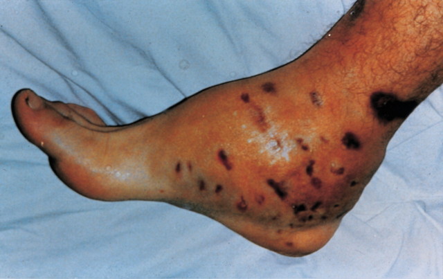
QUESTION: A 35-year-old agricultural worker from Albania has a 3-year history of recurrent, painless swellings with chronic purulent discharge from his right foot (see figure). He has no fever or systemic complaints. Physical examination reveals multiple painless swellings of the right foot and woody induration with sinus tracts discharging pus.
What is the diagnosis, differential diagnosis, and treatment for this condition?
ANSWER: The patient has mycetoma of the foot (Madura foot). The diagnosis was made on the basis of the characteristic clinical picture and the identification of typical grains in the purulent discharge. Gram staining of the affected tissues showed the fine branching hyphae of Actinomadura species within the actinomycetoma grains.
Mycetoma (Madura foot, maduromycosis) is a chronic, localized, subcutaneous infection caused by actinomycetes or fungi in which the organisms form aggregates (called grains or granules). These organisms elicit an inflammatory response in the deep dermis and subcutaneous tissue, leading to the development of draining sinuses communicating with the overlying skin and causing osteomyelitis.1,2 The disease was initially labeled Madura foot, named after the region in India where it was first identified. Mycetoma is common in the tropics or subtropics where people walk barefoot, although cases have occurred in other geographic zones.1 It typically occurs in areas where annual rainfall is low and in male agricultural workers between the ages of 20 and 50 years.3,4
More than 20 species of fungi and bacteria have been implicated as etiologic agents of mycetoma. Approximately 40% of cases are due to true fungi (eumycetoma), and 60% are caused by aerobic actinomycetes (actinomycetoma).5 The agents most often responsible for causing actinomycetoma are Nocardia brasiliensis, Actinomadura (Streptomyces) pelletieri, Streptomyces somaliensis, and Actinomadura madurae. Madurella mycetomatis and, in the United States, Pseudallescheria boydii are the most common cause of eumycetoma.4 The pathogens live in the soil and enter the skin where minor trauma has occurred. The most common site of infection is the foot.3,5
The infection runs a relentless course over many years, with destruction of contiguous bone and fascia. The infected site is characterized by painless swelling, woody induration, and sinus tracts that discharge pus intermittently. Systemic symptoms do not develop, and metastasis to distant sites in the body does not take place. Radiographs of bone should be performed to determine the presence and extent of bone involvement.
DIFFERENTIAL DIAGNOSIS
The differential diagnosis of mycetoma includes chronic osteomyelitis resulting from bacteria, mycobacteria, or actinomycosis; other deep fungal infections, such as blastomycosis, coccidioidomycosis, or sporotrichosis; botryomycosis; leishmaniasis; yaws; syphilis; and chromoblastomycosis.2,4 The diagnosis is determined on the basis of microscopic examination of pus and granules with potassium hydroxide preparation, Gram stain, and acid-fast stains of smears and by culture of the grains.4,5 The shape and color of the grains, along with their appearance in tissue subjected to skin biopsy, may be helpful in identifying the causative agent.2,4
TREATMENT
Treatment of mycetoma is difficult, in part because the condition is usually in an advanced stage by the time the diagnosis is made.
Treatment of actinomycetoma is surgical debridement, with prolonged treatment with antibiotics such as trimethoprim-sulfamethoxazole plus streptomycin, dapsone plus streptomycin, penicillin, tetracycline, and rifampin.4,6
Eumycetomas are unresponsive to antimicrobial therapy, although partial responses to amphotericin B, miconazole nitrate, ketoconazole, itraconazole, and thiabendazole have been reported.6 Fortunately, eumycetomas tend to be well circumscribed, yielding ready access to surgical approaches.
Figure 1.
Competing interests: None declared
References
- 1.Mariat F, Destombes P, Segretain G. The mycetomas: clinical features, pathology, etiology and epidemiology. Contrib Microbiol Immunol 1977;4: 1-39. [PubMed] [Google Scholar]
- 2.Hay RJ. Subcutaneous mycoses. In: Cook GC, ed. Manson's Tropical Diseases. London, England: WB Saunders; 1996: 1056-1062.
- 3.McGinnis MR. Mycetoma. Dermatol Clin 1996;14: 97-104. [DOI] [PubMed] [Google Scholar]
- 4.Rosenthal JR. Dematiaceous fungi. In: Gorbach SL, Bartlett JG, Blacklow NR, eds. Infectious Diseases. 2nd ed. Philadelphia, PA: WB Saunders; 1998: 2376-2381.
- 5.Saag MS. Mycetoma. In: Goldman L, Bennett JC, eds. Cecil Textbook of Medicine. 21st ed. Philadelphia, PA: WB Saunders; 2000: 1885-1887.
- 6.Welsh O, Salinas MC, Rodriguez MA. Treatment of eumycetoma and actinomycetoma. Curr Topics Med Mycol 1995;6: 47-71. [PubMed] [Google Scholar]


