Abstract
Rickettsia prowazekii, the causative agent of epidemic typhus, is an obligate intracellular parasitic bacterium that grows directly within the cytoplasm of the eucaryotic host cell. The absence of techniques for genetic manipulation hampers the study of this organism’s unique biology and pathogenic mechanisms. To establish the feasibility of genetic manipulation in this organism, we identified a specific mutation in the rickettsial rpoB gene that confers resistance to rifampin and used it to demonstrate allelic exchange in R. prowazekii. Comparison of the rpoB sequences from the rifampin-sensitive (Rifs) Madrid E strain and a rifampin-resistant (Rifr) mutant identified a single point mutation that results in an arginine-to-lysine change at position 546 of the R. prowazekii RNA polymerase β subunit. A plasmid containing this mutation and two additional silent mutations created in codons flanking the Lys-546 codon was introduced into the Rifs Madrid E strain of R. prowazekii by electroporation, and in the presence of rifampin, resistant rickettsiae were selected. Transformation, via homologous recombination, was demonstrated by DNA sequencing of PCR products containing the three mutations in the Rifr region of rickettsial rpoB. This is the first successful demonstration of genetic transformation of Rickettsia prowazekii and represents the initial step in the establishment of a genetic system in this obligate intracellular pathogen.
Members of the genus Rickettsia are unique bacterial pathogens that grow only within the cytoplasm or nucleoplasm of eucaryotic host cells (for a review, see reference 35). In this respect they differ from the other obligate intracellular bacteria of the genera Chlamydia, Coxiella, and Ehrlichia, which remain within intracytoplasmic vesicles. While they are restricted to this obligate intracytoplasmic existence, the rickettsiae are able to grow in an assortment of animal hosts, ranging from arthropods to humans. Arthropods serve as vectors for transmission of these bacteria to a variety of mammalian hosts, and for some species the arthropod can transmit the rickettsiae transovarially from generation to generation. While Rocky Mountain spotted fever and epidemic and endemic typhus, caused by Rickettsia rickettsii, Rickettsia prowazekii, and Rickettsia typhi, respectively, are the most well known of the rickettsial diseases, a variety of spotted fevers and rickettsial species are found worldwide (21).
The rickettsiae are well adapted for intracytoplasmic growth. They enter the cell by a process of induced phagocytosis and rapidly escape from the phagosome into the host cytoplasm by a process involving a phospholipase A2 activity (29, 37). Once in the cytoplasm, the rickettsiae are able to exploit the high-energy compounds found there by using specialized transport systems (35). Notable among these is an ATP/ADP translocase that can exchange rickettsial ADP for host ATP (34). However, the rickettsiae are not strict energy parasites and can generate ATP via an intact trichloroacetic acid cycle and oxidative phosphorylation (32, 35). Although the host cell cytoplasm is obviously a rich environment for an organism with such specialized transport systems, R. prowazekii maintains a relatively slow 8- to 12-h replication time (35). Such slow growth may maximize the number of rickettsiae produced within a host cell (36). Since the ability of R. prowazekii to invade, grow within, and ultimately destroy a host cell is the basis of its pathogenicity, an understanding of rickettsial intracytoplasmic growth mechanisms is crucial. Unfortunately, the lack of rickettsial mutants and a genetic system for manipulation of these novel parasitic bacteria has frustrated attempts to expand studies of its unique biology and mechanisms of pathogenesis.
Genetic analysis of R. prowazekii has been restricted to the isolation and characterization of rickettsial genes in Escherichia coli (1, 2, 9, 11, 16, 17, 33, 39–41), phylogenetic analyses based on selected gene sequences (22, 31), analysis of genome size by pulsed-field gel electrophoresis (12), and transcriptional analysis of selected R. prowazekii genes (7, 8, 20, 24). In addition, the R. prowazekii genome, with a size of 1,100 kb, is currently being sequenced. An initial report describing the sequence of 200 kb has been published (3). Despite these significant accomplishments, direct genetic manipulation of rickettsial genes has not been reported for any rickettsial species.
The barriers that must be overcome in order to establish a genetic transfer system in these obligate intracellular bacteria are significant. First, since these organisms grow only in the cytoplasm of a eucaryotic host cell, any manipulations designed to introduce DNA into the rickettsial cell must not prevent the organism from reinfecting a host cell. Second, unlike the other obligate intracellular bacteria, the Chlamydia and Coxiella, which have recently been transformed (27, 28), rickettsial species do not exhibit developmental stages and thus the production of more stable extracellular forms. Third, few rickettsial mutants have been identified that could be used for selection in a rickettsial system. Finally, the absence of identified rickettsial plasmids and bacteriophage limits the genetic systems available for manipulation. However, the recA gene of R. prowazekii has been identified, suggesting that standard homologous recombination systems should be operable in this organism (11). In addition, with the demonstration that R. prowazekii is extremely sensitive to rifampin (38) and the subsequent isolation of a rifampin-resistant mutant of R. prowazekii (5), a selectable phenotype has been identified that can be used to examine gene transfer in this organism. Rifampin binds to RNA polymerase and prevents transcription of the DNA (14, 30). Mutations that confer resistance to rifampin have been characterized in many bacteria, and most occur in the β subunit of RNA polymerase, the product of the rpoB gene (15, 23). To determine the feasibility of gene transfer in R. prowazekii, we identified a specific mutation in the R. prowazekii rpoB gene that confers resistance to rifampin and used this marker to establish, by using electroporation, the successful transformation of R. prowazekii by allelic exchange.
MATERIALS AND METHODS
Bacterial strains, plasmids, and oligonucleotides.
Bacterial strains, plasmids, and oligonucleotides used in this study are listed in Table 1. E. coli strains were grown on Luria-Bertani medium by standard techniques (4), and when required, the antibiotics ampicillin and tetracycline were added to a final concentration of 50 and 12.5 μg/ml, respectively. R. prowazekii Madrid E strain seed pool passage 282 was used for infecting mouse fibroblast L929 cells. Rickettsia-infected L929 cells were grown in an atmosphere of 5% CO2 at 34°C in modified Eagle’s medium (MEM) supplemented with 10% newborn-calf serum (Sigma, St. Louis, Mo.) and 1 mM l-glutamine (Sigma). For selection, rifampin (Sigma) was added to supplemented MEM at a final concentration of 200 ng/ml. Rickettsial growth was followed by microscopic examination of Gimenez-stained (13) infected cells growing on glass coverslips.
TABLE 1.
E. coli strains, plasmids, and oligonucleotides
| Strain, plasmid, or oligonucleotide | Description | Source and/or reference |
|---|---|---|
| Strains | ||
| R. prowazekii | ||
| Madrid E | Rifampin sensitive | (10) |
| Erifr1 | Rifampin resistant | N. Balayeva (5) |
| E. coli | ||
| JM109a | endA1 recA1 gyrA96 thi hsdR17 (rK− mK+) relA1 supE44 λ− Δ(lac-proAB) [F′ traD36 proA+B+ lacIqZΔM15] | Promega (42) |
| ES1301 mutS1 | lacZ53 mutS201::Tn5 thyA36 rha-5 metB1 deoC IN(rrnD-rrnE) | Promega |
| XL1-Blue | recA1 endA1 gyrA96 thi-1 hsdR17 supE44 relA1 lac [F′ proAB lacIqZΔM15 Tn10 (Tetr)] | Stratagene (6) |
| Plasmids | ||
| pAlter-1 | Amps Tetr | Promega |
| pBluescript II SK+ | Ampr | Stratagene |
| pMOB | Ampr | Gold BioTechnology (26)b |
| pMW489 | pBR322 with inserted 6.0-kb R. prowazekii HindIII fragment containing the sdhA gene | This study |
| pMW975 | pBluescript with inserted PCR fragment from Rifr rickettsia (DW89 and DW71), Ampr | This study |
| pMW982 | pBluescript with inserted PCR fragment from Rifr rickettsia (DW83 and DW92), Ampr | This study |
| pMW1000 | pBluescript with inserted PCR fragment from Rifs rickettsia (DW83 and DW92), Ampr | This study |
| pMW1004 | pBluescript with inserted PCR fragment from Rifs rickettsia (DW89 and DW71), Ampr | This study |
| pMW1022 | pAlter-1 with inserted PCR fragment from Rifr rickettsia (DW289 and DW260), Amps Tetr | This study |
| pMW1025 | pMW1022 mutagenized with DW298, ampicillin repair and tetracycline knockout oligos, Ampr Tets | This study |
| pMW1027 | pMOB with inserted 1.4-kb SacI-BamHI fragment from pMW1025 | This study |
| Deoxyoligonucleotidesc,d | ||
| DW71 | GCYTGNCKYTGCATRTT | DNAgency |
| DW83 | GATACCGTAATGCCTCATG | DNAgency |
| DW86 | ACTCTTCTATCGACTCTC | DNAgency |
| DW89 | TATTAAGAGTGCATAGAGCC | DNAgency |
| DW92 | CAACAAGACAAATTATGTACAAC | DNAgency |
| DW249 | CTGTACTAGGACCATCAGC | USA Biopolymer Laboratory |
| DW260 | CAAGCACCGAAGCACCAG | USA Biopolymer Laboratory |
| DW288 | TTTGGGATATAGACGAAGAT | Iowa State Sequencing Facility |
| DW289 | TTACTGAAAGCAGGACAAAA | Iowa State Sequencing Facility |
| DW298 | GCCCAAGGGCAGAAAGTTTTCTTTTATGAGTAAT | Oligo’s Etc. |
E. coli strains supplied with Altered Sites II in vitro mutagenesis system (Promega).
Gold BioTechnology, St. Louis, Mo.
All sequences are written 5′→3′.
DNAgency, Aston, Pa.; USA Biopolymer Laboratory, Mobile, Ala.; Iowa State Sequencing Facility, Ames, Iowa; Oligo’s Etc., Wilsonville, Oreg.
DNA techniques.
All DNA manipulations were performed by using standard techniques (4). Plasmid DNA was purified from E. coli by using the PERFECTprep or pZ523 plasmid purification columns purchased from 5 Prime→3 Prime, Inc. (Boulder, Colo.). Chromosomal DNA was isolated by the method of Marmur (18). Restriction enzymes were obtained from Life Technologies (Gaithersburg, Md.) and New England Biolabs (Beverly, Mass.) and used according to the manufacturer’s instructions. PCR amplifications of 100 μl of total volume containing 100 ng of template and a primer concentration of 1 μM were performed on a model 9600 thermal cycler (Perkin-Elmer Cetus, Norwalk, Conn.). PCR amplifications of rickettsial rpoB fragments were performed with the oligonucleotide primers listed in Table 1 and either Vent DNA polymerase (New England Biolabs) or AmpliTaq DNA polymerase and the GeneAmp PCR reagent kit purchased from Perkin-Elmer. A three-step cycle of 94°C for 1 min, 45 to 52°C for 1 to 2 min, and 72°C for 2 min was repeated for 25 to 30 cycles. A single 7-min extension at 72°C was included in the final cycle. For DNA sequencing, the PCR products obtained were purified using a GeneClean II kit (Bio 101, La Jolla, Calif.), sequenced directly or after ligation into the HincII site of pBluescript II SK+ (Stratagene, La Jolla, Calif.) and subsequent transformation into E. coli XL1-blue. Manual DNA sequencing was accomplished using the T7 Sequenase version 2.0 kit or the Thermo Sequenase cycle sequencing kit (Amersham Life Science, Inc., Cleveland, Ohio). Automated sequencing was performed at the University of South Alabama Biopolymer Center or at the DNA Sequencing Facility at Iowa State University (Ames, Iowa). For site-specific mutagenesis, PCR products were cloned into the SmaI site of pAlter-1 (Promega, Madison, Wis.), and mutations were introduced by using the Altered Sites II in vitro mutagenesis system (Promega). Probes for use in Southern hybridizations (25) were 32P labeled using the Multiprime DNA labelling system (Amersham) and [α-32P]dATP (3,000 Ci/mmol; ICN, Irvine, Calif.).
Electroporation.
R. prowazekii cells for electroporation were harvested from three 162-cm2 tissue culture flasks containing confluent monolayers of infected L929 cells (100% infected, approximately 200 to 300 rickettsiae/cell) as follows. The infected L929 cells were released from each flask surface with 5 ml of trypsin-EDTA (Sigma), diluted in 10 ml of supplemented MEM, and centrifuged in a clinical centrifuge at approximately 1,200 × g for 5 min at room temperature. The pellet from each flask was suspended in 3 ml of cold sucrose-phosphate-glutamic acid–Mg (220 mM sucrose, 3.8 mM KH2PO4, 8 mM K2HPO4, 5 mM potassium glutamate, 10 mM MgCl2), and the cell suspension was transferred to a precooled 15-ml conical centrifuge tube containing 2 g of 3-mm glass beads (Kimble Glass, Inc., Vineland, N.J.) and vortexed vigorously for 30 s in order to lyse the L929 cells. Vortexing was repeated two times with 30-s intervals of incubation on ice between each 30-s vortex. The three lysates, one from each of the original flasks, were combined into a single, precooled 50-ml conical centrifuge tube and centrifuged at approximately 1,200 × g in a clinical centrifuge to remove unbroken L929 cells and cellular debris. The supernatant, containing released R. prowazekii cells, was transferred to an Oakridge tube, and the rickettsiae were collected by centrifugation at 10,000 rpm for 10 min at 4°C by using a JA-20 rotor and a Beckman J2-21 refrigerated centrifuge (Beckman Instruments, Inc., Palo Alto, Calif.). The rickettsial pellet was washed twice with 5 ml of cold 250 mM sucrose and finally suspended in 200 μl of 250 mM sucrose (approximately 3 × 10¹⁰ bacterial cells per ml). A 50-μl sample of this rickettsial suspension was mixed with 1 to 20 μg of DNA and then transferred to a 0.1-cm gap electroporation cuvette (BTX Electronic Genetics, San Diego, Calif.) and chilled on ice for 10 min. The cuvette was placed in a BTX Electro Cell Manipulator (ECM 600) and electroporated (field strength = 17 kV/cm, pulse time = 4 to 5 ms, resistance = 129 Ω, capacitance = 50 μF). Immediately following electroporation, the rickettsiae were diluted into 1 ml of Hanks balanced salt solution (Sigma) supplemented with 5 mM glutamic acid and 0.1% gelatin, mixed with approximately 3 × 10⁷ L929 cells and incubated at 34°C for 1 h with continuous shaking. The infected L929 cells were harvested by centrifugation at approximately 1,200 × g, washed with 5 ml of supplemented MEM, suspended in 35 ml of supplemented MEM, and planted into two 162-cm2 flasks and one dish containing glass coverslips. For selection, rifampin was added to the flasks 24 h following infection to a final concentration of 200 ng/ml. The rifampin-containing medium was changed every 2 to 3 days in order to ensure continuous selection. Growth was followed by observation of Gimenez-stained cells from coverslips.
Nucleotide sequence accession number.
The sequence of the R. prowazekii rpoB gene is available in GenBank under accession no. AF034531.
RESULTS
Isolation and sequencing of the R. prowazekii rpoB gene.
One of the few mutants of R. prowazekii that has been identified is one that exhibits resistance to rifampin (5). This mutant, unlike the Madrid E parent strain, which is sensitive to extremely low levels of rifampin (20 ng/ml), is able to grow in the presence of 200 ng of rifampin per ml, suggesting that this antimicrobial agent could provide the selection needed for genetic transfer experiments. The molecular basis for this resistance in R. prowazekii was hypothesized to be an altered RNA polymerase, since rifampin could inhibit rickettsial RNA polymerase purified from the rifampin-sensitive Madrid E strain but not that purified from the resistant mutant in in vitro assays (10a). Since rifampin resistance in bacteria usually results from mutations within the rpoB gene encoding the β subunit of RNA polymerase (23), we focused our efforts on identifying an rpoB mutation associated with the resistant phenotype. Characterization of such a mutation would identify a DNA fragment with a selectable marker for use in transformation experiments.
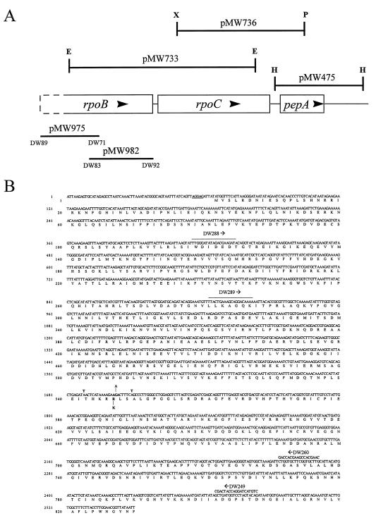
Isolation of the rpoB genes from the rifampin-sensitive and rifampin-resistant R. prowazekii strains was accomplished by using a PCR approach. The 3′ end of the R. prowazekii rpoB gene had previously been isolated in our laboratory as one of several linked genes identified in three overlapping recombinant plasmids (Fig. 1A). Sequence immediately upstream of the rpoB start codon was kindly provided by Siv Andersson prior to publication of a rickettsial genome survey (3). Primers (DW89 and DW92) generated based upon these sequences, when used in a PCR, amplified a predicted fragment of approximately 4.2 kb that contained the entire rickettsial rpoB gene (data not shown). However, repeated attempts to clone this fragment into E. coli were unsuccessful. To overcome this problem, two overlapping fragments encompassing the entire rpoB gene were generated by using primers DW89 and DW83 in conjunction with DW71 and DW92 (Fig. 1A). DW71, which was synthesized prior to our obtaining the complete rpoB sequence, is a degenerate primer to the conserved amino acid sequence, NMQRQA, while DW83 was obtained from DNA sequence contained in pMW975. The resulting clones have an overlapping region of 617 bp and together cover the entire rpoB gene from 71 bases upstream of the start codon to 34 bases downstream of the stop codon. To ensure fidelity of the PCR and accuracy of the sequence, two independently generated PCR fragments were cloned for both the sensitive and resistant strains, and sequence was obtained for both strands. Comparison of the rifampin-sensitive and rifampin-resistant rpoB sequences revealed only one difference. The sequence of this region of the R. prowazekii rpoB gene from the rifampin-sensitive Madrid E strain is presented in Fig. 1B. The single difference, a mutation of G to A at position 1727 that results in a change of lysine to arginine at residue 546 within the translated protein, is indicated. This putative rifampin resistance mutation is located within a highly conserved region of the β subunit where most of the mutations characterized in other bacteria have been found. Indeed, mutation at this codon has been shown to confer rifampin resistance in E. coli (23). However, the rickettsial mutation is unique since none of the E. coli mutations resulted in the substitution of lysine for arginine at this conserved site.
FIG. 1.
(A) Schematic map of the R. prowazekii rpoB gene region. The orientation of transcription is indicated by arrows. Plasmids containing the rickettsial fragments are indicated by horizontal lines. The lengths of the lines indicate the corresponding segment of the gene map contained within the recombinant plasmid. DW89, DW71, DW83, and DW92 mark the positions of the specific oligonucleotides used to amplify the inserts of the indicated plasmids. E, EcoRI; H, HindIII; P, PstI; X, XbaI. (B) Nucleotide sequence of the 5′ half of the R. prowazekii rpoB gene. The DNA sequence of the noncoding strand is shown. The predicted amino acid sequence is given in the single-letter code. The putative Shine-Dalgarno-like sequence is doubly underlined. Relevant oligonucleotide sequences discussed in the text are indicated, and the direction of DNA synthesis is indicated by arrows. Mutations are indicated at the appropriate positions above and below the sequence.
Electroporation of R. prowazekii.
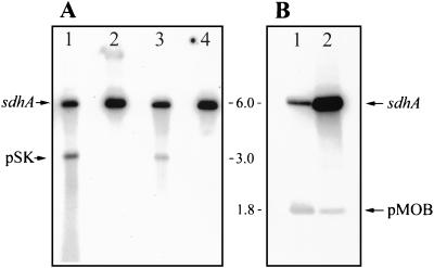
The first step in achieving R. prowazekii transformation was to identify conditions that would permit extracellular DNA to gain entrance into the rickettsial cell. Due to the efficiency and broad applicability of electroporation (19), we chose this method for our studies. Electroporation of R. prowazekii cells was tested at field strengths of 9 to 20 kV/cm. There was no decrease in viability at any of these field strengths, and when plasmid DNA (pBluescript II SK+) was added, no difference in the uptake of the DNA was noted at the different field strengths (data not shown). Except where indicated, a field strength of 17 kV/cm was chosen as the standard field strength for the electroporations conducted in this study. To confirm that DNA was entering the rickettsial cells, rickettsiae were electroporated in the presence of pBluescript II SK+ plasmid and intracellular plasmid detected by Southern hybridization. To ensure that extracellular plasmid was not detected, the rickettsiae were harvested and treated with RQ1 DNase (Promega) prior to lysis of the bacterial cells. The DNA was extracted and digested with HindIII, and the fragments were separated by agarose gel electrophoresis, transferred to a nylon membrane, and probed with labeled pMW489 (a recombinant plasmid consisting of pBR322 and a 6.0-kb R. prowazekii HindIII fragment containing the sdhA gene). As shown in Fig. 2A, electroporation allowed the plasmid DNA to enter a DNase-resistant site at detectable levels.
FIG. 2.
Detection of plasmid DNA in electroporated rickettsiae. The sdhA gene and pBluescript II SK+ (pSK) and pMOB plasmids are indicated. Numbers are sizes in kilobases. (A) Autoradiograph of DNA extracted from DNase-treated R. prowazekii cells immediately following electroporation, digested with HindIII, and probed with pMW489, a plasmid containing the R. prowazekii sdhA gene on a 6.0-kb HindIII fragment. Lanes 1 and 3, R. prowazekii cells electroporated in the presence of 1 μg of pBluescript II SK+ plasmid; lanes 2 and 4, R. prowazekii cells mixed with 1 μg of pBluescript II SK+ plasmid but not electroporated. (B) Detection of electroporated DNA after infection of host cells. R. prowazekii cells were electroporated with 10 μg of pMOB and the electroporated rickettsiae were allowed to infect L929 cells. At 1 h (lane 1) and 48 h (lane 2) after infection, the rickettsiae were harvested and treated with DNase. After inactivating the DNase, rickettsial DNA was extracted, digested with HindIII, and probed with pMW489.
To assess plasmid stability, R. prowazekii cells were electroporated in the presence of pMOB plasmid (26), chosen for its small size, and the rickettsiae were then allowed to infect L929 cells prior to harvesting at selected times after infection. Once again, intracellular plasmid was detected by hybridization as described above. The results of this experiment (Fig. 2B) demonstrated that small amounts of plasmid DNA could be detected for at least 48 h after electroporation. The small amounts of plasmid detected and the decrease over time indicate that much of the plasmid DNA is degraded and that the plasmid is not replicating (Fig. 2B). These data indicate that the E. coli ColE1-specific origin found on pMOB does not function in R. prowazekii. In contrast, the increase in the number of rickettsiae, as evidenced by the increased sdhA signal, can easily be detected.
Construction of the transforming DNA.
To prepare a transforming DNA molecule that would permit us to differentiate a transformant from a spontaneously occurring rifampin-resistant mutant, a marked fragment of the resistance gene was engineered. We first amplified a 1,390-bp fragment of the rifampin-resistant rpoB gene, by using primers DW289 and DW260 (Fig. 1B) to place the Lys-546 mutation at the center of this DNA fragment. The fragment was cloned into the pAlter-1 vector, and two silent mutations were generated, one on either side of the Rifr mutation (Fig. 1). The fragment was then excised from pAlter-1 and inserted into pMOB. The rickettsial insert of the pMOB-based clone was sequenced and shown to be identical to the original rpoB Rifr gene sequence with the exception of the two modified bases. This plasmid, designated pMW1027, was used in the transformation experiments.
Transformation of R. prowazekii to rifampin resistance.
The Madrid E strain of R. prowazekii was electroporated in the presence of 20 μg of pMW1027, and the rickettsiae were allowed to infect L929 cells. Twenty-four hours after infection, 100% of the L929 cells were infected with R. prowazekii and each cell contained approximately 50 to 70 rickettsiae. Rifampin (200 ng/ml) was added, and incubation was continued. Rickettsiae were rapidly cleared from the host cells following rifampin addition. At 7 days postinfection, rickettsiae could not be detected on the stained coverslips. In the cultures of R. prowazekii electroporated with pMW1027, rickettsial growth was again detected at 11 days postinfection, with approximately 1% of the host cells containing 200 to 300 rickettsiae/cell. These culture flasks were incubated for three more days before harvesting the rickettsiae, at which time approximately 20% of the host cells were infected. A control culture infected with rickettsiae electroporated with pMOB alone showed no evidence of rickettsial growth until 18 days postinfection. This culture was continued to 24 days postinfection before harvesting, at which time 15% of the host cells were infected with approximately 200 to 300 rickettsiae/cell. The rickettsiae were harvested from these cultures, and chromosomal DNA was isolated for use as template DNA in a PCR.
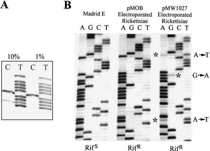
Due to the difficulty associated with isolating individual Rifr clones by plaque purification or limited dilution techniques, we chose to identify the presence of Rifr transformants by DNA sequencing of PCR products. In order to determine the level of detection by using this method, we mixed two template DNAs differing at a single base at varying ratios and sequenced the mixture. The results (Fig. 3A) reveal that a mutation accounting for no more than 10% of the total template DNA in the sequencing reaction could be easily detected.
FIG. 3.
(A) Sequencing reactions (only C and T lanes are shown) comparing different ratios of templates that differ in sequence at one position. The template present at the indicated percentages contains two C residues. (B) DNA sequence of PCR products from Rifs and Rifr rickettsiae. The mutations described in the text are indicated on the right. Asterisks identify the sequence residues of the minor background Rifr population.
To ensure that we would not amplify any clones that were generated over the course of these studies, new primers (DW288 and DW249) (Fig. 1B), whose sequences have never been present on the same plasmid construct, were chosen. Thus, amplification would be specific for chromosomal DNA. Using these primers, a 2.1-kb fragment was amplified by using template DNA obtained from both the pMW1027-electroporated rickettsiae and the control pMOB-electroporated rickettsiae. Direct sequencing of these PCR fragments revealed the presence of the Rifr G-to-A mutation in both the experimental (pMW1027) and control (pMOB) fragments (Fig. 3B). The presence of this mutation in the pMOB electroporated cells, an independently isolated, spontaneously occurring Rifr mutant, supported our initial hypothesis that this site is important in rifampin resistance. More importantly, the two nonselected mutations were present only in the PCR product amplified from template DNA isolated from the rickettsiae electroporated with pMW1027 (Fig. 3B). While it is possible to detect a minor population which had not undergone allelic exchange, represented by the bands marked with asterisks in Fig. 3B, the intensities of these bands in comparison to the major band at each site indicates that transformants greatly outnumber spontaneous mutants. This background population of rifampin-resistant rickettsiae that does not contain the G-to-A mutation at position 1727 presumably results from spontaneous mutations at other sites within rpoB. A repeat experiment achieved a comparable percentage of transformants, illustrating that transformation occurs at a frequency higher than the spontaneous mutation rate. These data demonstrate the successful introduction of DNA into this obligate intracellular parasite and subsequent recombination of this DNA into the rickettsial genome, presumably by homologous recombination mechanisms.
DISCUSSION
The cloning of R. prowazekii genes into E. coli has provided important information on rickettsial gene structure and function, and the completion of the R. prowazekii genome sequencing project will identify its genetic capabilities. However, an ability to manipulate these genes in a rickettsial background is needed to understand their importance to rickettsial growth and pathogenicity. In this report we have established that electroporation can be used to introduce DNA into R. prowazekii cells and that this organism has the capability of incorporating this DNA, presumably by homologous recombination mechanisms, into its genome.
Introduction of DNA into the rickettsial cells by electroporation could be accomplished at several field strengths ranging from 9 kV/cm to higher than 17 kV/cm. Interestingly, we could detect no loss of viability under the conditions used even at the higher field strengths. Accordingly, since rickettsial cells are approximately 10 times smaller than E. coli cells, we chose one of the higher field strengths (17 kV/cm) for our standard electroporation conditions.
Important to successful rickettsial transformation was the identification of an appropriate selectable marker. While chloramphenicol and tetracycline would be preferred markers, because of rickettsial sensitivity to these drugs and the existence of vectors that contain these resistance genes, these antibiotics are clinically important in the treatment of rickettsial diseases and thus are unavailable for genetic studies in these bacteria. However, the existence of a rifampin-resistant R. prowazekii mutant, the association of rifampin resistance and mutations within the rpoB gene in other bacteria, and the sensitivity of R. prowazekii RNA polymerase to rifampin in vitro (10a) suggested that rifampin would provide an excellent selection for studies to determine the feasibility of rickettsial gene transfer.
We established the existence of a rifampin resistance mutation within the R. prowazekii rpoB gene, the gene that codes for the β subunit of DNA-dependent RNA polymerase. This mutation falls within a region of the rickettsial protein that corresponds to the rifampin resistance region of E. coli (23). Surprisingly, the mutation results in a very conservative substitution, a lysine for an arginine at residue 546. Although, this is a frequently mutated codon in other bacteria (23), this specific amino acid substitution has not been described. During these studies, we obtained an additional spontaneous rifampin-resistant R. prowazekii mutant which exhibited the same mutation of Arg to Lys, suggesting that this is a common site for rickettsial rifampin resistance mutations. However, other mutations may be possible within this region. For example, we identified a mutation in another independently isolated rifampin-resistant mutant that results in a change of amino acid 533 (Asp→Tyr). Since spontaneous mutants occurred at a detectable frequency, it was necessary to introduce marker mutations that would permit us to differentiate transformants from these mutants. This was easily accomplished by introducing silent mutations in codons surrounding the Lys-546 mutation. Because the sensitivity provided by direct sequencing of PCR products permitted the detection of transformants among spontaneous mutants, the laborious techniques for isolating single rickettsial clones were unnecessary for demonstrating rickettsial transformation. Indeed, by observing the intensities of the bands on the DNA sequence autoradiographs it was possible to directly observe that transformants accounted for the majority of the rifampin-resistant mutants.
In conclusion, this study has established that R. prowazekii can survive electroporation conditions, that DNA can be transferred into rickettsial cells during electroporation, that this DNA can recombine into the rickettsial genome, and that transformants can be detected over background spontaneous mutants. While obstacles remain to be overcome before genetic manipulation of rickettsial species becomes commonplace, the success of these experiments is a crucial first step. Studies can now be directed toward identifying additional selection mechanisms that will permit the genetic manipulation of any rickettsial gene and the identification of plasmids that will function in this organism. The successful transformation of R. prowazekii also provides a model for other bacteria that cannot be grown outside the eucaryotic host cell. The availability of genetic tools will finally provide the means for investigating the unique biological and pathogenic mechanisms of these obligate intracellular bacteria.
ACKNOWLEDGMENTS
We thank Andria Hines and Marie Solomon for expert technical assistance.
This work was supported by NIH grant AI20384 to D.O.W.
REFERENCES
- 1.Aliabadi Z, Winkler H H, Wood D O. Isolation and characterization of the Rickettsia prowazekii gene encoding the flavoprotein subunit of succinate dehydrogenase. Gene. 1993;133:135–140. doi: 10.1016/0378-1119(93)90238-x. [DOI] [PubMed] [Google Scholar]
- 2.Anderson B E, Tzianabos T. Comparative sequence analysis of a genus-common rickettsial antigen gene. J Bacteriol. 1989;171:5199–5201. doi: 10.1128/jb.171.9.5199-5201.1989. [DOI] [PMC free article] [PubMed] [Google Scholar]
- 3.Andersson S G E, Eriksson A-S, Näslund A K, Andersen M S, Kurland C G. The Rickettsia prowazekii genome: a random sequence analysis. Microb Comp Genomics. 1997;1:293–315. [PubMed] [Google Scholar]
- 4.Ausubel F, Brent R, Kingston R E, Moore D D, Seidman J G, Smith J A, Struhl K, editors. Current protocols in molecular biology. 1, 2, and 3. New York, N.Y: John Wiley & Sons, Inc.; 1997. [Google Scholar]
- 5.Balayeva N M, Frolova O M, Genig V A, Nikolskaya V N. Some biological properties of antibiotic resistant mutants of Rickettsia prowazekii strain E induced by nitroso-guanidine. In: Kazár J, editor. Rickettsiae and rickettsial diseases. Bratislava, Slovakia: Publishing House of the Slovak Academy of Sciences; 1985. pp. 85–91. [Google Scholar]
- 6.Bullock W O, Fernandez J M, Short J M. A high efficiency plasmid transforming recA Escherichia coli strain with beta-galactosidase selection. BioTechniques. 1987;5:376–379. [Google Scholar]
- 7.Cai J, Pang H, Wood D O, Winkler H H. The citrate synthase-encoding gene of Rickettsia prowazekii is controlled by two promoters. Gene. 1995;163:115–119. doi: 10.1016/0378-1119(95)00365-d. [DOI] [PubMed] [Google Scholar]
- 8.Cai J, Winkler H H. Identification of tlc and gltA mRNAs and determination of in situ RNA half-life in Rickettsia prowazekii. J Bacteriol. 1993;175:5725–5727. doi: 10.1128/jb.175.17.5725-5727.1993. [DOI] [PMC free article] [PubMed] [Google Scholar]
- 9.Carl M, Dobson M E, Ching W-M, Dasch G A. Characterization of the gene encoding the protective paracrystalline-surface-layer protein of Rickettsia prowazekii: presence of a truncated identical homolog in Rickettsia typhi. Proc Natl Acad Sci USA. 1990;87:8237–8241. doi: 10.1073/pnas.87.21.8237. [DOI] [PMC free article] [PubMed] [Google Scholar]
- 10.Clavero G, Perez Gallardo F. Estudio experimental de una cepa apatogena e inmunizante de Rickettsia Prowazeki Cepa E. Rev Sanid Hig Publica. 1943;17:1–27. [Google Scholar]
- 10a.Ding, H. F., and H. H. Winkler. Unpublished data.
- 11.Dunkin S M, Wood D O. Isolation and characterization of the Rickettsia prowazekii recA gene. J Bacteriol. 1994;176:1777–1781. doi: 10.1128/jb.176.6.1777-1781.1994. [DOI] [PMC free article] [PubMed] [Google Scholar]
- 12.Eremeeva M E, Roux V, Raoult D. Determination of genome size and restriction pattern polymorphism of Rickettsia prowazekii and Rickettsia typhi by pulsed field gel electrophoresis. FEMS Microbiol Lett. 1993;112:105–112. doi: 10.1111/j.1574-6968.1993.tb06431.x. [DOI] [PubMed] [Google Scholar]
- 13.Gimenez D F. Staining rickettsiae in yolk-sac cultures. Stain Technol. 1964;39:135–140. doi: 10.3109/10520296409061219. [DOI] [PubMed] [Google Scholar]
- 14.Hartmann G, Honikel K O, Knusel F, Nuesch J. The specific inhibition of the DNA-directed RNA synthesis by rifamycin. Biochim Biophys Acta. 1967;145:843–844. doi: 10.1016/0005-2787(67)90147-5. [DOI] [PubMed] [Google Scholar]
- 15.Jin D J, Gross C A. Mapping and sequencing of mutations in the Escherichia coli rpoB gene that lead to rifampin resistance. J Mol Biol. 1988;202:45–58. doi: 10.1016/0022-2836(88)90517-7. [DOI] [PubMed] [Google Scholar]
- 16.Marks G L, Winkler H H, Wood D O. Isolation and characterization of the gene coding for the major sigma factor of Rickettsia prowazekii DNA-dependent RNA polymerase. Gene. 1992;121:155–160. doi: 10.1016/0378-1119(92)90175-o. [DOI] [PubMed] [Google Scholar]
- 17.Marks G L, Wood D O. Characterization of the gene coding for the Rickettsia prowazekii DNA primase analogue. Gene. 1993;123:121–125. doi: 10.1016/0378-1119(93)90550-m. [DOI] [PubMed] [Google Scholar]
- 18.Marmur J. A procedure for the isolation of deoxyribonucleic acid from microorganisms. J Mol Biol. 1961;3:208–218. [Google Scholar]
- 19.Nickoloff J A, editor. Electroporation protocols for microorganisms. 47 of methods in molecular biology. Totowa, N.J: Humana Press, Inc.; 1995. [Google Scholar]
- 20.Pang H, Winkler H H. Transcriptional analysis of the 16S rRNA gene in Rickettsia prowazekii. J Bacteriol. 1996;178:1750–1755. doi: 10.1128/jb.178.6.1750-1755.1996. [DOI] [PMC free article] [PubMed] [Google Scholar]
- 21.Raoult D, Roux V. Rickettsioses as paradigms of new or emerging infectious diseases. Clin Microbiol Rev. 1997;10:694–719. doi: 10.1128/cmr.10.4.694. [DOI] [PMC free article] [PubMed] [Google Scholar]
- 22.Roux V, Rydkina E, Eremeeva M, Raoult D. Citrate synthase gene comparison, a new tool for phylogenetic analysis, and its application for the rickettsiae. Int J Syst Bacteriol. 1997;47:252–261. doi: 10.1099/00207713-47-2-252. [DOI] [PubMed] [Google Scholar]
- 23.Severinov K, Soushko M, Goldfarb A, Nikiforov V. Rifampicin region revisited. New rifampicin-resistant and streptolydigin-resistant mutants in the β subunit of Escherichia coli RNA polymerase. J Biol Chem. 1993;268:14820–14825. [PubMed] [Google Scholar]
- 24.Shaw E I, Marks G L, Winkler H H, Wood D O. Transcriptional characterization of the Rickettsia prowazekii major macromolecular synthesis operon. J Bacteriol. 1997;179:6448–6452. doi: 10.1128/jb.179.20.6448-6452.1997. [DOI] [PMC free article] [PubMed] [Google Scholar]
- 25.Southern E M. Detection of specific sequences among DNA fragments separated by gel electrophoresis. J Mol Biol. 1975;98:503–517. doi: 10.1016/s0022-2836(75)80083-0. [DOI] [PubMed] [Google Scholar]
- 26.Strathmann M, Hamilton B A, Mayeda C A, Simon M I, Meyerowitz E M, Palazzolo M J. Transposon-facilitated DNA sequencing. Proc Natl Acad Sci USA. 1991;88:1247–1250. doi: 10.1073/pnas.88.4.1247. [DOI] [PMC free article] [PubMed] [Google Scholar]
- 27.Suhan M L, Chen S-Y, Thompson H A. Transformation of Coxiella burnetii to ampicillin resistance. J Bacteriol. 1996;178:2701–2708. doi: 10.1128/jb.178.9.2701-2708.1996. [DOI] [PMC free article] [PubMed] [Google Scholar]
- 28.Tam J E, Davis C H, Wyrick P B. Expression of recombinant DNA introduced into Chlamydia trachomatis by electroporation. Can J Microbiol. 1994;40:583–591. doi: 10.1139/m94-093. [DOI] [PubMed] [Google Scholar]
- 29.Walker T S, Winkler H H. Penetration of cultured mouse fibroblasts (L cells) by Rickettsia prowazekii. Infect Immun. 1978;22:200–208. doi: 10.1128/iai.22.1.200-208.1978. [DOI] [PMC free article] [PubMed] [Google Scholar]
- 30.Wehrli W, Handschin J, Wunderli W. Interaction between rifampicin and DNA-dependent RNA polymerase of E. coli. In: Losick R, Chamberlin M, editors. RNA polymerase. Cold Spring Harbor, N.Y: Cold Spring Harbor Laboratory; 1976. pp. 397–412. [Google Scholar]
- 31.Weisburg W G, Dobson M E, Samuel J E, Dasch G A, Mallavia L P, Baca O, Mandelco L, Sechrest J E, Weiss E, Woese C R. Phylogenetic diversity of the rickettsiae. J Bacteriol. 1989;171:4202–4206. doi: 10.1128/jb.171.8.4202-4206.1989. [DOI] [PMC free article] [PubMed] [Google Scholar]
- 32.Weiss E. The biology of rickettsiae. Annu Rev Microbiol. 1982;36:345–370. doi: 10.1146/annurev.mi.36.100182.002021. [DOI] [PubMed] [Google Scholar]
- 33.Williamson L R, Plano G V, Winkler H H, Krause D C, Wood D O. Nucleotide sequence of the Rickettsia prowazekii ATP/ADP translocase-encoding gene. Gene. 1989;80:269–278. doi: 10.1016/0378-1119(89)90291-6. [DOI] [PubMed] [Google Scholar]
- 34.Winkler H H. Rickettsial permeability: an ADP-ATP transport system. J Biol Chem. 1976;251:389–396. [PubMed] [Google Scholar]
- 35.Winkler H H. Rickettsia species (as organisms) Annu Rev Microbiol. 1990;44:131–153. doi: 10.1146/annurev.mi.44.100190.001023. [DOI] [PubMed] [Google Scholar]
- 36.Winkler H H. Rickettsia prowazekii, ribosomes and slow growth. Trends Microbiol. 1995;3:196–198. doi: 10.1016/s0966-842x(00)88920-9. [DOI] [PubMed] [Google Scholar]
- 37.Winkler H H, Daugherty R M. Phospholipase A activity associated with the growth of Rickettsia prowazekii in L929 cells. Infect Immun. 1989;57:36–40. doi: 10.1128/iai.57.1.36-40.1989. [DOI] [PMC free article] [PubMed] [Google Scholar]
- 38.Wisseman C L, Jr, Waddell A D, Walsh W T. In vitro studies of the action of antibiotics on Rickettsia prowazekii by two basic methods of cell culture. J Infect Dis. 1974;130:564–574. doi: 10.1093/infdis/130.6.564. [DOI] [PubMed] [Google Scholar]
- 39.Wood D O, Solomon M J, Speed R R. Characterization of the Rickettsia prowazekii pepA gene encoding leucine aminopeptidase. J Bacteriol. 1993;175:159–165. doi: 10.1128/jb.175.1.159-165.1993. [DOI] [PMC free article] [PubMed] [Google Scholar]
- 40.Wood D O, Waite R T. Sequence analysis of the Rickettsia prowazekii gyrA gene. Gene. 1994;151:191–196. doi: 10.1016/0378-1119(94)90655-6. [DOI] [PubMed] [Google Scholar]
- 41.Wood D O, Williamson L R, Winkler H H, Krause D C. Nucleotide sequence of the Rickettsia prowazekii citrate synthase gene. J Bacteriol. 1987;169:3564–3572. doi: 10.1128/jb.169.8.3564-3572.1987. [DOI] [PMC free article] [PubMed] [Google Scholar]
- 42.Yanisch-Perron C, Vieira J, Messing J. Improved M13 phage cloning vectors and host strains: nucleotide sequences of the M13mp18 and pUC19 vectors. Gene. 1985;33:103–119. doi: 10.1016/0378-1119(85)90120-9. [DOI] [PubMed] [Google Scholar]


