Abstract
Few studies have examined the basilar venous plexus (BVP) and to our knowledge, no previous study has described its histology. The present anatomical study was performed to better elucidate these structures. In ten cadavers, the BVP was dissected. The anatomical and histological evaluation of the intraluminal trabeculae within this sinus were evaluated. Once all gross measurements were made, the clivus and overlying BVP were harvested and submitted for histological analysis. A BVP was identified in all specimens and in each of these, intraluminal trabeculae were identified. The mean number of trabeculae per plexus was five. These were most concentrated in the upper half of the clivus and were more often centrally located. These septations traveled in a posterior to anterior direction and usually, from inferiorly to superiorly however some were noted to travel horizontally. In a few specimens the trabeculae had wider bases, especially on the posterior attachment to the meningeal layer of dura mater. More commonly, the trabeculae ended in a denticulate form at their two terminal ends. The trabeculae were on average were 0.85 mm in length. The mean width of the trabeculae was 0.35 mm. These septations were consistent with the cords of Willis as are found in the lumen of some of the other intradural venous sinuses. An understanding of the internal anatomy of the BVP can aid in our understanding of venous pathology. Furthermore, this knowledge will benefit patients undergoing interventional treatments that involve the BVP.
Keywords: Plexus, Intravascular procedures, Sinus, Anatomy, Clivus
Introduction
The basilar venous plexus (Figs. 1, 2) is a variable structure originating between the meningeal and endosteal layers of the dura overlying the dorsal surface of the clivus. It has been referred to as the plexus basilaris of Virchow, the posterior clinoid sinus, the anterior occipital sinus, and the sinus of Littré, among other names [1-3]. The plexus connects the dural venous sinuses of the posterior cranial fossa [2, 4]. Laterally, the plexus connects the inferior petrosal sinuses with the internal vertebral venous plexus inferiorly [5]. Posteriorly, the plexus joins with the marginal sinus, while it connects anteriorly with the anterior condylar veins [2, 6, 7]. Anastomoses with small veins from the brainstem and cerebellum have also been described [8, 9]. Understanding the anatomy of this venous structure and its complexity isimportant for clinicians treating patients with neurological diseases of the venous system and cavernous sinus, as well as for interpreting imaging results of these structures.
Fig. 1.
Schematic drawings of the basilar venous plexus (red arrow) and surrounding anatomy. *Marginal sinus.
Fig. 2.
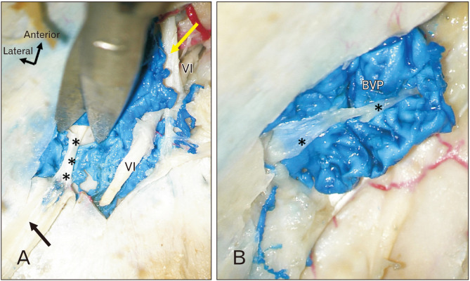
(A) Gross cadaveric dissection inside the basilar venous plexus (blue) after opening a window into the dura mater (black arrow) over the clivus. Note the septation (*) within the basilar venous plexus. The septation is seen attaching to the anterior and posterior walls of the plexus. For reference, laterally, note the right abducens nerve (VI) traveling through the dura (Dorello’s canal) over the clivus and continuing anteriorly under the petroclinoid (clival) ligament (yellow arrow) to enter the cavernous sinus with the inferior petrosal sinus. (B) Gross dissection noting a more or less horizontally positioned intraluminal septation (*) within the basilar venous plexus (BVP).
In addition to the variable anastomoses, the central location of the basilar venous plexus dorsal to the clivus, is notable [10-12]. The clivus is associated with numerous pathologies including neoplasm, inflammatory reactions, and trauma, all of which can affect the overlying basilar venous plexus [13]. Although the clivus has been widely studied, the basilar venous plexus remains poorly understood. Therefore, further investigation into its anatomy and variants is valuable for managing disease in and around the clivus. We report our findings of the anatomical and histological findings of the intraluminal trabeculae of the basilar venous plexus.
Materials and Methods
In ten adult cadaveric specimens, the basilar venous plexus was studied. Specifically, the anatomical and histological evaluation of the intraluminal trabeculae were evaluated. Five specimens were latex injected and five were freshly frozen. Six males and four females made up the cohort with a mean age at death of 80.1 years (range 65 to 101 years). In the supine position, the calvaria was removed from each specimen using an oscillating bone saw. The dura mater was opened, and the brain removed. Using a surgical microscope (Zeiss), the meningeal layer of dura mater over the clivus was carefully opened and the underlying trabeculae dissected. When found, the length and width of the trabeculae were measured and the orientation of these septations documented. All measurements were made using microcalipers (Mitutoyo). Once the gross measurements were made, the clivus and overlying basilar venous plexus were harvested and fixed in 10% formalin. Specimens were decalcified in ethylenediaminetetraacetic acid for one month and then submitted for histological analysis (H&E, Masson’s trichrome). The authors state that every effort was made to follow all local and international ethical guidelines and laws that pertain to the use of human cadaveric donors in anatomical research [14].
Results
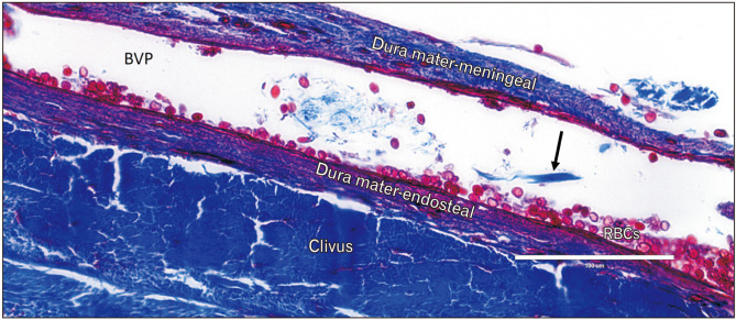
A basilar venous plexus was identified in all specimens and in all of these, intraluminal trabeculae were identified (Fig. 2). The number of trabeculae per plexus ranged from 4 to 8 (mean 5). These were most concentrated in the upper half of the clivus and were more often centrally located. These septations traveled in a posterior to anterior direction and usually, from inferiorly to superiorly. Some trabeculae had wider bases, especially on the more posterior attachment to the meningeal layer of the dura mater (Fig. 2). Otherwise, most trabeculae ended in a denticulate form at the two terminal ends. Uncommonly (approximately 10% of specimens), some trabeculae traveled in a more or less horizontal direction (Fig. 2). The trabeculae varied in size but were on average, 0.85 mm (range 0.5 to 1.45 mm) in length. The mean width of the trabeculae was 0.35 mm (range 0.24 to 0.48 mm). Grossly, these septations were consistent with the cords of Willis as are found in the lumen of some of the other intradural venous sinuses e.g., superior sagittal sinus. Histologically, these were consistent with meningeal tissue and connected two endothelial surfaces of the basilar venous plexus (Figs. 3, 4). None of the specimens was found to have signs of previous surgery to the cranium and specifically, the skull base.
Fig. 3.
Histological sections (Masson’s trichrome) the basilar venous plexus. (A) The plexus (arrows) is seen within the dura mater overlying the clivus. The trabecular portion of the clivus is labeled with black font and the cortical surface of the clivus is labeled with white font. (B) The plexus (BVP) is seen at greater magnification (portion within the yellow box on the A) with the presence of an intraluminal septation (*). Also, note the blood cells (BCs) within the basilar venous plexus.
Fig. 4.
Histological section (Masson’s trichrome) through the clivus noting the basilar venous plexus (BVP). An intraluminal septation is seen at the arrow. Also, note the red blood cells (RBCs) within the lumen of the basilar venous plexus.
Discussion
Few publications have addressed the various relationships of the basilar venous plexus to surrounding venous structures. Renn and Rhoton [15] reported that the plexus was the widest and most constant intercavernous connection and was present in 82% of specimens. More recently, Aquini et al. [16] examined 32 cadavers using electrolytic decalcification, vascular filling, X-rays and serial anatomical sections and found that 100% had a basilar venous plexus, and in 23 (71.9%), it was the widest at the intercavernous connection.
The pathology of the basilar venous plexus has not been well documented; however, the venous interconnections provide collateral venous drainage in the event of dural arteriovenous and carotid cavernous fistula formation1. Coskun et al. [17] reported four cases of carotid-cavernous fistulae diagnosed using computerized tomography angiography, three involving the basilar venous plexus. Halbach et al. [18] described thirteen patients with cavernous dural fistulas who underwent transvenous embolization to promote fistula closure. In one case, the basilar venous plexus was used for initial embolization by inserting a small catheter, followed by two more embolization procedures to cross the midline through a trans-sellar vein to reach the sinus. Takahashi et al. [19] described a single case of a patient presenting with a dural arteriovenous fistula and venous reflux due to bridging vein connections between the basilar venous plexus, the cerebellar hemispheric veins, and the lateral mesencephalic vein. Difficulty in navigating catheters inside the basilar venous plexus may be due to the trabeculae identified in the present study. Therefore, these structures should be kept in mind during such intravascular procedures. Further study is required to optimize catheter manipulation within these venous structures.
Pathological engorgement of the basilar venous plexus has also been discussed in clinical cases with various presenting neurologic deficits. Choi et al. [20] described a case of such engorgement resulting in right abducens nerve palsy. A 60-year-old woman presented with horizontal diplopia lasting seven days with recurrence one year later, involving multiple diplopia episodes [20]. Noblett et al. [13] also reported a case of a 56-year-old woman who presented with right-sided, pulsatile headaches associated with intermittent vision changes in the right eye. The imaging findings suggested that her symptoms were due to a basilar venous plexus anomaly, highlighting the importance of distinguishing between venous enlargement and clival pathology.
The location of the basilar venous plexus must also be considered during skull base surgery, especially during endoscopic transsphenoidal and transclival approaches, which allow surgeons access to the ventral brainstem [21]. In a recent cadaveric study of the inferior intercavernous sinus and its applications to trans-sphenoidal pituitary surgery, Wahl et al. [22] described a connection between the basilar venous plexus and the inferior intercavernous sinus. This connection was found on the inferior surface via a diploic vein in one specimen among 50, highlighting the variability in venous drainage of this area and the importance of considering it prior to surgery.
Several case studies have demonstrated the importance of surgical preparation when both the posterior intercavernous sinus and the basilar venous plexus are transected [23, 24]. Complications during endoscopic transclival surgery can result in bleeding from the basilar venous plexus, and management during surgery has included hemostatic agents, compression, and platinum coils, all of which must be readily available to the surgeon [25]. Additionally, Mizutani et al. [21] cautioned against disruption of the venous drainage of the brainstem via bridging veins draining into the basilar venous plexus.
Trabeculations or septations of the dural venous sinuses have been reported, mainly within the superior sagittal or transverse sinus, often referenced as the “cords of Willis.” Farb [26] identified the cords of Willis in the superior sagittal sinus of all 56 patients studied and in 58 of 112 transverse sinuses examined with contrast-enhanced magnetic resonance venography. Strydom et al. [27] studied the transverse sinuses of both cadavers and patients and found that 29.4% of the 102 subjects had septations in them. Altafulla et al. [28] also found a total of 42 septations in 24 cadaveric transverse sinuses studied (12 cadavers). In the present study, all basilar venous plexuses were found to contain such trabeculae.
Pathological conditions involving trabeculae of the dural venous sinuses have been mentioned in relation to both the generation of turbulent blood flow and potential thrombosis or thromboembolism [27, 29]. Stenosis of the transverse sinus is also well known to contribute idiopathic intracranial hypertension [27, 29]. The possible benefits of dural sinus septations were discussed by Altafulla et al. [28] in their study of the architecture of transverse sinus trabeculations and their tensile strength. The authors suggested that these intraluminal septations help maintain the three-dimensional shape of the dural sinuses where hemodynamic stress was maximal, for instance where the direction of blood flow changes and in areas of maximum curvature of the sinuses. Although speculative, these structures in the basilar venous plexus may have a similar function and require further investigation.
In conclusion, to our knowledge, septations within the basilar venous plexus have not been previously mentioned in the literature. Further understanding of the internal anatomy of this plexus will aid in our treatment of any associated pathology. Furthermore, this knowledge could benefit patients undergoing open, endoscopic and interventional treatments that involve the basilar venous plexus and its surrounding structures.
Acknowledgements
The authors sincerely thank those who donated their bodies to science so that anatomical research could be performed. Results from such research can potentially increase mankind’s overall knowledge that can then improve patient care. Therefore, these donors and their families deserve our highest gratitude [30].
Funding Statement
Funding None.
Footnotes
Author Contributions
Conceptualization: VSG, RST. Data acquisition: EPM, KJ. Data analysis or interpretation: SK, JI. Drafting of the manuscript: MC, ASD. Critical revision of the manuscript: AC (Arada Chaiyamoon), JJC, FR, AC (Ana Carrera). Approval of the final version of the manuscript: all authors.
Conflicts of Interest
No potential conflict of interest relevant to this article was reported.
References
- 1.Standring S. Gray's anatomy: the anatomical basis of clinical practice. 42nd ed. Elsevier; 2020. [DOI] [Google Scholar]
- 2.Tubbs RS, Hansasuta A, Loukas M, Louis RG, Jr, Shoja MM, Salter EG, Oakes WJ. The basilar venous plexus. Clin Anat. 2007;20:755–9. doi: 10.1002/ca.20494. [DOI] [PubMed] [Google Scholar]
- 3.Patel C, Tubbs RS. In: Anatomy, Imaging and Surgery of the Intracranial Dural Venous Sinuses. Tubbs RS, editor. Elsevier; 2020. The basilar plexus; pp. 125–30. [DOI] [Google Scholar]
- 4.Arnautović KI, al-Mefty O, Pait TG, Krisht AF, Husain MM. The suboccipital cavernous sinus. J Neurosurg. 1997;86:252–62. doi: 10.3171/jns.1997.86.2.0252. [DOI] [PubMed] [Google Scholar]
- 5.Cunningham DJ, Robinson A. Cunningham's manual of practical anatomy. 8th ed. Oxford University Press; 1927. [DOI] [Google Scholar]
- 6.San Millán Ruíz D, Gailloud P, Rüfenacht DA, Delavelle J, Henry F, Fasel JH. The craniocervical venous system in relation to cerebral venous drainage. AJNR Am J Neuroradiol. 2002;23:1500–8. [PMC free article] [PubMed] [Google Scholar]
- 7.Mizutani K, Toda M, Kurasawa J, Akiyama T, Fujiwara H, Jinzaki M, Yoshida K. Analysis of the venous channel within the clivus using multidetector computed tomography digital subtraction venography. Neuroradiology. 2017;59:213–9. doi: 10.1007/s00234-017-1784-4. [DOI] [PubMed] [Google Scholar]
- 8.Hollinshead WH, Rosse C. Textbook of anatomy. 4th ed. Harper and Row; 1985. [DOI] [Google Scholar]
- 9.Wackenheim A, Braun JP. The veins of the posterior fossa: normal and pathologic findings. Springer-Verlag; 1978. [DOI] [Google Scholar]
- 10.Dichiro G, Anderson WB. The clivus. Clin Radiol. 1965;16:211–23. doi: 10.1016/S0009-9260(65)80046-0. [DOI] [PubMed] [Google Scholar]
- 11.Pagella F, Ugolini S, Zoia C, Matti E, Carena P, Lizzio R, Benazzo M. Clivus pathologies from diagnosis to surgical multidisciplinary treatment. Review of the literature. Acta Otorhinolaryngol Ital. 2021;41(Suppl. 1):S42–50. doi: 10.14639/0392-100X-suppl.1-41-2021-04.d30a3ca7aa4f4b1cae0136b666bac95d [DOI] [PMC free article] [PubMed] [Google Scholar]
- 12.Rai R, Iwanaga J, Shokouhi G, Loukas M, Mortazavi MM, Oskouian RJ, Tubbs RS. A comprehensive review of the clivus: anatomy, embryology, variants, pathology, and surgical approaches. Childs Nerv Syst. 2018;34:1451–8. doi: 10.1007/s00381-018-3875-x. [DOI] [PubMed] [Google Scholar]
- 13.Noblett D, Chang J, Shahlaie K. Prominent retroclival venous plexus, normal anatomic variant. Appl Radiol. 2018;47:42–4. doi: 10.37549/AR2500. [DOI] [PMC free article] [PubMed] [Google Scholar]
- 14.Iwanaga J, Singh V, Ohtsuka A, Hwang Y, Kim HJ, Moryś J, Ravi KS, Ribatti D, Trainor PA, Sañudo JR, Apaydin N, Şengül G, Albertine KH, Walocha JA, Loukas M, Duparc F, Paulsen F, Del Sol M, Adds P, Hegazy A, Tubbs RS. Acknowledging the use of human cadaveric tissues in research papers: recommendations from anatomical journal editors. Clin Anat. 2021;34:2–4. doi: 10.1002/ca.23671. [DOI] [PubMed] [Google Scholar]
- 15.Renn WH, Rhoton AL., Jr Microsurgical anatomy of the sellar region. J Neurosurg. 1975;43:288–98. doi: 10.3171/jns.1975.43.3.0288. [DOI] [PubMed] [Google Scholar]
- 16.Aquini MG, Marrone AC, Schneider FL. Intercavernous venous communications in the human skull base. Skull Base Surg. 1994;4:145–50. doi: 10.1055/s-2008-1058966. [DOI] [PMC free article] [PubMed] [Google Scholar]
- 17.Coskun O, Hamon M, Catroux G, Gosme L, Courthéoux P, Théron J. Carotid-cavernous fistulas: diagnosis with spiral CT angiography. AJNR Am J Neuroradiol. 2000;21:712–6. [PMC free article] [PubMed] [Google Scholar]
- 18.Halbach VV, Higashida RT, Hieshima GB, Hardin CW, Pribram H. Transvenous embolization of dural fistulas involving the cavernous sinus. AJNR Am J Neuroradiol. 1989;10:377–83. [PMC free article] [PubMed] [Google Scholar]
- 19.Takahashi S, Kato K, Tomura N, Watarai J, Mizoi K. Dural arteriovenous fistula of the cavernous sinus with cortical venous reflux of the posterior fossa via a bridging vein. Radiat Med. 2001;19:219–22. [PubMed] [Google Scholar]
- 20.Choi HY, Kim SB, Park S. Recurrent abducens nerve palsy due to basilar venous plexus engorgement. Neurol Sci. 2015;36:1913–5. doi: 10.1007/s10072-015-2271-5. [DOI] [PubMed] [Google Scholar]
- 21.Mizutani K, Akiyama T, Yoshida K, Toda M. Skull base venous anatomy associated with endoscopic skull base neurosurgery: a literature review. World Neurosurg. 2018;120:405–14. doi: 10.1016/j.wneu.2018.09.067. [DOI] [PubMed] [Google Scholar]
- 22.Wahl L, Lockwood JD, Keet K, Henry BM, Gielecki J, Iwanaga J, Bui CJ, Dumont AS, Tubbs RS. The inferior intercavernous sinus: an anatomical study with application to trans-sphenoidal approaches to the pituitary gland. Clin Neurol Neurosurg. 2020;196:106000. doi: 10.1016/j.clineuro.2020.106000. [DOI] [PubMed] [Google Scholar]
- 23.Silva D, Attia M, Kandasamy J, Alimi M, Anand VK, Schwartz TH. Endoscopic endonasal transsphenoidal "above and below" approach to the retroinfundibular area and interpeduncular cistern--cadaveric study and case illustrations. World Neurosurg. 2014;81:374–84. doi: 10.1016/j.wneu.2012.08.013. [DOI] [PubMed] [Google Scholar]
- 24.Fraser JF, Nyquist GG, Moore N, Anand VK, Schwartz TH. Endoscopic endonasal minimal access approach to the clivus: case series and technical nuances. Neurosurgery. 2010;67(3 Suppl Operative):ons150–8. discussion ons158. doi: 10.1227/01.NEU.0000383130.80179.41. [DOI] [PubMed] [Google Scholar]
- 25.Mizutani K, Akiyama T, Toda M, Kanazawa T, Kamamoto D, Yoshida K. Transvenous embolization of the basilar plexus before endoscopic transsphenoidal transclival surgery. World Neurosurg. 2017;97:756.e13–8. doi: 10.1016/j.wneu.2016.10.022. [DOI] [PubMed] [Google Scholar]
- 26.Farb RI. The dural venous sinuses: normal intraluminal architecture defined on contrast-enhanced MR venography. Neuroradiology. 2007;49:727–32. doi: 10.1007/s00234-007-0250-0. [DOI] [PubMed] [Google Scholar]
- 27.Strydom MA, Briers N, Bosman MC, Steyn S. The anatomical basis of venographic filling defects of the transverse sinus. Clin Anat. 2010;23:153–9. doi: 10.1002/ca.20911. [DOI] [PubMed] [Google Scholar]
- 28.Altafulla JJ, Prickett J, Iwanaga J, Dumont AS, Tubbs RS. Intraluminal anatomy of the transverse sinus: implications for endovascular therapy. Anat Cell Biol. 2020;53:393–7. doi: 10.5115/acb.20.041. [DOI] [PMC free article] [PubMed] [Google Scholar]
- 29.Subramaniam RM, Tress BM, King JO, Eizenberg N, Mitchell PJ. Transverse sinus septum: a new aetiology of idiopathic intracranial hypertension? Australas Radiol. 2004;48:114–6. doi: 10.1111/j.1440-1673.2004.01269.x. [DOI] [PubMed] [Google Scholar]
- 30.Iwanaga J, Singh V, Takeda S, Ogeng'o J, Kim HJ, Moryś J, Ravi KS, Ribatti D, Trainor PA, Sañudo JR, Apaydin N, Sharma A, Smith HF, Walocha JA, Hegazy AMS, Duparc F, Paulsen F, Del Sol M, Adds P, Louryan S, Fazan VPS, Boddeti RK, Tubbs RS. Standardized statement for the ethical use of human cadaveric tissues in anatomy research papers: recommendations from Anatomical Journal Editors-in-Chief. Clin Anat. 2022;35:526–8. doi: 10.1002/ca.23849. [DOI] [PubMed] [Google Scholar]


