Abstract
We investigated the effects of 6 weeks of detraining on muscle size and strength in young men who had previously participated in 6 weeks (3 days/week) of 30 % of concentric one-repetition maximal (1-RM) dumbbell curl training [one arm: concentric blood flow restricted (BFR) exercise (CON-BFR); the other arm: eccentric BFR exercise (ECC-BFR)]. MRI-measured muscle cross-sectional area (CSA) at 10 cm above the elbow joint increased from pre to post (p < 0.01), and the muscle CSA following detraining remained greater than pre (p < 0.01) but was similar to that observed at post. Maximal voluntary contraction (MVC) increased from pre to post (p < 0.05), and the MVC following detraining remained greater than pre (p < 0.05) but was similar to that observed at post. The ECC-BFR did not produce any changes across time. Increased muscle strength following 6 weeks of CON-BFR was well preserved at 6 weeks of detraining, which may be primarily related to muscle hypertrophy.
Keywords: Cessation, Vascular occlusion, Muscle hypertrophy, Strength gain
Introduction
Dynamic high-load resistance training (HRT)-induced increases in muscle size and strength are important fundamental factors for improving and maintaining physical function and sports performance regardless of age [1]. Nevertheless, after cessation of training (detraining), muscle size and strength progressively return toward baseline levels [2, 3]. However, previous studies have reported that the increased muscle strength/size following HRT is retained for the same period of the training [2, 4], suggesting that the effect of HRT is extended for a lengthy detraining period.
Dynamic resistance training programs consist of concentric and/or eccentric muscle actions. It is well established that most studies investigating HRT demonstrate that eccentric training is more effective than repetition matched concentric isokinetic training for muscle hypertrophy [5, 6]. Additionally, muscle hypertrophy following HRT was well preserved following the detraining period with eccentric muscle actions [7]. This suggests that eccentric muscle actions during HRT may play an important role in muscle adaptation during training and detraining.
In the past decade, several studies have reported that low-load dynamic resistance training [20–30 % one-repetition maximum (1-RM)] with blood flow restriction (BFR) elicits similar muscle hypertrophy as HRT regardless of age [8–10]. This technique may be an alternative training method to improve muscle size and strength in healthy individuals or older adults and patients who may be unable to perform high-intensity exercise. Recently, Yasuda et al. [11] revealed that muscle hypertrophy and strength gain from BFR in combination with dynamic resistance training predominately occur from concentric BFR (CON-BFR) but not eccentric BFR (ECC-BFR). Therefore, the mechanisms underlying muscle hypertrophy and strength gain may differ between BFR resistance training and HRT resistance training. Similarly to HRT, BFR studies [12] report that muscle strength is preserved following a 24-week detraining period. In general, muscle size and/or strength gain is retained at a higher degree following detraining because of the greater improvements achieved during the training period [13, 14]. Although currently unexplored, there is a possibility that muscle adaptation following detraining would be maintained at a higher degree for CON-BFR compared with ECC-BFR. Thus, the purpose of this study was to investigate the effects of detraining after CON-BFR or ECC-BFR training on muscle size and strength.
Materials and methods
Subjects
Ten healthy men (age 20–28 years, standing height 160–176 cm, body mass 50.9–70.1 kg) who had previously completed 6 weeks (3 days/week) of arm curl training [11] completed 6 weeks of detraining. During the training period, training intensity and volume were set at 30 % of concentric 1-RM and 75 repetitions (30 repetitions and the next 3 sets each consisting of 15 repetitions, with 30 s of rest between sets) for each arm, respectively. One arm was randomly chosen to perform concentric exercise, while the other arm performed eccentric exercise at the same exercise load. In a randomized order, either concentric or eccentric exercise was performed first followed by the other exercise completed on the same day. During these protocols, subjects performed their respective action with a cadence of 1.5 s for concentric or 1.5 s for eccentric exercise using a metronome, and the investigators manually performed the opposite muscle action. During the training sessions, CON-BFR and ECC-BFR wore elastic cuffs around the most proximal region of the upper arm. On the first day of training, the cuff was set at 100 mmHg. The pressure was increased by 10 mmHg at each subsequent training session until a pressure of 160 mmHg was reached. The pressure intensity was the same between concentric and eccentric exercises at every session [11]. Both arms were reevaluated for arm curl 1-RM strength, maximal voluntary contraction (MVC) and muscle CSA of the biceps brachii. All subjects were fully informed of the risks associated with the experimental procedures and gave their written informed consent before participation. During the detraining period, subjects stopped resistance training and returned to the normal daily activities they completed prior to the resistance training period. The principles of the World Medical Association Declaration of Helsinki were followed, and the study was approved by the Ethics Committee for Human Experiments, University of Tokyo.
Measurement
Maximal dynamic strength (concentric 1-RM) was assessed using a dumbbell on the arm curl bench [11, 15]. After a warm-up, the intensity was set at approximately 80 % of the predicted concentric 1-RM. Following each successful lift, the intensity was increased by ~5 % until the subjects failed to lift the load through the entire range of motion. A test was considered valid only when the subjects used proper form and completed the entire lift in a controlled manner without assistance. On average, 5–6 trials were required to complete a 1-RM test. Approximately 3–5 min of rest were allotted between each attempt to ensure recovery [16].
MVC of the elbow flexors was measured by a dynamometer (Taiyo Kogyo Co., Tokyo, Japan). Each subject was comfortably seated on an adjustable chair, with the arm positioned on a stable table at chest level with the elbow bent at an angle of 90° (0° at full extension). The upper arm was maintained in the horizontal plane (at 90°), while the wrist was fixed at the end of the dynamometer lever arm in a position of supination for elbow flexion. The elbow flexion force was measured with a transducer while the subjects performed two trials separated by a 60-s rest interval [11, 15]. Subjects were instructed to perform an MVC as quickly as possible during a period of about 3 s. The recorded value for the MVC was taken as the highest and most stable 1 s of the 3-s contraction. The highest MVC value was used for data analysis. The test-retest reliability of MVC measurements using the standard error of measurement (SEM) and minimal difference was previously determined from all subjects (0.62 and 1.71 Nm).
Muscle CSA was obtained using a magnetic resonance imaging (MRI) scanner (0.2-T Open MRI, Hitachi, Tokyo, Japan). A T-1 weighted, spin-echo, axial plane sequence was performed with a 500-ms repetition time and a 23-ms echo time. Subjects rested quietly in the magnet bore in a supine position, with their arms extended along their trunk. Continuous transverse images with 10-mm slice thickness were obtained from both upper arms of the body. All MRI data were transferred to a personal computer for analysis using specially designed image analysis software (sliceOmatic, Tomovision Inc., QC, Canada). Muscle tissue cross-sectional area (CSA) data for elbow flexors (biceps brachii and brachialis) at 10 cm above the elbow joint and at the mid-upper arm were digitized. Test–retest reliability of CSA measurements using SEM and minimal difference had been previously determined from seven subjects (0.38 and 1.0 cm2 for mid-upper arm and 0.27 and 0.74 cm2 for 10 cm above the elbow joint).
All measurements were completed before (Pre), within 3–4 days after training (Post) and following 6 weeks of detraining.
Statistical analyses
Results are mean ± standard deviation (SD). Two-way analysis of variance (ANOVA) with repeated measures (condition [CON-BFR, ECC-BFR]) × time [pre, post, detraining] was used to evaluate training effects for all dependent variables. When a significant interaction was observed, post hoc testing was performed with a one-way repeated measures ANOVA across time within each condition. In addition, a paired sample’s t test was used to determine whether differences existed between conditions within each time point. If there was no significant interaction, the main effects were analyzed. The family-wise error rate was maintained by Bonferroni correction of the p value. Statistical significance was set at p ≤ 0.05. Pre/post and post/detraining effect sizes (ESs, Cohen’s d) for 1-RM, muscle CSA and 1-RM/muscle CSA were calculated with the following formula: [post mean − pre mean]/pre SD, [detraining mean − post mean]/post SD; d < 0.2 is a trivial effect, d = 0.2–0.5 is a small effect, d = 0.5–0.8 is a moderate effect, and d > 0.8 is a large effect [17].
Results
In this study, pre and post data concerning muscle CSA, muscle performance, relative performance and effect sizes were based on data presented in a previous study [11].
Muscle CSA
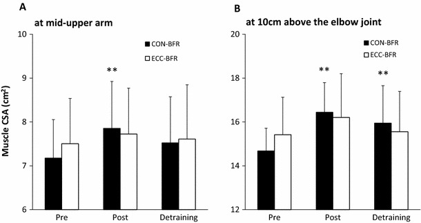
A condition × time interaction was found for muscle CSA at the mid-upper arm (p = 0.01). Follow-up one-way repeated measures ANOVA found significant differences across time for the concentric condition (p < 0.001) with muscle CSA increasing from pre to post (p = 0.003). In addition, muscle CSA following 6 weeks of detraining was significantly less than post (p = 0.03) but not different from baseline (p = 0.084). The eccentric condition did not produce any changes across time (p = 0.431). There were no significant simple effects between conditions within each time point (p > 0.999) (Fig. 1a).
Fig. 1.

Muscle cross-sectional area (CSA) in elbow flexors at the pre, post and detraining period. In this study, pre and post data concerning muscle CSA were based on data presented in a previous study [11]. ** different from pre-training, p < 0.01
A condition × time interaction was found for muscle CSA at 10 cm above the elbow joint (p = 0.034). Follow-up one-way repeated measures ANOVA found significant differences across time for the concentric condition (p < 0.001) with muscle CSA increasing from pre to post (p = 0.003). In addition, muscle CSA following 6 weeks of detraining remained significantly greater than baseline (p = 0.003) but was similar to that observed at post (p = 0.192). The eccentric condition did not produce any changes across time (p = 0.058). There were no significant simple effects between conditions within each time point (p ≥ 0.363) (Fig. 1b).
Muscle performance
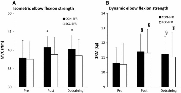
A condition × time interaction was found for MVC (p = 0.013). Follow-up one-way repeated measures ANOVA found significant differences across time for the concentric condition (p = 0.005) with MVC increasing from pre to post (p = 0.033). In addition, MVC following 6 weeks of detraining remained significantly greater than pre values (p = 0.048) but was similar to that observed at post (p = 0.999). The eccentric condition did not produce any changes across time (p = 0.397). There were no significant simple effects between conditions within each time point (p ≥ 0.816) (Fig. 2a).
Fig. 2.

Maximum strength in elbow flexors at the pre, post and detraining periods. In this study, pre and post data concerning muscle performance were based on data presented in a previous study [11]. §Different from pre-training, p < 0.01; * different from pre-training, p < 0.05
A condition × time interaction was not found for 1-RM (p = 0.818); however, there was a significant main effect for time (p < 0.001). When conditions were collapsed across each time point, 1-RM increased from pre to post (p = 0.003). In addition, 1-RM following 6 weeks of detraining remained significantly greater than pre values (p = 0.003) but was similar to that observed at post (p = 0.081) (Fig. 2b).
Relative performance (muscle strength/muscle size)
There were no significant interactions or main effects for MVC divided by muscle CSA of the mid-upper arm (p ≥ 0.396) or 10 cm above the elbow joint (p ≥ 0.314). In addition, there were no significant interactions or main effects for 1-RM divided by muscle CSA of the mid-upper arm (p ≥ 0.132) or 10 cm above the elbow joint (p ≥ 0.100).
Effect sizes (ESs)
The magnitude of change in muscle size and strength between pre- and post-training were always larger for CON-BFR (moderate or large) than they were for ECC-BFR (small or moderate). The ESs for muscle size from post to detraining were small, and the ESs from post- to detraining for muscle strength were trivial. For ECC-BFR, the ESs for muscle size and strength from post to detraining were trivial except for the CSA 10 cm above the elbow joint where they were small (Table 1).
Table 1.
Effect size in muscle size and muscle strength
| CON-BFR | ECC-BFR | |||
|---|---|---|---|---|
| Pre to post | Post to detraining | Pre to post | Post to detraining | |
| CSA at mid-upper arm | 0.77 | −0.31 | 0.21 | −0.11 |
| CSA 10 cm above the elbow joint | 1.70 | −0.36 | 0.46 | 0.33 |
| MVC | 0.60 | −0.16 | 0.26 | −0.06 |
| 1-RM | 0.75 | −0.13 | 0.53 | −0.17 |
Pre and post data concerning muscle CON-BFR concentric blood flow restriction, CSA cross-sectional area, ECC-BFR eccentric blood flow restriction, MVC maximal voluntary contraction and 1-RM were based on previous study [11]
Discussion
The primary finding of this study was that the pronounced muscle hypertrophy (10 cm above the elbow joint) and strength gain following 6 weeks of CON-BFR training were retained after 6 weeks of detraining. Although no published studies have investigated the effect of detraining after CON-BFR resistance training on muscle adaptation, our results were similar to those observed with detraining (4-6 weeks) following traditional HRT [2, 3, 18].
Muscle CSA at 10 cm above the elbow joint was retained following detraining, unlike the muscle CSA at the mid-upper arm for CON-BFR. The ES (pre to post) in muscle CSA was greater for 10 cm above the elbow joint (large: 1.70) compared with the mid-upper arm (moderate: 0.77), although the ES (post to detraining) in muscle CSA was similar between the two groups (small: −0.36 and −0.31, respectively) (Table 1). Consequently, muscle CSA following detraining was retained at a high level at 10 cm above the elbow joint for CON-BFR (Fig. 1).
Our results are in agreement with those of a previous BFR study [12], which demonstrated that muscle strength after BFR training remained elevated above pre-training levels after detraining (Fig. 2a, Table 1). Recently, Yasuda et al. [12] reported that increased muscle strength following 12 weeks of BFR resistance training was well preserved at 24 weeks of detraining, which appeared to be due mainly to neural adaptation in older adults. In this study, however, the relative dynamic strength (1-RM divided by muscle CSA of the mid-upper arm or 10 cm above the elbow joint) for both CON-BFR and ECC-BFR did not increase over the duration of the experiment. Previously, Bemben et al. [19] demonstrated that low-load (40 % 1-RM) resistance training showed greater improvements in 1-RM strength for the leg press and knee extension (34 and 21 %, respectively) than for the biceps curl (14 %), whereas improvement in muscle CSA was similar between the rectus femoris and biceps brachii muscles (20 and 28 %, respectively). Thus, it appears that the relative dynamic strength (biceps curl) in this study did not change, unlike in the previous BFR study (leg press and knee extension) [12].
In this study, dynamic strength (concentric 1-RM) following training for ECC-BFR also remained elevated above the pre-training level after detraining (Fig. 2b; Table 1). On the other hand, MVC strength for ECC-BFR did not increase over the duration of the experiment, unlike the CON-BFR (Fig. 2a). It is well known that the strength gain is confined to the homologous muscle of the opposite untrained limb, and the increase in strength is the greatest during the same movement task performed by the trained limb (‘cross-education’ effect) [20]. Although speculative, we wish to suggest the increase and maintenance of dynamic concentric 1-RM strength in the eccentric arm was due to the cross-education effect from the concentric arm. This is based on the eccentric arm working at 30 % of the concentric 1-RM, which was estimated to be only ~10 % of the maximal eccentric effort [11]. Given the low workload completed by the eccentric arm, it is unlikely that this eccentric action was affecting the concentric arm. We feel the lack of change for the eccentric arm in the more sensitive isometric muscle contraction corroborates this hypothesis.
Some limitations of this study should be discussed. First, the cross-education effect for muscle strength may have influenced both arms. Second, because the training and detraining period was only 6 weeks, it is uncertain whether the results pertain to longer time periods (i.e., 6 months or a year). Lastly, because our subjects were untrained, it is uncertain whether the results pertain to trained subjects. Additional research into these issues is needed.
In conclusion, increased muscle strength following 6 weeks of CON-BFR was well preserved at 6 weeks of detraining, which may be related primarily to muscle hypertrophy. Also, when performing a pre-set number of repetitions using low-intensity CON-BFR or ECC-BFR exercise, CON-BFR exercise did not produce significant muscle soreness [21]. Given the potential health risks for those with contraindications to completing higher intensity resistance training, CON-BFR may be an effective training program for promoting muscle size and strength in a clinical setting.
Acknowledgments
The authors thank the students who participated in this study. We also thank Toshiaki Nakajima, MD, PhD, the University of Tokyo School of Medicine, for helpful discussion and technical support. This study was supported in part by grants-in-aid (#23700713 and #25750288 to TY) from the Japan Ministry of Education, Culture, Sports, Science and Technology.
Conflict of interest
The authors declare no conflict of interest.
References
- 1.American College of Sports Medicine American College of Sports Medicine position stand. Progression models in resistance training for healthy adults. Med Sci Sports Exerc. 2009;41:687–708. doi: 10.1249/MSS.0b013e3181915670. [DOI] [PubMed] [Google Scholar]
- 2.Lovell DI, Cuneo R, Gass GC. The effect of strength training and short-term detraining on maximum force and the rate of force development of older men. Eur J ApplPhysiol. 2010;109:429–435. doi: 10.1007/s00421-010-1375-0. [DOI] [PubMed] [Google Scholar]
- 3.Narici MV, Roi GS, Landoni L, Minetti AE, Cerretelli P. Changes in force, cross-sectional area and neural activation during strength training and detraining of the human quadriceps. Eur J ApplPhysiolOccupPhysiol. 1989;59:310–319. doi: 10.1007/BF02388334. [DOI] [PubMed] [Google Scholar]
- 4.Hakkinen K, Alen M, Kallinen M, Newton RU, Kraemer WJ. Neuromuscular adaptation during prolonged strength training, detraining and re-strength-training in middle-aged and elderly people. Eur J ApplPhysiol. 2000;83:51–62. doi: 10.1007/s004210000248. [DOI] [PubMed] [Google Scholar]
- 5.Farthing JP, Chilibeck PD. The effects of eccentric and concentric training at different velocities on muscle hypertrophy. Eur J Appl Physiol. 2003;89:578–586. doi: 10.1007/s00421-003-0842-2. [DOI] [PubMed] [Google Scholar]
- 6.Higbie EJ, Cureton KJ, Warren GL, 3rd, Prior BM. Effects of concentric and eccentric training on muscle strength, cross-sectional area, and neural activation. J ApplPhysiol. 1996;81:2173–2181. doi: 10.1152/jappl.1996.81.5.2173. [DOI] [PubMed] [Google Scholar]
- 7.Hather BM, Tesch PA, Buchanan P, Dudley GA. Influence of eccentric actions on skeletal muscle adaptations to resistance training. Acta PhysiolScand. 1991;143:177–185. doi: 10.1111/j.1748-1716.1991.tb09219.x. [DOI] [PubMed] [Google Scholar]
- 8.Abe T, Yasuda T, Midorikawa T, Sato Y, Kerns CF, Inoue K, Koizumi K, Ishii N. Skeletal muscle size and circulating IGF-1 are increased after two weeks of twice daily “KAATSU” resistance training. Int. J. Kaatsu Training Res. 2005;1:6–12. doi: 10.3806/ijktr.1.6. [DOI] [Google Scholar]
- 9.Loenneke JP, Wilson JM, Marin PJ, Zourdos MC, Bemben MG. Low intensity blood flow restriction training: a meta-analysis. Eur J Appl Physiol. 2012;112:1849–1859. doi: 10.1007/s00421-011-2167-x. [DOI] [PubMed] [Google Scholar]
- 10.Yasuda T, Fukumura K, Fukuda T, Uchida Y, Iida H, Meguro M, Sato Y, Yamasoba T, Nakajima T. Muscle size and arterial compliance after blood flow-restricted low-intensity resistance training in older adults. Scand J Med Sci Sports. 2013;24:799–806. doi: 10.1111/sms.12087. [DOI] [PubMed] [Google Scholar]
- 11.Yasuda T, Loenneke JP, Thiebaud RS, Abe T. Effects of low-intensity blood flow restricted concentric or eccentric training on muscle size and strength. PLoS ONE. 2012;12:e52843. doi: 10.1371/journal.pone.0052843. [DOI] [PMC free article] [PubMed] [Google Scholar]
- 12.Yasuda T, Fukumura K, Sato Y, Yamasoba T, Nakajima T. Effects of detraining after blood flow restricted low-intensity training on muscle size and strength in older adults. Aging Clinical and Experimental Research. 2014;26:561–564. doi: 10.1007/s40520-014-0208-0. [DOI] [PubMed] [Google Scholar]
- 13.FatourosIG Kambas A, Katrabasas I, Nikoladis K, Chatzinikolaou A, Leontsini D, Taxildaris K. Strength training and detraining effects on muscular strength, anaerobic power, and mobility of inactive older men are intensity dependent. Br J Sports Med. 2005;39:776–780. doi: 10.1136/bjsm.2005.019117. [DOI] [PMC free article] [PubMed] [Google Scholar]
- 14.Tokmakidis SP, Kalapotharakos VI, Smilios I, Parlavantzas A. Effects of detraining on muscle strength and mass after high or moderate intensity of resistance training in older adults. ClinPhysiolFunct Imaging. 2009;29:316–319. doi: 10.1111/j.1475-097X.2009.00866.x. [DOI] [PubMed] [Google Scholar]
- 15.Yasuda T, Brechue WF, Fujita T, Shirakawa J, Sato Y, Abe T. Muscle activation during low-intensity muscle contractions with restricted blood flow. J Sports Sci. 2009;27:479–489. doi: 10.1080/02640410802626567. [DOI] [PubMed] [Google Scholar]
- 16.Abe T, deHoyos DV, Pollock ML, Garzarella L. Time course for strength and muscle thickness changes following upper and lower body resistance training in men and women. Eur J App lPhysiol. 2000;81:174–180. doi: 10.1007/s004210050027. [DOI] [PubMed] [Google Scholar]
- 17.Cohen J. Statistical power analysis for the behavioral sciences. 2. Hillsdale: Lawrence Erlbaum Associates Inc.; 1988. pp. 19–39. [Google Scholar]
- 18.Houston ME, Froese EA, Valeriote SP, Green HJ, Ranney DA. Muscle performance, morphology and metabolic capacity during strength training and detraining: a one leg model. Eur J ApplPhysiolOccupPhysiol. 1983;51:25–35. doi: 10.1007/BF00952534. [DOI] [PubMed] [Google Scholar]
- 19.Bemben DA, Fetters NL, Bemben MG, Nabavi N, Koh ET. Musculoskeletal responses to high- and low-intensity resistance training in early postmenopausal women. Med Sci Sports Exerc. 2000;32:1949–1957. doi: 10.1097/00005768-200011000-00020. [DOI] [PubMed] [Google Scholar]
- 20.Lee M, Carroll TJ. Cross education: possible mechanisms for the contralateral effects of unilateral resistance training. Sports Med. 2007;37:1–14. doi: 10.2165/00007256-200737010-00001. [DOI] [PubMed] [Google Scholar]
- 21.Thiebaud RS, Yasuda T, Loenneke JP, Abe T. Indirect markers of muscle damage induced by low-intensity blood flow restricted concentric or eccentric muscle action. Interv Med ApplSci. 2013;5:53–59. doi: 10.1556/IMAS.5.2013.2.1. [DOI] [PMC free article] [PubMed] [Google Scholar]


