Abstract
Accumulating evidence has revealed pivotal roles of glycogen synthase kinase-3β (GSK3β) inactivation on cardiac protection. Because the precise mechanisms of cardiac protection against ischemia/reperfusion (I/R) injury by GSK3β-inactivation remain elusive, we investigated the relationship between GSK3β-mediated mitochondrial hexokinase II (mitoHK-II; a downstream target of GSK3β) dissociation and mitochondrial permeability transition pore (mPTP) opening. In Langendorff-perfused hearts, GSK3β inactivation by SB216763 improved the left ventricular-developed pressure and retained mitoHK-II binding after I/R. In permeabilized myocytes, GSK3β depolarized mitochondrial membrane potential with accelerated mitochondrial calcein release (suggesting GSK3β-mediated mPTP opening) and decreased mitoHK-II bindings. GSK3β-mediated mPTP opening depended on mitoHK-II binding, i.e., it was accelerated by dissociation of mitoHK-II (dicyclohexylcarbodiimide) and attenuated by enhancement of mitoHK-II binding (dextran). However, inactivation of mitoHK-II by glucose-depletion or glucose-6-phosphate inhibited the GSK3β-mediated mPTP opening. We conclude that GSK3β-mediated mPTP opening may be involved in I/R injury and regulated by mitoHK-II binding and activity.
Electronic supplementary material
The online version of this article (10.1007/s12576-018-0611-y) contains supplementary material, which is available to authorized users.
Keywords: Glycogen synthase kinase-3β, Mitochondrial permeability transition pore, Mitochondrial hexokinase II, Ischemia–reperfusion
Introduction
Mitochondria play pivotal roles not only manipulating cellular function through ATP production and Ca2+ regulation [1] but also determining myocardial cell fate during ischemia/reperfusion (I/R) [2]. Accumulating evidence indicates that the inhibition of mitochondrial permeability transition pores (mPTP) contributes to the core mechanism of cardiac protection including pro-survival PI3 K-Akt kinase cascades, which are activated by preconditioning to initiate myocardial protection [2, 3].
Glycogen synthase kinase 3β (GSK3β), which is inactivated by its phosphorylation, is a constitutive multifunctional serine/threonine kinase [4, 5] and manipulates cellular glycogen synthase activity and multiple protective pro-survival signaling pathways [4]. Recent investigations suggested that the mitochondria-bound hexokinase II (mitoHK-II) is a downstream target of GSK3β [6, 7]. MitoHK-II is reported to promote neuronal survival in human neuron-like cells [8], and dissociation of HK-II from mitochondria increased the chemotherapy-induced lethal cell damage in HeLa cells [6]. In addition, ischemic preconditioning is associated with reduced cytosolic HK-II activity during ischemia and biphasic induction of mitoHK-II activity before and after ischemia [9].
Despite intensive efforts, the precise mechanisms of myocardial survival against I/R by GSK3β inactivation have not been fully elucidated. Thus, in this study we aimed to investigate (1) the effects of GSK3β inactivation on cardiac function during and after I/R, (2) the relationship between GSK3β-mediated mPTP opening and mitoHK-II binding, and (3) the impact of HK-II inactivation by manipulating glucose metabolism on GSK3β-mediated mPTP opening.
Methods
This investigation conforms to the Guide for the Care and Use of Laboratory Animals published by the US National Institutes of Health (NIH Publication, revised 2011) and the Hamamatsu University School of Medicine Animal Care and Use Committee. Heart isolation and obtaining hemodynamic measurements in Langendorff-perfused hearts was described previously [10]. Isolated hearts were subjected to 35 min of global ischemia followed by 40 min of reperfusion with and without SB216763 (a GSK3β inhibitor 3 μmol/l) treatment. In SB216763-treated hearts, SB216763 was pretreated for 25 min before ischemia, and then hearts were subjected with I/R without SB216763. To monitor LV pressure, a water-filled latex balloon connected to a pressure transducer and polygraph (Nihon Kohden Co., Japan), was inserted into the LV from the left atrium, and the hearts were electrically paced at 5 Hz.
Myocytes were isolated from male Sprague–Dawley rats and sarcolemmal membrane was permeabilized as described previously [10, 11]. After sarcolemmal membrane permeabilization, the concentration of free calcium in the solution was changed to 177 nmol/l. Western-blot analysis in mitochondrial fractions was performed as previously described [10]. Permeabilized myocytes were treated with drugs according to the protocol, and then homogenized using a ProteoExtract® Cytosol/Mitochondria Fractionation Kit (Merck Bioscience, Bad Soden, Germany). Densitometric analysis was performed using the Molecular Imager ChemiDocTM system (Bio-Rad Laboratories, Hercules, CA, USA). Fluorescence measurements were performed with a laser scanning confocal microscope (LSM5 PASCAL, Carl Zeiss AG, Oberkochen, Germany) coupled to an inverted microscope with a 63 × water-immersion objective lens. Mitochondrial membrane potential (ΔΨm) was measured with tetramethylrhodamine ethyl ester (TMRE 10 nmol/l), and opening of mPTP was measured with calcein-AM (1 μmol/l), as described previously [11].
A recombinant active form of GSK3β was purchased from Millipore (Billerica, MA, USA). Cyclosporin A (CsA), protein kinase A catalytic subunit (PKAcat), dextran, and alamethicin were purchased from Sigma–Aldrich (St. Louis, MO, USA). SB216763 was purchased from Tocris Bioscience (Ellisville, MO, USA). Dicyclohexylcarbodiimide (DCCD) was purchased from Wako Chemicals (Richmond, VA, USA). Fluorescent dyes were purchased from ThermoFisher Scientific (Waltham, MA, USA).
Data are presented as mean ± SEM, and the number of cells or experiments is shown as n. Statistical analyses were performed using one-way ANOVA followed by Bonferroni’s test or by Kruskal–Wallis’s test, and two-way ANOVA followed by Bonferroni’s test, according to the study protocol. P < 0.05 was accepted as statistically significant.
Results
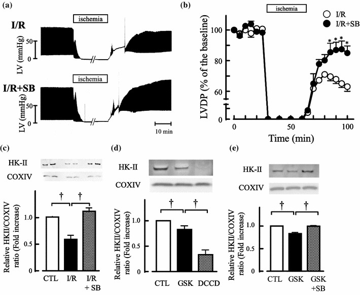
We first investigated the effects of GSK3β inactivation on I/R injury using SB216763. As shown in Fig. 1a, b, recovery of hemodynamics after reperfusion was significantly improved in SB216763-treated hearts (LVDP at 100 min 85.0 ± 4.6% of I/R + SB, P < 0.05 vs. 63.1 ± 3.0% of I/R). As a downstream target of GSK3β, expression level of mitochondrial hexokinase II (mitoHK-II) was assessed in SB216763-treated hearts. MitoHK-II was significantly reduced after I/R, and SB216763 prevented the reduction of mitoHK-II by I/R (Fig. 1c).
Fig. 1.

GSK3β inactivation by SB216763 protects hearts and retains mitoHK-II. a Representative recording of left ventricular (LV) pressure in Langendorff-perfused hearts. The isolated hearts were subjected to I/R in the absence (I/R) or presence of SB216763 (I/R + SB 3 μmol/l). In SB216763-treated hearts, SB216763 was pretreated for 25 min before ischemia, and then hearts were subjected to I/R without SB216763. b Time course of changes in left ventricular developed pressure (LVDP) during I/R in the absence (I/R ○) and presence of SB216763 (I/R + SB ●). The values are mean ± SEM from 6 to 8 experiments. *P < 0.05 vs. I/R by two-way ANOVA followed with Bonferroni’s test. c Western-blot analysis of mitoHK-II in hearts from a, b and non-ischemic control (CTL). Data are presented as fold-increase of mitoHK-II/COXIV ratio from CTL, and values are mean ± SEM from six independent experiments. †P < 0.01 by one-way ANOVA followed with Bonferroni’s test. d, e Western-blot analysis of mitoHK-II in permeabilized myocytes. Cells were exposed to GSK3β (GSK; 10 nmol/l), GSK3β plus SB216763 (GSK + SB 3 μmol/l), and DCCD (1 μmol/l) for 1 h, and the mitochondrial fraction was then extracted. Data are presented as fold-increase of mitoHK-II/COXIV from control (CTL) and values are mean ± SEM from seven experiments. †P < 0.05 by one-way ANOVA followed by Bonferroni’s test
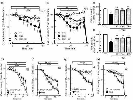
The causal relationship between GSK3β and mitoHK-II binding was further investigated using recombinant (active form) GSK3β and permeabilized myocytes. GSK3β significantly reduced the expression level of mitoHK-II similar to DCCD (Fig. 1d an agent to dissociate HK-II from mitochondria). In addition, GSK3β exhibited the dose-dependent mitoHK-II reduction (Supplemental fig. 1) and SB216763 inhibited the GSK3β-mediated mitoHK-II reduction (Fig. 1e). We next examined the effects of GSK3β (active form) on mPTP opening in permeabilized myocytes. As shown in Fig. 2a, the entrapped mitochondrial calcein release was accelerated by GSK3β (10 nmol/l calcein intensity at 35 min 79.2 ± 1.3% of the baseline, P < 0.05 vs. 94.2 ± 1.4% CTL) in a SB216763-sensitive manner (3 μmol/l 92.1 ± 1.2% P < 0.01 vs. GSK). In Fig. 2b, GSK3β significantly reduced TMRE intensity (at 25 min 78.3 ± 2.4% of the baseline, P < 0.05 vs. 100 ± 1.4% of CTL, suggesting depolarized ΔΨm), and SB216763 attenuated the GSK3β-mediated TMRE reduction (89.3 ± 4.3%, P < 0.01 vs. GSK, suggesting protection of ΔΨm). In addition, when cells were pretreated with cyclosporine A (CsA, an inhibitor of mPTP 0.1 mmol/l), the GSK3β-mediated mitochondrial calcein release (Fig. 2c 90.4 ± 1.8% of the baseline, P < 0.01 vs. GSK) and TMRE reduction (Fig. 2d 89.7 ± 3.6% of the baseline, P < 0.01 vs. GSK) were significantly inhibited. Because protein kinase A is known to inactivate GSK3β, the effects of protein kinase A catalytic subunit (PKAcat) on GSK3β-mediated mPTP opening were evaluated. In our previous report, PKAcat alone (< 15 U/ml) did not alter mPTP opening [11], here we treated permeabilized myocytes with 10 U/ml of PKAcat prior to GSK3β. As shown in Fig. 2c, d, PKAcat inhibited the GSK3β-mediated mitochondrial calcein release (94.5 ± 1.5%, P < 0.01 vs. GSK) and TMRE reduction (91.5 ± 2.1%, P < 0.01 vs. GSK, suggesting ΔΨm protection). These results suggest that the active from of GSK3β opens mPTP.
Fig. 2.

GSK3β promotes mPTP opening. a, b Time course of changes in calcein a and TMRE b intensity in permeabilized myocytes, which were perfused with control internal solution (CTL; ○) and GSK3β (10 nmol/l) in the absence (GSK; ●) or presence of SB216763 (3 μmol/l, GSK + SB; △). Alamethicin (a pore-forming antibiotic Ala) was applied to obtain maximal calcein release from the mitochondrial matrix in calcein experiments, and DNP, an uncoupler, was applied at the end of TMRE experiments. Data are presented as the % of intensity at 0 min, and the values are mean ± SEM from 13 to 21 experiments. †P < 0.05 vs. CTL, *P < 0.05 vs. GSK by two-way ANOVA followed with Bonferroni’s test. c, d Summarized data of calcein c and TMRE d intensities after 30 min perfusion of GSK3β (GSK), GSK3β plus SB216763 (GSK + SB), GSK3β plus cyclosporine A (GSK + CsA; 0.1 μmol/l), and GSK3β plus PKA catalytic subunit (GSK + PKA; 10 U/ml). Some cells were pretreated with SB, CsA, or PKA for 5 min, and then GSK3β was perfused with them for 30 min. Values are mean ± SEM from 11 to 18 experiments. #P < 0.01 vs. control, **P < 0.01 vs. GSK by one-way ANOVA with Bonferroni’s test. e, f Time course of changes in calcein e and TMRE f intensities during and after perfusion with GSK3β (●; 10 nmol/l), and GSK3β plus DCCD (○; 1 μmol/l). Data are presented as the % of intensity at 0 min, and the values are mean ± SEM from 5 to 16 independent experiments. *P < 0.05 vs. GSK3β by two-way ANOVA. g, h Time course of changes in calcein g and TMRE h intensities during and after perfusion with GSK3β (●), and GSK3β plus dextran (○; 1%). Permeabilized myocytes were perfused with dextran for 20 min, and then GSK3β was applied. Values are mean ± SEM from 5 to 19 independent experiments. *P < 0.05 vs. GSK3β by two-way ANOVA followed by Bonferroni’s test
GSK3β-mediated mPTP opening was further investigated by accelerating the mitoHK-II dissociation using DCCD (1 μmol/l). As shown in Fig. 2e, f, DCCD enhanced the GSK3β-mediated mitochondrial calcein release (Fig. 2e calcein intensity at 40 min 59.3 ± 1.5% of the baseline, P < 0.05 vs. GSK), as well as TMRE reduction (Fig. 2f TMRE intensity at 40 min 59.5 ± 1.7% of base line, P < 0.05 vs. GSK). Next, GSK3β-mediated mPTP opening was evaluated by accelerating the mitoHK-II binding using dextran [12]. We examined the GSK3β-mediated mPTP opening by perfusing 1% of dextran, which did not change either the osmolality of medium or the fluorescence intensities (calcein or TMRE) in our experimental condition. In contrast to DCCD, dextran attenuated both calcein release from mitochondria (calcein intensity at 40 min 96.7 ± 1.3% of the baseline, P < 0.05 vs. GSK) and TMRE reduction (89.4 ± 3.7% of the baseline, P < 0.05 vs. GSK, suggesting ΔΨm depolarization) by GSK3β. Thus, our results suggest that the sensitivity of GSK3β-mediated mPTP opening was regulated by mitoHK-II binding.
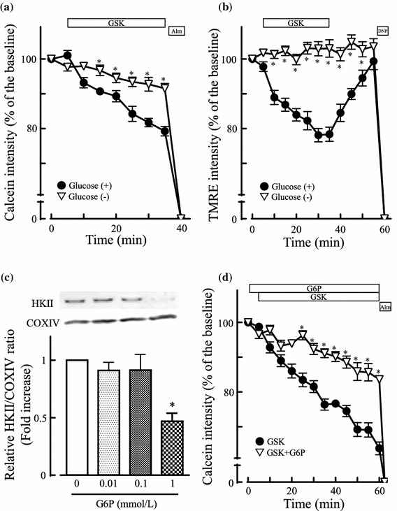
We finally explored the contribution of enzymatic activity of mitoHK-II on the GSK3β-mediated mPTP opening. Because the activity of mitoHK-II depends on its substrates (glucose Km = 0.3 mmol/l) and products (glucose-6-phosphate G6P, Ki = 0.02 mmol/l) [13], an inactivation of mitoHK-II was first facilitated by substrate (glucose) depletion. Under the glucose depletion where mitoHK-II was inactivated (Supplemental fig. 4a, b), not only the mitochondrial calcein release by GSK3β (Fig. 3a calcein intensity at 35 min 91.5 ± 1.5%, P < 0.05 vs. with glucose), but also the TMRE reduction ΔΨm depolarization by GSK3β (Fig. 3b TMRE intensity at 30 min 103.2 ± 2.3%, P < 0.05 vs. with glucose) was attenuated. The inactivation of mitoHK-II was also examined by treating cells with G6P. Although it is reported that a high concentration of G6P itself dissociates HK-II from mitochondria [14], G6P did not alter mitoHK-II binding under the concentration of 0.01 and 0.1 mmol/l in our experimental condition (Fig. 3c). When permeabilized myocytes were exposed to 0.01 mmol/l of G6P, which could inactivate HK-II [13] without affecting mitochondrial binding, the GSK3β-mediated mitochondrial calcein release was attenuated (Fig. 3d, calcein intensity at 35 min 83.6 ± 0.9% of baseline, P < 0.05 vs. 63.8 ± 1.8% of baseline of GSK). In addition, while Azoulay-Zohar et al. reported the G6P-induced mPTP opening via mitoHK-II dissociation (half maximal concentration of G6P to open mPTP is 5 mmol/l), higher concentration of G6P (5.8 mmol/l) did not either accelerate the GSK3β-mediated mitochondrial calcein release or reduce TMRE intensity in our experimental condition (Supplemental fig. 3). We could not ensure the inactivation of mitoHK-II by G6P (0.01 mmol/l) in our experimental condition because the HK-II activity is measured by the HK-II’s ability to convert glucose into G6P and application of G6P (0.01 mmol/l) exceeded the limit of assay measurement (Supplemental fig. 4a, b). However, we considered that the HK-II inactivation by G6P may attenuate the GSK3β-mediated mPTP opening because similar results were obtained with glucose depletion.
Fig. 3.

MitoHK-II inactivation inhibits GSK3β-induced mPTP opening. a, b Time course of changes in calcein a and TMRE b intensities in GSK3β-treated permeabilized myocytes with and without glucose [glucose (–); ▽, glucose (+); ●]. Data are presented as the % of intensity at 0 min, and the values are mean ± SEM from 15 to 21 experiments. *P < 0.05 vs. GSK3β with glucose by two-way ANOVA followed with Bonferroni’s test. c Western-blot analysis of mitoHK-II after glucose-6-phosphate (G6P; 0, 0.01, 0.1, and 1 mmol/l) treatment in permeabilized myocytes. Data are presented as fold-increase of mitoHK-II/COXIV ratio from non-treated control (0 mmol/l), and the values are mean ± SEM from four independent experiments. *P < 0.05 vs. 0 mmol/l of G6P by one-way ANOVA with Kruskal–Wallis. d Time course of changes in mitochondrial calcein intensities. The permeabilized myocytes were perfused with GSK3β (●) or GSK3β plus G6P (▽, 0.01 mmol/l). Data are presented as the % of intensity at 0 min, and the values are mean ± SEM from 9 to 15 experiments. *P < 0.05 by two-way ANOVA followed with Bonferroni’s test
Thus, in contrast to mitoHK-II dissociation, inactivation of mitoHK-II (through glucose depletion or G6P) attenuated the GSK3β-mediated mPTP opening.
Discussion
In this study, we studied the cardioprotective effects by GSK3β inactivation against I/R injury and investigated the roles of mitoHK-II binding on the mPTP opening. The main findings of this study are as follows: (1) Pharmacological inhibition of GSK3β prevented dissociation of mitoHK-II and improved the recovery of cardiac performance after reperfusion, (2) active form of GSK3β dissociated mitoHK-II and opened mPTP, and (3) inactivation of mitoHK-II by manipulating glucose metabolism attenuated GSK3β-mediated mPTP opening in permeabilized myocytes.
GSK3β is a constitutive serine–threonine kinase and involved in cellular energy metabolism [4, 5, 15]. Many investigations suggested that inactivation of GSK3β plays key roles in the protective effects of ischemic and pharmacological preconditioning [4, 5], post-conditioning [16], and chemical cardioprotective interventions [17, 18]. As compatible with previous investigations, our results revealed that inactivation of GSK3β by SB216763 exhibited cardiac protection against I/R injury in Langendorff-perfused hearts (Fig. 1a, b). Although our results revealed short time effects by pharmacological inhibition, Woulfe et al. also have shown less cardiac dilatation and preserved LV function up to 8 weeks after myocardial infarction in inducible GSK-3β knockdown mice [19]. It has already been reported that the active form of GSK3β opens mPTP in response to ROS or Ca2+ overload in isolated mitochondria [16], and the inhibition of GSK3β acts as a master switch to limit the mPTP opening in cardiac myocytes [4]. As compatible with previous reports, we showed that the recombinant (active form) GSK3β opened mPTP (Fig. 2a, b) in a dose-dependent manner and half maximal concentration of mPTP opening was 2.5 nmol/l in our experimental condition (Supplemental fig. 2). In addition, the inactivation of GSK3β by SB216763 or protein kinase A catalytic subunit inhibited the GSK3β-mediated mPTP opening (Fig. 2c, d). Although we did not show direct evidence that the phosphorylated GSK3β (inactive form) inhibited mPTP opening during I/R in isolated hearts, the GSK3β-inactivation appears to suppress the susceptibility of mPTP through preventing mitoHK-II dissociation [6], attenuating the affinity of adenine nucleotide translocase (ANT, located in the inner mitochondrial membrane) to cyclophilin D (an mPTP regulatory protein located in the inner mitochondrial membrane), suppressing mitochondrial Ca2+ overload [20], and reducing ATP hydrolysis during ischemia [5]. Further investigations are required to reveal the relationship between GSK3β inactivation and mPTP inhibition in I/R injury.
HK-II is heterogeneously distributed in the mitochondria and cytosol, and mitoHK-II is physically binds external surface of outer mitochondrial membrane (OMM) through porin or voltage-dependent anion channel (VDAC; located in the OMM), which enables mitoHK-II to use mitochondrial ATP to catalyze the phosphorylation of glucose to yield G6P [6]. We showed that the GSK3β inactivation (by SB216763) attenuated the mitoHK-II dissociation by I/R in Langendorff-perfused hearts (Fig. 1), and that recombinant GSK3β (activated form) dissociated HK-II from mitochondria in a SB216763-sensitive manner in permeabilized myocytes (Fig. 2), suggesting that GSK3β dissociates mitoHK-II. Because our results exhibited net positive of total mitoHK-II by SB216763 in isolated hearts, we cannot completely deny the possibility of contribution of translocated cytosolic HK-II to mitochondria, by which total amount of mitoHK-II increased rather than the inhibition of mitoHK-II dissociation. However, previous investigations have supported our results and phosphorylated GSK3β (inactive form) can stabilize mitoHK-II binding through the suppression of VDAC phosphorylation [6, 7, 21].
We showed that the chemical stimuli (DCCD or dextran) modulating mitoHK-II bindings altered the GSK3β-mediated mPTP opening (Fig. 2e–h). Although the precise mechanisms by which mitoHK-II dissociation induces mPTP opening have not been completely understood, the instability of VDAC by mitoHK-II dissociation may be responsible [22]. In addition to its dissociation, mitoHK-II also regulated the GSK3β-mediated mPTP opening by its inactivation (Fig. 3). Because the mitoHK-II phosphorylates glucose by preferentially utilizing the intra-mitochondrial ATP [23], inactivation of mitoHK-II may suppress matrix ATP consumption, which is required for glucose metabolism, and may result in the inhibition of GSK3β-mediated mPTP opening. It would be difficult to apply our results to common pathophysiological conditions because we could estimate the effects of mitoHK-II inactivation only in the extreme experimental conditions, where permeabilized myocytes perfused with no glucose or abundant G6P solution. However, at least in our experimental condition, mitoHK-II regulated GSK3β-mediated mPTP not only by its dissociation but also by inactivation. Our investigation has not sufficiently revealed the relationship between the extent of HK-II inactivation and mPTP inhibition because we could not ensure the inactivation of mitoHK-II by G6P due to the technical issue (Supplemental fig. 4a, b). In fact, 0.1 mmol/l of G6P, which is expected the better HK-II inactivation than 0.01 mmol/l without affecting mitochondrial HK-II binding, failed to restore the GSK3β-mediated TMRE suppression in our experimental condition (Supplemental fig. 3a). Further investigation will be required to explore the contribution of mitoHK-II on mPTP opening under I/R.
Electronic supplementary material
Below is the link to the electronic supplementary material.
Supplemental Figure 1: Dose and time dependency of mitoHK-II reduction by GSK3β. Western blot analysis of mitoHK-II after GSK3β treatments (GSK; 1, 10, and 50 nmol/L for 1 or 2 hr). Data are presented as fold-increase of mitoHK-II/COXIV from non-treated control (CTL) and values are mean ± SEM from 4 experiments. †P < 0.05 by one-way ANOVA followed with Bonferroni’s test.
Supplemental Figure 2: Dose dependency of GSK3β-mediated mPTP opening. a Summarized data of calcein intensities after 30 min perfusion of GSK3β (0.5–100 nmo/L). Values are mean ± SEM from 6 to 16 experiments. *P < 0.01 vs. control by one-way ANOVA with Bonferroni’s test. b The correlation of GSK3β concentration and mitochondrial calcein release (at 30 min of perfusion) was analyzed. The estimated half maximal concentration of GSK3β was 2.5 nmol/L in our experimental condition.
Supplemental Figure 3: Effect of high concentration of G6P on the GSK3β-mediated calcein release from mitochondria. a Summarized data of TMRE intensities after 30 min perfusion of GSK3β (10 nmo/L) with absence and presence of glucose-6-phosphate (G6P; 0.1, 1, 5.8 mmol/L). Values are mean ± SEM from 4 to 8 experiments. *P < 0.01 vs. control by one-way ANOVA with Bonferroni’s test. b Time course of changes in calcein intensities. The permeabilized myocytes were perfused with GSK3β (●) or GSK3β plus G6P (○, 5.8 mmol/L). Data are presented as the % of intensity at 0 min, and the values are mean ± SEM from 8 experiments.
Supplemental Figure 4: MitoHK-II activities under the condition G6P or glucose depletion. a Representative time courses in NADH generation by mitoHK-II. The mitochondria were obtained from rat hearts and divided with non-treated (○), G6P-treated (●, 0.01 mmol/L), and glucose depletion (▼) group. The NADH generation was assessed by an enzyme-coupled assays kit (Hexokinase Colorimetric Assay Kit: Bio Vision Inc, USA) and a multi plate reader Synergy™ HT (Biotek Instruments) with the measurement of absorbance at 450 nm in 96-well plates according to the manufacturer’s protocol. b Summarized data of mitoHK-II activities. The mitoHK-II activities were calculated by the NADH generation (nmol/mL) per 1 min during measurements (30 min) per 1 mg of rat hearts. Data are mean ± SEM from 6 experiments, and †P < 0.01 by paired t test vs non-treated control (CTL).
Funding
This work was supported by the JSPS KAKENHI Grants JP16K09428 (to M. S) and JP15K09073 (to H. K).
Compliance with ethical standards
Conflict of interest
The authors declare that they have no conflicts of interest.
Ethical Approval
All applicable international, national, and/or institutional guidelines for the care and use of animals in the field of Physiology Science have been followed, and studies have been approved by a research ethics committee at the Hamamatsu University School of Medicine.
References
- 1.Takeuchi A, Kim B, Matsuoka S. The destiny of Ca(2+) released by mitochondria. J Physiol Sci. 2015;65:11–24. doi: 10.1007/s12576-014-0326-7. [DOI] [PMC free article] [PubMed] [Google Scholar]
- 2.Minamino T. Cardioprotection from ischemia/reperfusion injury: basic and translational research. Circ J. 2012;76:1074–1082. doi: 10.1253/circj.CJ-12-0132. [DOI] [PubMed] [Google Scholar]
- 3.Bu HM, Yang CY, Wang ML, Ma HJ, Sun H, Zhang Y. K(ATP) channels and MPTP are involved in the cardioprotection bestowed by chronic intermittent hypobaric hypoxia in the developing rat. J Physiol Sci. 2015;65:367–376. doi: 10.1007/s12576-015-0376-5. [DOI] [PMC free article] [PubMed] [Google Scholar]
- 4.Juhaszova M, Zorov DB, Kim S-H, Pepe S, Fu Q, Fishbein KW, Ziman BD, Wang S, Ytrehus K, Antos CL, Olson EN, Sollott SJ. Glycogen synthase kinase-3β mediates convergence of protection signaling to inhibit the mitochondrial permeability transition pore. J Clin Invest. 2004;113:1535–1549. doi: 10.1172/JCI19906. [DOI] [PMC free article] [PubMed] [Google Scholar]
- 5.Nishihara M, Miura T, Miki T, Tanno M, Yano T, Naitoh K, Ohori K, Hotta H, Terashima Y, Shimamoto K. Modulation of the mitochondrial permeability transition pore complex in GSK-3beta-mediated myocardial protection. J Mol Cell Cardiol. 2007;43:564–570. doi: 10.1016/j.yjmcc.2007.08.010. [DOI] [PubMed] [Google Scholar]
- 6.Pastorino JG, Hoek JB. Regulation of hexokinase binding to VDAC. J Bioenerg Biomembr. 2008;40:171–182. doi: 10.1007/s10863-008-9148-8. [DOI] [PMC free article] [PubMed] [Google Scholar]
- 7.Sun L, Shukair S, Naik TJ, Moazed F, Ardehali H. Glucose phosphorylation and mitochondrial binding are required for the protective effects of hexokinases I and II. Mol Cell Biol. 2008;28:1007–1017. doi: 10.1128/MCB.00224-07. [DOI] [PMC free article] [PubMed] [Google Scholar]
- 8.Gimenez-Cassina A, Lim F, Cerrato T, Palomo GM, Diaz-Nido J. Mitochondrial hexokinase II promotes neuronal survival and acts downstream of glycogen synthase kinase-3. J Biol Chem. 2009;284:3001–3011. doi: 10.1074/jbc.M808698200. [DOI] [PubMed] [Google Scholar]
- 9.Gurel E, Smeele KM, Eerbeek O, Koeman A, Demirci C, Hollmann MW, Zuurbier CJ. Ischemic preconditioning affects hexokinase activity and HKII in different subcellular compartments throughout cardiac ischemia–reperfusion. J Appl Physiol. 2009;1985(106):1909–1916. doi: 10.1152/japplphysiol.90537.2008. [DOI] [PubMed] [Google Scholar]
- 10.Nonaka D, Katoh H, Kumazawa A, Satoh T, Saotome M, Urushida T, Satoh H, Hayashi H (2015) Intracellular renin protects cardiomyocytes from ischemic injury in diabetic heart. J Cardiovasc Dis Diag 3
- 11.Nagasaka S, Katoh H, Niu CF, Matsui S, Urushida T, Satoh H, Watanabe Y, Hayashi H. Protein kinase A catalytic subunit alters cardiac mitochondrial redox state and membrane potential via the formation of reactive oxygen species. Circ J. 2007;71:429–436. doi: 10.1253/circj.71.429. [DOI] [PubMed] [Google Scholar]
- 12.Aflalo C, Azoulay H. Binding of rat brain hexokinase to recombinant yeast mitochondria: effect of environmental factors and the source of porin. J Bioenerg Biomembr. 1998;30:245–255. doi: 10.1023/A:1020544803475. [DOI] [PubMed] [Google Scholar]
- 13.Robey RB, Hay N. Mitochondrial hexokinases, novel mediators of the antiapoptotic effects of growth factors and Akt. Oncogene. 2006;25:4683–4696. doi: 10.1038/sj.onc.1209595. [DOI] [PubMed] [Google Scholar]
- 14.Azoulay-Zohar H, Israelson A, Abu-Hamad S, Shoshan-Barmatz V. In self-defence: hexokinase promotes voltage-dependent anion channel closure and prevents mitochondria-mediated apoptotic cell death. Biochem J. 2004;377:347–355. doi: 10.1042/bj20031465. [DOI] [PMC free article] [PubMed] [Google Scholar]
- 15.Amin H, Vachris J, Hamilton A, Steuerwald N, Howden R, Arthur ST. GSK3beta inhibition and LEF1 upregulation in skeletal muscle following a bout of downhill running. J Physiol Sci. 2014;64:1–11. doi: 10.1007/s12576-013-0284-5. [DOI] [PMC free article] [PubMed] [Google Scholar]
- 16.Gomez L, Paillard M, Thibault H, Derumeaux G, Ovize M. Inhibition of GSK3beta by postconditioning is required to prevent opening of the mitochondrial permeability transition pore during reperfusion. Circulation. 2008;117:2761–2768. doi: 10.1161/CIRCULATIONAHA.107.755066. [DOI] [PubMed] [Google Scholar]
- 17.Kostyak JC, Hunter JC, Korzick DH. Acute PKCdelta inhibition limits ischaemia–reperfusion injury in the aged rat heart: role of GSK-3beta. Cardiovasc Res. 2006;70:325–334. doi: 10.1016/j.cardiores.2006.02.009. [DOI] [PubMed] [Google Scholar]
- 18.Park SS, Zhao H, Mueller RA, Xu Z. Bradykinin prevents reperfusion injury by targeting mitochondrial permeability transition pore through glycogen synthase kinase 3beta. J Mol Cell Cardiol. 2006;40:708–716. doi: 10.1016/j.yjmcc.2006.01.024. [DOI] [PubMed] [Google Scholar]
- 19.Woulfe KC, Gao E, Lal H, Harris D, Fan Q, Vagnozzi R, DeCaul M, Shang X, Patel S, Woodgett JR, Force T, Zhou J. Glycogen synthase kinase-3beta regulates post-myocardial infarction remodeling and stress-induced cardiomyocyte proliferation in vivo. Circ Res. 2010;106:1635–1645. doi: 10.1161/CIRCRESAHA.109.211482. [DOI] [PMC free article] [PubMed] [Google Scholar]
- 20.Gomez L, Thiebaut PA, Paillard M, Ducreux S, Abrial M, Crola Da Silva C, Durand A, Alam MR, Van Coppenolle F, Sheu SS, Ovize M. The SR/ER-mitochondria calcium crosstalk is regulated by GSK3beta during reperfusion injury. Cell Death Differ. 2016;23:313–322. doi: 10.1038/cdd.2015.101. [DOI] [PMC free article] [PubMed] [Google Scholar]
- 21.Pastorino JG, Hoek JB, Shulga N. Activation of glycogen synthase kinase 3beta disrupts the binding of hexokinase II to mitochondria by phosphorylating voltage-dependent anion channel and potentiates chemotherapy-induced cytotoxicity. Cancer Res. 2005;65:10545–10554. doi: 10.1158/0008-5472.CAN-05-1925. [DOI] [PubMed] [Google Scholar]
- 22.Zuurbier CJ, Smeele KM, Eerbeek O. Mitochondrial hexokinase and cardioprotection of the intact heart. J Bioenerg Biomembr. 2009;41:181–185. doi: 10.1007/s10863-009-9209-7. [DOI] [PubMed] [Google Scholar]
- 23.Wilson JE. Isozymes of mammalian hexokinase: structure, subcellular localization and metabolic function. J Exp Biol. 2003;206:2049–2057. doi: 10.1242/jeb.00241. [DOI] [PubMed] [Google Scholar]
Associated Data
This section collects any data citations, data availability statements, or supplementary materials included in this article.


