Abstract
The purpose of this study was to investigate whether vitamin C supplementation prevents high-intensity intermittent endurance training-induced mitochondrial biogenesis in the skeletal muscle. Male Wistar-strain rats were assigned to one of five groups: a control group, training group, small dose vitamin C supplemented training group, middle dose vitamin C supplemented training group, and large dose vitamin C supplemented training group. The rats of the trained groups were subjected to intense intermittent swimming training. The vitamin C supplemented groups were administrated vitamin C for the pretraining and training periods. High-intensity intermittent swimming training without vitamin C supplementation significantly increased peroxisome proliferator-activated receptor-γ coactivator-1α protein content and citrate synthase activity in the epitrochlearis muscle. The vitamin C supplementation did not alter the training-induced increase of these regardless of the dose of vitamin C supplementation. The results demonstrate that vitamin C supplementation does not prevent high-intensity intermittent training-induced mitochondrial biogenesis in the skeletal muscle.
Keywords: Antioxidant, Reactive oxygen species, High intensity intermittent exercise, Peroxisome proliferator-activated receptor γ coactivator-1α
Introduction
Endurance training induces several adaptations in skeletal muscle such as mitochondrial biogenesis, glucose transporter 4 (GLUT4) expression, and angiogenesis [19]. The coactivator peroxisome proliferator-activated receptor-γ coactivator-1α (PGC-1α) is involved in these adaptations [2, 19, 21, 23, 24, 32]. The expression and/or activation of PGC-1α are regulated by multiple intracellular signaling, including AMP-activated kinase (AMPK), p38 MAPK, and Ca2+ [1, 10, 15]. Reactive oxygen species (ROS) also regulate the PGC-1α expression in vitro [14, 27, 29]. The effects of ROS on endurance training-induced adaptation of skeletal muscle have been evaluated in vivo by antioxidant supplementation. Gomez-Cabrera et al. [8] reported that vitamin C supplementation prevents the increase in endurance training-induced protein and gene expression of PGC-1α, nuclear respiratory factor-1 (NRF-1), and mitochondrial transcription factor A (mTFA) and protein expression of cytochrome c. On the other hand, it was also reported that antioxidant supplementation did not prevent the endurance training-induced adaptation in skeletal muscle. Strobel et al. [30] demonstrated that supplementation of vitamin E and α-lipoic acid does not prevent the endurance training-induced mitochondrial biogenesis but decreases the baseline levels of mitochondrial biogenesis markers. Higashida et al. [12] also reported that vitamin C and E supplementation did not attenuate the swimming training-induced increase in the expression of PGC-1α, GLUT4, and mitochondrial proteins such as ATP synthase, citrate synthase (CS), and cytochrome oxidase IV (COXIV). Therefore, the effects of antioxidant supplementation on endurance training-induced adaptation in skeletal muscle have not been elucidated because of differences in the study design, such as the mode, intensity, or duration of the training program, and the amount or type of the antioxidants used and the length of supplementation.
High intensity intermittent exercise training increases the CS and β-hydroxyacyl-CoA dehydrogenase (β-HAD) activity to the same level as that obtained by low intensity prolonged exercise training in humans and rodents [4, 32]. Furthermore, acute high intensity intermittent swimming exercise increases the PGC-1α protein content to the same level as acute low intensity prolonged exercise [34]. Terada et al. [34] also demonstrated that AMPK activity increased more by acute high intensity intermittent exercise than by low intensity prolonged exercise. These findings suggest that the cellular signaling pathways are not activated to the same level even if high intensity intermittent training and low intensity prolonged training increase mitochondrial enzyme activities to the same level. However, there are currently no studies investigating the effects of antioxidant supplementation on high intensity intermittent training-induced mitochondrial biogenesis.
The purpose of this study was therefore to investigate whether vitamin C supplementation prevents high-intensity intermittent endurance training-induced mitochondrial biogenesis.
Methods
Animals
Five-week-old male Wistar-strain rats (n = 34) were purchased from CLEA Japan (Osaka, Japan). The animals were housed in a room with an 0800–2000/2000–0800 hours light/dark cycle and fed a chow (MF; Oriental Yeast) that contains vitamin C (4 mg/100 g) and water ad libitum. The rats took about 0.76 mg vitamin C per day from the diet. Room temperature was maintained at 26 ± 2 °C. The animal use protocol was approved by Animal Studies Committee of The University of Tokushima. The rats were assigned to one of five groups: a control group (Con; n = 6), training group (Tr; n = 7), small dose vitamin C supplemented training group (T + 250VC; n = 6), middle dose vitamin C supplemented training group (T + 500VC; n = 7), and large dose vitamin C supplemented training group (T + 750VC; n = 7).
Vitamin C supplementation
The rats in the T + 250VC, T + 500VC, and T + 750VC groups were given 250, 500, and 750 mg ascorbic acid/kg body weight/day, respectively. The dose of 500 mg ascorbic acid/kg/day has been reported to prevent the training-induced mitochondrial biogenesis [8]. The rats in the vitamin C supplementation groups were given the ascorbic acid for a total of 6 weeks. During a 2-week pre-training period, they were given ascorbic acid dissolved in their drinking water. In the 4-week training period, they were administered ascorbic acid solution with a feeding needle 1 h before each training session. They were given the ascorbic acid until 24 h before the dissection. They were also allowed to have free access to drinking water containing no ascorbic acid. The rats of the Con and Tr groups were given water ad libitum for the 6-week period. Furthermore, in the training period, they were administered water with a feeding needle 1 h before each training session.
Training protocol
High intensity intermittent swimming training was employed for this study. The study used the training protocol described by Terada et al. [33]. The T + 250VC, T + 500VC, and T + 750VC groups repeated a 20-s swimming bout which progressively increased from 3 to 12 times, with a weight equivalent to 8 % of their body weight. Ten seconds for recovery was allowed between each swimming bout. Each of the trained rats swam alone in a barrel filled with water to a depth of 50 cm and the water temperature was maintained at 35 °C. The training lasted for 4 weeks with a frequency of 5 days per week. For the first 2 weeks, the exercise bouts were increased by 1 time/day from 3 up to 12 times. For the last 2 weeks, the exercise bouts of 12 times/day were maintained.
Tissue preparation
Blood samples were collected from tail vein 2 weeks after the onset of vitamin C supplementation. In addition, blood samples were obtained from the tail vein immediately after the last exercise bout on the 11th training session, to evaluate the effect of the acute high-intensity intermittent exercises on plasma lactate. Blood samples were centrifuged at 980 ×g for 3 min. Then, plasma samples were stored at −34 °C for later analysis.
All rats were anesthetized with the sodium pentobarbital (40 mg/kg) 48 h after the last training session, to avoid the effect of the acute exercise. The epitrochlearis muscle was excised. The muscle was frozen in liquid nitrogen and stored at −50 °C until analysis.
Analysis of blood samples
Vitamin C concentration of the plasma obtained after 2 weeks vitamin C supplementation was measured using the method described by Benzie and Strain [3]. Plasma lactate concentration was measured using Lactate Reagent (Trinity Biotech).
Measurement of skeletal muscle enzyme activity
The frozen epitrochlearis muscles were homogenized in ice-cold 0.17M phosphate buffer (pH 7.4) containing 0.05 % BSA. Citrate synthase (CS) and lactate dehydrogenase (LDH) activity of the epitrochlearis muscle were measured according to the methods of Srere [28] and Green et al. [9], respectively.
Western blotting
The frozen muscle samples were homogenized in ice-cold homogenizing buffer (25 mM HEPES, 250 mM sucrose, 2 mM EDTA, 0.1 % Triton X-100, and 1 tablet per 50 mL Complete Protein Inhibitor Cocktail Tablet, pH 7.4) as described by Suwa et al. [31]. The homogenate was centrifuged at 400 ×g for 10 min at 4 °C. The protein content of supernatants was measured by using the Pierce® BCA Protein Assay (Thermo). Aliquots were diluted with homogenizing buffer to 5.5 mg/mL protein. Samples were then mixed with sample buffer (137.7 mM Tris–HCl, 2.754 % SDS, 27.54 % glycerol, 16.52 % 2-mercaptoethanol, and 0.0055 % bromophenol blue, pH 6.8) to 3.5 mg protein/mL and heated at 60 °C for 10 min. Aliquots of samples were separated by electrophoresis on 10 % SDS–polyacrylamide gels and then transferred to polyvinyl difluoride (PVDF) membranes. The membranes were blocked for 1 h at room temperature in 1× blocking buffer (Sigma). Blots were then incubated in the blocking buffer containing rabbit polyclonal primary antibody against PGC-1α (AB3242; Millipore). The blots were washed with a wash buffer (PBS-0.1 % Tween 20) at room temperature and further incubated for 30 min with a secondary alkaline phosphatase (AP)-conjugated goat anti-rabbit antibody (Sigma-Aldrich). The blots were washed and developed by using the alkaline phosphatase conjugate substrate kit (Bio-Rad) and the density of the bands was determined using ImageJ software.
Statistical analysis
The data are presented as mean ± SE. A one-way analysis of variance (ANOVA) was used (Jandel Sigma Stat) to compare the difference among the groups. Bonferroni’s post hoc test was conducted to determine the significance among the means if ANOVA indicated a significant difference. Statistical significance was defined as P < 0.05.
Results
Plasma ascorbic acid concentration and nonenzymatic antioxidant capacity after 2 weeks vitamin C supplementation
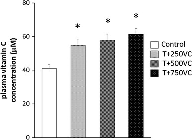
The plasma ascorbic acid concentration in the rats of all of the vitamin C supplemented groups was higher in comparison to that of no vitamin C supplemented group (P < 0.05; Fig. 1).
Fig. 1.

Plasma vitamin C concentration 2 weeks after the onset of vitamin C supplementation. Values are presented as the mean ± SE. *P < 0.05 significantly different from the control group
PGC-1α protein content after training
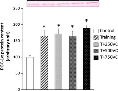
The training significantly increased the PGC-1α protein content in the epitrochlearis muscle (P < 0.05; Fig. 2). However, vitamin C supplementation did not alter the training-induced increase of PGC-1α protein content regardless of the dose of vitamin C supplementation (Fig. 2).
Fig. 2.

Peroxisome proliferator-activated receptor-γ coactivator-1α (PGC-1α) protein content in rat epitrochlearis muscles taken 48 h after the last training session. Values are presented as the mean ± SE. *P < 0.05 significantly different from the control group
Skeletal muscle enzyme activity
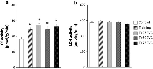
The training significantly increased CS activity of the epitrochlearis muscle (P < 0.05; Fig. 3a). However, vitamin C supplementation did not alter the training-induced increase in CS activity regardless of the dose of vitamin C supplementation (Fig. 3a). Neither the training nor vitamin C supplementation altered LDH activity of the skeletal muscle (Fig. 3b).
Fig. 3.

The citrate synthase (CS) activity in rat epitrochlearis muscle (a) and lactate dehydrogenase (LDH) activity in rat epitrochlearis muscles (b) taken 48 h after the last training session. Values are presented as the mean ± SE. *P < 0.05 significantly different from the control group
Blood lactate concentration after acute exercise
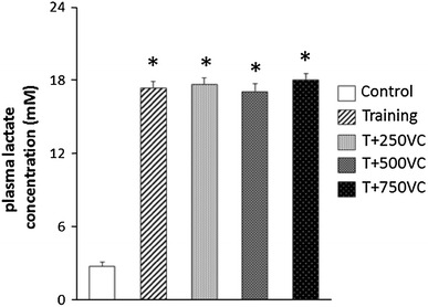
The plasma lactate concentration was elevated to 17.6 mM immediately after the acute high-intensity intermittent exercise in all the training groups. The plasma lactate concentration of all the training groups was significantly higher in comparison to that of the control group (P < 0.05; Fig. 4).
Fig. 4.

Plasma lactate concentration immediately after the last exercise bout on the 11th training session. Values are presented as the mean ± SE. *P < 0.05 significantly different from the control group
Discussion
This study attempted to reveal the effects of vitamin C supplementation on high intensity intermittent swimming training-induced mitochondrial biogenesis in the skeletal muscle. The main findings of this study were that vitamin C supplementation did not prevent high intensity intermittent training-induced increase of PGC-1α protein content and CS activity regardless of the dose of vitamin C supplementation.
The characteristics of high-intensity intermittent swimming training
The rats of the training groups in the current study worked on high intensity swimming training. It has been reported that this training model induces increase in PGC-1α protein content, CS and β-HAD activity, and GLUT4 protein content in rat skeletal muscle [4, 7, 33]. ROS generation is suggested to increase with the exercise employed for the present training study. The following findings obtained support that suggestion: the acute high intensity intermittent swimming exercise elevated the plasma lactate concentration to 17.6 mM. Exogenously added lactate accelerates H2O2 production in vitro [11]. Recently, Higashida et al. [12] evaluated the effects of antioxidant supplementation on low intensity prolonged swimming training-induced adaptation in rat skeletal muscle. However, low intensity swimming exercise differs from high intensity swimming exercise in the ROS production or the increase of oxidative stress during the exercise. Low intensity prolonged swimming exercise does not alter the blood lactate concentration (data not shown). Furthermore, Higashida et al. [12] showed that increased plasma malondialdehyde (MDA) concentration following low intensity swimming exercise is inhibited by antioxidant supplementation. These findings suggested that ROS production during low intensity swimming exercise is smaller than the production during high intensity swimming exercise.
The effects of vitamin C supplementation on training-induced adaptation in skeletal muscle
The results of this study indicate that vitamin C supplementation, irrespective of doses used, does not alter the training-induced increases in PGC-1α protein content and CS activity. The increase in CS activity is suggested to be attributable to an augmented mitochondrial biogenesis, because CS activity can be employed as a marker of mitochondrial volume density of the skeletal muscle [13, 25].
PGC-1α is a master regulator of mitochondrial biogenesis [17, 36]. PGC-1α binds transcription factors and enhances their transcriptional activity for both nuclear-encoded and mitochondria-encoded mitochondrial proteins [35]. PGC-1α also enhances the transcriptional activity of myocyte enhancer factor 2 (MEF2), which is transcriptional factor for PGC-1α itself [10, 37]. Moreover, PGC-1α is activated by phosphorylation with AMPK, p38MAPK, or deacetylation with SIRT1 [1, 15, 18].
ROS may be involved in the activation and/or expression of PGC-1α via AMPK or p38 MAPK activation [14, 16]. Therefore, it is likely that the significantly increased CS activities observed in all the training groups of the current study arose from the augmentation of the activity and protein content of PGC-1α via the activation of AMPK or p38MAPK by ROS.
It is also likely that the signaling pathway involving ROS activated PGC-1α or increased PGC-1α protein content in the skeletal muscle of the trained rats during the high intensity intermittent training with or without vitamin C supplementation, thus leading to an augmentation of the mitochondrial biogenesis reflected as CS activities over the control. It should be emphasized that the co-involvement of ROS independent signaling cannot be ruled out, considering the importance of redundancy in the adaptation mechanism for survival.
The reason why vitamin C supplementation of any doses had no significant influence on the training-induced increase in mitochondrial biogenesis is not known. However, it is suggested that even the highest dose of vitamin C supplementation could not provide sufficient antioxidative protection on the epitrochlearis muscle from incurring oxidative stress. Therefore, ROS produced seem to stimulate intracellular signalings leading to mitochondrial biogenesis under vitamin C supplementation. The insufficient antioxidant protection may arise from the difficulty in raising intramuscular vitamin C concentration to sufficiently high level for suppressing oxidative stress. This contention is supported by the finding of this study shown in Fig. 1 that the vitamin C supplementation significantly elevated plasma vitamin C level over the control at low dose (250 mg/kg/day); however, at higher doses (500, 750 mg/kg/day) it did not further elevate the vitamin C level beyond that attained with low dose. No significant further elevation of plasma vitamin C level at higher doses is thought to be due to increased excretion of vitamin C into urine [5]. Furthermore, vitamin C is required to be transported from extracellular sources into muscle cells by two types of transporters: sodium-dependent vitamin C co-transporter SVCT2 for ascorbate transport and facilitative hexose transporter GLUT-4 for dehydroascorbic acid transport [26]. These transporters are expressed higher in slow-twitch fibers than in fast-twitch fibers [6, 20]. The reported studies suggest that the epitrochlearis muscle, a typical fast-twitch muscle composed of 85 % of fast-twitch fibers and 15 % of slow-twitch fibers, is poorly equipped with the vitamin C transporters [22].
Acknowledgment
This research was supported by Grants-in-Aid for Scientific Research from the Japan Society for the Promotion of Science (Project Number: 21500631).
Conflict of interest
None.
References
- 1.Akimoto T, Pohnert SC, Li P, Zhang M, Gumbs C, Rosenberg PB, Williams RS, Yan Z. Exercise stimulates Pgc-1α transcription in skeletal muscle through activation of the p38 MAPK pathway. J Biol Chem. 2005;280:19587–19593. doi: 10.1074/jbc.M408862200. [DOI] [PubMed] [Google Scholar]
- 2.Baar K, Wende AR, Jones TE, Marison M, Nolte LA, Chen M, Kelly DP, Holloszy JO. Adaptations of skeletal muscle to exercise: rapid increase in the transcriptional coactivator PGC-1α. FASEB J. 2002;16:1879–1886. doi: 10.1096/fj.02-0367com. [DOI] [PubMed] [Google Scholar]
- 3.Benzie IFF, Strain JJ. Ferric reducing/antioxidant power assay: direct measure of total antioxidant activity of biological fluids and modified version for simultaneous measurement of total antioxidant power and ascorbic acid concentration. Methods Enzymol. 1999;299:15–27. doi: 10.1016/S0076-6879(99)99005-5. [DOI] [PubMed] [Google Scholar]
- 4.Burgomaster KA, Howarth KR, Phillips SM, Rakobowchuk M, Macdonald MJ, McGee SL, Gibala MJ. Similar metabolic adaptations during exercise after low volume sprint interval and traditional endurance training in humans. J Physiol. 2008;586:151–160. doi: 10.1113/jphysiol.2007.142109. [DOI] [PMC free article] [PubMed] [Google Scholar]
- 5.Carr AC, Bozonet SM, Pullar JM, Simcock JW, Vissers MC. Human skeletal muscle ascorbate is highly responsive to changes in vitamin C intake and plasma concentrations. Am J Clin Nutr. 2013;97:800–807. doi: 10.3945/ajcn.112.053207. [DOI] [PMC free article] [PubMed] [Google Scholar]
- 6.Dohm GL. Invited review: regulation of skeletal muscle GLUT-4 expression by exercise. J Appl Physiol. 2002;93:782–787. doi: 10.1152/japplphysiol.01266.2001. [DOI] [PubMed] [Google Scholar]
- 7.Fujimoto E, Machida S, Higuchi M, Tabata I. Effects of nonexhaustive bouts of high-intensity intermittent swimming training on GLUT-4 expression in rat skeletal muscle. J Physiol Sci. 2010;60:95–101. doi: 10.1007/s12576-009-0072-4. [DOI] [PMC free article] [PubMed] [Google Scholar]
- 8.Gomez-Cabrera MC, Domenech E, Romagnoli M, Arduini A, Borras C, Pallardo FV, Sastre J, Viña J. Oral administration of vitamin C decreases muscle mitochondrial biogenesis and hampers training-induced adaptations in endurance performance. Am J Clin Nutr. 2008;87:142–149. doi: 10.1093/ajcn/87.1.142. [DOI] [PubMed] [Google Scholar]
- 9.Green HJ, Fraser IG, Ranney DA. Male and female differences in enzyme activities of energy metabolism in vastus lateralis muscle. J Neurol Sci. 1984;65:323–331. doi: 10.1016/0022-510X(84)90095-9. [DOI] [PubMed] [Google Scholar]
- 10.Handschin C, Rhee J, Lin J, Tam PT, Spiegelman BM. An autoregulatory loop controls peroxisome proliferator-activated receptor γ coactivator 1α expression in muscle. Proc Natl Acad Sci USA. 2003;100:7111–7116. doi: 10.1073/pnas.1232352100. [DOI] [PMC free article] [PubMed] [Google Scholar]
- 11.Hashimoto T, Hussien R, Oommen S, Gohil K, Brooks GA. Lactate sensitive transcription factor network in L6 myocytes: activation of MCT1 expression and mitochondrial biogenesis. FASEB J. 2007;21:2602–2612. doi: 10.1096/fj.07-8174com. [DOI] [PubMed] [Google Scholar]
- 12.Higashida K, Kim SH, Higuchi M, Holloszy JO, Han DH. Normal adaptations to exercise despite protection against oxidative stress. Am J Physiol: Endocrinol Metab. 2011;301:E779–E784. doi: 10.1152/ajpendo.00655.2010. [DOI] [PMC free article] [PubMed] [Google Scholar]
- 13.Hood DA. Contractile activity-induced mitochondrial biogenesis in skeletal muscle. J Appl Physiol. 2001;90:1137–1157. doi: 10.1152/jappl.2001.90.3.1137. [DOI] [PubMed] [Google Scholar]
- 14.Irrcher I, Ljubicic V, Hood DA. Interactions between ROS and AMP kinase activity in the regulation of PGC-1α transcription in skeletal muscle cells. Am J Physiol Cell Physiol. 2009;296:C116–C123. doi: 10.1152/ajpcell.00267.2007. [DOI] [PubMed] [Google Scholar]
- 15.Jäger S, Handschin C, St-Pierre J, Spiegelman BM. AMP-activated protein kinase (AMPK) action in skeletal muscle via direct phosphorylation of PGC-1alpha. Proc Natl Acad Sci USA. 2007;104:12017–12022. doi: 10.1073/pnas.0705070104. [DOI] [PMC free article] [PubMed] [Google Scholar]
- 16.Kang C, O’Moore KM, Dickman JR, Ji LL. Exercise activation of muscle peroxisome proliferator-activated receptor-gamma coactivator-1alpha signaling is redox sensitive. Free Radic Biol Med. 2009;47:1394–1400. doi: 10.1016/j.freeradbiomed.2009.08.007. [DOI] [PubMed] [Google Scholar]
- 17.Kelly DP, Scarpulla RC. Transcriptional regulatory circuits controlling mitochondrial biogenesis and function. Genes Dev. 2004;18:357–368. doi: 10.1101/gad.1177604. [DOI] [PubMed] [Google Scholar]
- 18.Lagouge M, Argmann C, Gerhart-Hines Z, Meziane H, Lerin C, Daussin F, Messadeq N, Milne J, Lambert P, Elliott P, Geny B, Laakso M, Puigserver P, Auwerx J. Resveratrol improves mitochondrial function and protects against metabolic disease by activating SIRT1 and PGC-1α. Cell. 2006;127:1109–1122. doi: 10.1016/j.cell.2006.11.013. [DOI] [PubMed] [Google Scholar]
- 19.Lira VA, Benton CR, Yan Z, Bonen A. PGC-1alpha regulation by exercise training and its influences on muscle function and insulin sensitivity. Am J Physiol Endocrinol Metab. 2010;299:E145–E161. doi: 10.1152/ajpendo.00755.2009. [DOI] [PMC free article] [PubMed] [Google Scholar]
- 20.Low M, Sandoval D, Avilés E, Pérez F, Nualart F, Henríquez JP. The ascorbic acid transporter SVCT2 is expressed in slow-twitch skeletal muscle fibres. Histochem Cell Biol. 2009;131(5):565–574. doi: 10.1007/s00418-008-0552-2. [DOI] [PubMed] [Google Scholar]
- 21.Nagatomo F, Fujino H, Kondo H, Kouzaki M, Gu N, Takeda I, Tsuda K, Ishihara A. The effects of running exercise on oxidative capacity and PGC-1 alpha mRNA levels in the soleus muscle of rats with metabolic syndrome. J Physiol Sci. 2012;62:105–114. doi: 10.1007/s12576-011-0188-1. [DOI] [PMC free article] [PubMed] [Google Scholar]
- 22.Nesher R, Karl IE, Kaiser KE, Kipnis DM. Epitrochlearis muscle. I. Mechanical performance, energetics, and fiber composition. Am J Physiol. 1980;239:E454–E460. doi: 10.1152/ajpendo.1980.239.6.E454. [DOI] [PubMed] [Google Scholar]
- 23.Norrbom J, Sundberg CJ, Ameln H, Kraus WE, Jansson E, Gustafsson T. PGC-1alpha mRNA expression is influenced by metabolic perturbation in exercising human skeletal muscle. J Appl Physiol. 2004;96:189–194. doi: 10.1152/japplphysiol.00765.2003. [DOI] [PubMed] [Google Scholar]
- 24.Pilegaard H, Saltin B, Neufer PD. Exercise induces transient transcriptional activation of the PGC-1α gene in human skeletal muscle. J Physiol. 2003;546:851–858. doi: 10.1113/jphysiol.2002.034850. [DOI] [PMC free article] [PubMed] [Google Scholar]
- 25.Reichmann H, Hoppeler H, Mathieu-Costello O, von Bergen F, Pette D. Biochemical and ultrastructural changes of skeletal muscle mitochondria after chronic electrical stimulation in rabbits. Pflugers Arch. 1985;404:1–9. doi: 10.1007/BF00581484. [DOI] [PubMed] [Google Scholar]
- 26.Savini I, Rossi A, Pierro C, Avigliano L, Catani MV. SVCT1 and SVCT2: key proteins for vitamin C uptake. Amino Acids. 2008;34:347–355. doi: 10.1007/s00726-007-0555-7. [DOI] [PubMed] [Google Scholar]
- 27.Silveira LR, Pilegaard H, Kusuhara K, Curi R, Hellsten Y. The contraction induced increase in gene expression of peroxisome proliferator-activated receptor (PPAR)-gamma coactivator 1alpha (PGC-1alpha), mitochondrial uncoupling protein 3 (UCP3) and hexokinase II (HKII) in primary rat skeletal muscle cells is dependent on reactive oxygen species. Biochim Biophys Acta. 2006;1763:969–976. doi: 10.1016/j.bbamcr.2006.06.010. [DOI] [PubMed] [Google Scholar]
- 28.Srere PA. Citrate synthase. Methods Enzymol. 1969;13:3–11. doi: 10.1016/0076-6879(69)13005-0. [DOI] [Google Scholar]
- 29.St Pierre J, Drori S, Uldry M, Silvaggi JM, Rhee J, Jager S, Handschin C, Zheng K, Lin J, Yang W, Simon DK, Bachoo R, Spiegelman BM. Suppression of reactive oxygen species and neurodegeneration by the PGC-1 transcriptional coactivators. Cell. 2006;127:397–408. doi: 10.1016/j.cell.2006.09.024. [DOI] [PubMed] [Google Scholar]
- 30.Strobel NA, Peake JM, Matsumoto A, Marsh SA, Coombes JS, Wadley GD. Antioxidant supplementation reduces skeletal muscle mitochondrial biogenesis. Med Sci Sports Exerc. 2011;43:1017–1024. doi: 10.1249/MSS.0b013e318203afa3. [DOI] [PubMed] [Google Scholar]
- 31.Suwa M, Nakano H, Radak Z, Kumagai S. Endurance exercise increases the SIRT1 and peroxisome proliferatoractivated receptor γ coactivation-1α protein expressions in rat skeletal muscle. Metabolism. 2008;57:986–998. doi: 10.1016/j.metabol.2008.02.017. [DOI] [PubMed] [Google Scholar]
- 32.Terada S, Goto M, Kato M, Kawanaka K, Shimokawa T, Tabata I. Effects of low-intensity prolonged exercise on PGC-1 mRNA expression in rat epitrochlearis muscle. Biochem Biophys Res Commun. 2002;296:350–354. doi: 10.1016/S0006-291X(02)00881-1. [DOI] [PubMed] [Google Scholar]
- 33.Terada S, Tabata I, Higuchi M. Effect of high-intensity intermittent swimming training on fatty acid oxidation enzyme activity in rat skeletal muscle. Jpn J Physiol. 2004;54:47–52. doi: 10.2170/jjphysiol.54.47. [DOI] [PubMed] [Google Scholar]
- 34.Terada S, Kawanaka K, Goto M, Shimokawa T, Tabata I. Effects of high-intensity intermittent swimming on PGC-1α protein expression in rat skeletal muscle. Acta Physiol Scand. 2005;184:59–65. doi: 10.1111/j.1365-201X.2005.01423.x. [DOI] [PubMed] [Google Scholar]
- 35.Uguccioni G, D’souza D, Hood DA (2010) Regulation of PPARγ coactivator-1α function and expression in muscle: effect of exercise. PPAR Res. doi:10.1155/2010/937123 [DOI] [PMC free article] [PubMed]
- 36.Wende A, Schaeffer PJ, Parker GJ, Zechner C, Han DH, Chen MM, Hancock CR, Lehman JJ, Huss JM, McClain DA, Holloszy JO, Kelly DP. A role for the transcriptional coactivator PGC-1alpha in muscle refueling. J Biol Chem. 2007;282:36642–36651. doi: 10.1074/jbc.M707006200. [DOI] [PubMed] [Google Scholar]
- 37.Yan Z, Li P, Akimoto T. Transcriptional control of the Pgc-1alpha gene in skeletal muscle in vivo. Exerc Sport Sci Rev. 2007;35:97–101. doi: 10.1097/JES.0b013e3180a03169. [DOI] [PubMed] [Google Scholar]


