Abstract
The effect of static exercise on neurovascular coupling (NVC) was investigated by measuring the blood flow velocity in the posterior cerebral artery (PCAv) during 2-min static handgrip exercises (HG) at 30 % of the maximum voluntary contraction in 17 healthy males. NVC was estimated as the relative change in PCAv from eye closing to a peak response to looking at a reversed checkerboard. The conductance index (CI) was calculated by dividing PCAv by the mean arterial pressure (MAP). HG significantly increased PCAv from the resting baseline, with an increase in MAP and a reduction in CI, whereas NVC did not differ significantly between the resting and HG. Compared to the resting baseline, HG significantly increased the pressor response to visual stimulation by 5.6 ± 1.1 (mean ± SE) mmHg, while the CI response was significantly inhibited by −7.0 ± 1.5 %. These results indicate that NVC was maintained during HG via contributions from both the pressor response and vasodilatation.
Keywords: Posterior cerebral artery, Static handgrip exercise, Exercise pressor reflex, Post-exercise muscle ischaemia
Introduction
Nutrition and blood flow is secured in the brain, particularly in the working region of the brain, by neurovascular coupling (NVC), which is defined as adjusting the local cerebral blood flow (CBF) according to the underlying cortical neural activity [1]. In fact, transcranial Doppler (TCD) ultrasound flowmetry studies have revealed the existence of visually evoked CBF velocity responses (VEFR) [2]. Subjects looking at a reversed checkerboard, which is used widely as a visual stimulant, was shown to induce an 18 % increase in the blood flow velocity (PCAv) in the posterior cerebral artery (PCA), which supplies blood to the visual cortex [3]. The magnitude of this VEFR has been used to assess NVC.
It is important to meet a metabolic demand increased by visual information processing in the visual cortex under any physiological conditions. Nevertheless, a previous study has indicated that an increase in mean arterial pressure (MAP) modified the magnitude of the NVC. The magnitude of the NVC increased during the cold pressor test (CPT), accompanied by an increase in the baseline PCAv with pressor response [3]. Thus, there is a possibility that the NVC is not always maintained, but modified when the baseline PCAv increases under the influence of pressor response with sympathetic activation. In the present study, to investigate an effect of pressor response on NVC, static handgrip exercise (HG) and post-exercise muscle ischaemia (PEMI) were applied as stimulations which cause an acute elevation in MAP induced by an activation of the sympathetic nerve.
In addition to the effect of MAP at baseline, i.e., before visual stimulation, on the magnitude of the NVC, a possible MAP increase in response to visual stimulation could affect the NVC. No study observed has as yet the response of MAP against visual stimulation.
The PCAv response itself to static exercise has not yet been studied. Ogoh et al. [4] reported an increase in the blood flow velocity (MCAv) in the middle cerebral artery (MCA) during HG at 30 % of the maximum voluntary contraction (MVC). A regional difference between the carotid and vertebral arteries during dynamic exercise was revealed [5]. Although the NVC is reportedly maintained, in spite of the increased baseline PCAv, during aerobic exercise at 70 % of the maximum heart rate (HR) [6], it is still unclear whether the magnitude of the NVC and PCAv increases during HG and activation of the exercise pressor reflex, which originates from mechano- and metabosensitive receptors activated by mechano- and metaboreflexes in active muscles [7].
Previous studies reported an absence of vasoconstriction in MCA and anterior cerebral artery (ACA) with sympathetic activation induced by metaboreflex after rhythmic handgrip exercise at 20 % MVC and isometric calf exercise at 35 % MVC [8, 9]. In turn, it is uncertain whether the NVC and PCAv are affected by metaboreflex activation after HG as well as MCA and ACA because of different branches of these cerebral arteries.
In the present study, we describe the response of PCAv to a visual stimulation during HG and PEMI. Then, we examined hypotheses: (1) HG increases the magnitude of the PCAv response to visual stimulation due to possible effects of PCAv and MAP at baseline and during visual stimulation on NVC; and (2) pressor response accompanied by metaboreflex modifies the NVC. These hypotheses were tested by evaluating changes in NVC and PCAv during HG and PEMI after HG to isolate metaboreflex activation. No study using the CPT [3] and dynamic cycling exercise [6] has revealed the role of pressor response on NVC and PCAv. The present study investigated the effect of acute elevation in MAP on NVC and PCAv.
Methods
Subjects
Seventeen males (age 25 ± 1.1 years, mean ± SE; height 172 ± 1 cm; weight 65 ± 2 kg) volunteered for this study. All subjects were non-smokers, normotensive, and free from any known autonomic dysfunction and cardiovascular disease, and were not taking any medication. The Ethics Committee of the Institution of Health Science, Kyushu University, Japan, approved the experimental protocol, and each subject provided written informed consent to participate prior to the commencement of the study. All protocols conformed to the Declaration of Helsinki. Before the experiments were performed, each subject visited the laboratory for familiarization with the techniques and procedures of the protocol.
Protocol
The subjects arrived at the laboratory after having abstained from caffeinated beverages and strenuous exercise for 6 h, and from eating for 2 h; they had not experienced sleep loss during the previous night. All studies were performed in a darkened and quiet room at an ambient temperature of 23 °C. The MVC was taken as the greatest force produced during a maximal effort with the subject’s left hand using a load cell attached to a modified HG dynamometer at least 10 min before starting the measurement. These data were used to determine the 30 % MVC for each subject. The subjects were instructed to keep their eyes closed at all times except for periods when visual stimulation was applied. Subjects who wore glasses took them off for the duration of the experiment.
After a 2-min resting period, subjects performed 2 min of HG at an intensity of 30 % MVC with auditory feedback from an experimenter. They were instructed not to hold their breath so as to avoid a Valsalva maneuver during the HG. A cuff placed on the left upper arm was rapidly inflated to suprasystolic pressure (220 mmHg) 5 s before cessation of the HG (PEMI trial). The cuff remained inflated for 2 min to activate the metaboreflex. In the control trial, PEMI was not performed and a 2-min recovery period was allowed after the exercise period. Subjects received visual stimulation for 6 min in both the PEMI and control trials, and rested for at least 15 min between trials. The order of the trials was randomized.
Measurements
Blood pressure (BP), HR, PCAv, and the end-tidal carbon dioxide partial pressure (P ETCO2) were measured continuously in all trials. The beat-by-beat BP was recorded with a continuous finger photoplethysmography attached to the right middle finger (Finometer; Finapres Medical Systems, Amsterdam, The Netherlands). The continuous HR was determined from a standard electrocardiogram (ECG; MEG2100; Nihon-Kohden, Tokyo, Japan). The analogue signals were sampled at 1 kHz using an A/D converter (PowerLab 8/30; ADInstruments, Colorado Springs, CO, USA). The minute-by-minute HR and mean arterial pressure (MAP) were calculated from the ECG and BP recordings, respectively. The exerted force was measured by a load cell (LTZ-200KA; Kyowa Dengyo, Tokyo, Japan) in the HG dynamometer. The analogue signal of the force was produced by an instrumentation amplifier (WGA-650A; Kyowa Dengyo). P ETCO2 was monitored with a gas analyser (AE-310s; Minato Medical Science, Tokyo, Japan).
The mean PCAv was obtained by transcranial ultrasonography (WAKI; Atys Medical, St-Genis-Laval, France). A 2-MHz Doppler probe was placed at the right temporal window and fixed using a head band. The vessel was identified by TCD ultrasonography according to standard criteria [10]. The MCA was insonated at a depth of 50–60 mm with a standard hand probe, followed by insonation of the PCA at a depth of 60–70 mm. To confirm the accurate insonation of the PCA, we performed ipsilateral carotid compression, which increased the PCAv and decreased the MCAv [2]. The length of the sample volume was set at 6 mm.
Visual stimulation
The stimulus session consisted of six repetitions of 20-s periods of eye closing and 40-s periods of visual stimulation. The visual stimulant was a reversed checkerboard. Subjects were seated 0.5 m from the front of a 24-inch flat computer screen (visual angle of 25°). During the stimulation period, subjects were asked to gaze at a small red spot at the center of the computer screen. During the eye-closing period, the screen was black and the subjects were asked to close their eyes. The checkerboard pattern comprised black-and-white squares arranged with a spatial frequency of 1.6 cycles/degree. The black-and-white squares were alternated at a frequency of 2 Hz.
Data analysis
We estimated the NVC as the averaged relative change in PCAv between the mean value obtained during the 20 s of eye closing and the peak response obtained during the 40 s of visual stimulation. In the 2-min resting baseline period, the peak velocity to visual stimulation was identified for each trial, and this was averaged across two repetitions in each individual. During the 2-min HG and PEMI or the corresponding recovery periods, NVC was determined during the last 1 min in order to remove MAP fluctuations. The conductance index (CI) of the cerebral vessel was calculated by dividing the PCAv by the MAP.
Data were expressed as mean ± SE values. The effects of trial and time on the PCAv, the MAP, and the CI responses to visual stimulation were examined by two-way repeated-measures ANOVA. When a significant F value was detected, the data were further analyzed using Bonferroni’s post hoc test. The HR, MAP, P ETCO2, PCAv, and CI of non-stimulation (eye closing) during HG were compared to their resting baseline counterparts using a paired t test. The PCAv, MAP, and CI responses to visual stimulation were compared to the prestimulation (eye-closing) baseline data using a paired t test. The level of statistical significance was set at p < 0.05. The statistical analyses were performed with SPSS (PASW statistics 18; SPSS, IL, USA).
Results
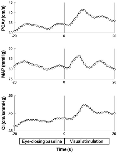
When a subject looked at a reversed checkerboard at rest, the PCAv increased (Fig. 1). The stimulation by checkerboard also increased MAP whereas HR showed no change at rest. Similar responses were observed during HG and recovery period.
Fig. 1.

The time series of PCAv, MAP, and CI immediately before and after visual stimulation during resting baseline in the control trial in a subject. Looking at a reversed checkerboard increased the PCAv, accompanied by an increase in MAP and PCA CI
Systemic changes
HG significantly increased HR and MAP from the resting baseline values in both the control and PEMI trials (p < 0.05; Table 1). HR returned to the resting baseline level during PEMI and the corresponding recovery period (p > 0.05). MAP was not significantly different during the recovery period of the control trial, compared to the baseline (p > 0.05). The HR and MAP at rest and during HG did not significantly differ between the control and PEMI trials (p > 0.05). P ETCO2 did not change significantly from the resting level during either HG or the recovery periods (39.2 ± 0.6, 39.7 ± 0.7, and 39.5 ± 0.7 mmHg, respectively, p > 0.05).
Table 1.
Heart rate (HR), mean arterial pressure (MAP), and posterior cerebral artery blood flow velocity (PCAv) at resting baseline, and during static handgrip exercise and recovery or post-exercise muscle ischaemia (PEMI)
| Control trial | PEMI | |||||
|---|---|---|---|---|---|---|
| Rest | Handgrip | Recovery | Rest | Handgrip | PEMI | |
| HR (bpm) | 65.0 ± 2.0 | 76.2 ± 2.6*† | 63.9 ± 2.1 | 63.8 ± 1.8 | 77.5 ± 2.5*† | 67.1 ± 2.6 |
| MAP (mmHg) | 86.6 ± 2.8 | 105.3 ± 4.0*† | 89.4 ± 3.2 | 88.0 ± 3.3 | 105.6 ± 4.4*† | 99.2 ± 3.4†‡ |
| PCAv (cm/s) | 32.2 ± 2.0 | 36.0 ± 2.4*† | 33.0 ± 2.1 | 33.3 ± 1.7 | 37.1 ± 2.1*† | 33.7 ± 2.1 |
Data are mean ± SE values
* p < 0.05 vs. resting baseline; † p < 0.05 vs. recovery period; ‡ p < 0.05 vs. control trial
PCAv during HG and PEMI
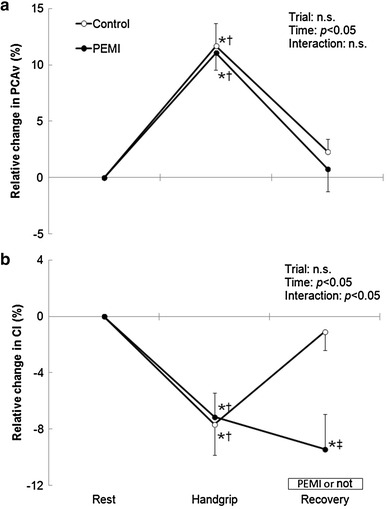
HG increased PCAv by 11.7 ± 2.0 and 11.1 ± 1.5 %, while it decreased CI in PCA by −7.6 ± 2.2 and −7.1 ± 1.7 % from the resting baseline in the control and PEMI trials, respectively (p < 0.05; Table 1; Fig. 2a, b). PCAv did not change significantly from the resting baseline during PEMI or the corresponding recovery periods (0.8 ± 2.0 and 2.3 ± 1.1 %, respectively, p > 0.05). PCAv and CI at rest and during HG did not differ significantly between the control and PEMI trials (p > 0.05). Conversely, CI decreased significantly from the resting baseline during PEMI (−9.4 ± 2.5 %, p < 0.05) but not during the recovery period of the control trial (−1.0 ± 1.3 %, p > 0.05).
Fig. 2.

Relative changes in a the blood flow velocity in the posterior cerebral artery (PCAv) and b the conductance index (CI) from resting baseline to handgrip exercise, and recovery or post-exercise muscle ischaemia (PEMI). n.s. not significant; *p < 0.05 vs. resting baseline; † p < 0.05 vs. recovery; ‡ p < 0.05 vs. control
NVC during HG and PEMI
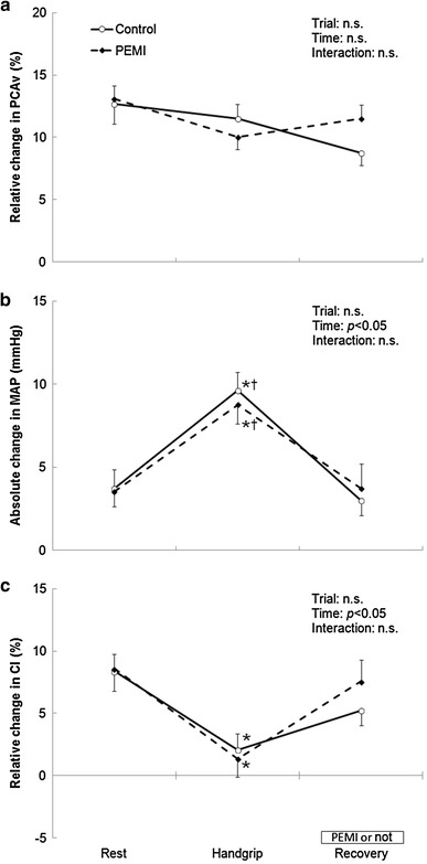
Compared to the resting baseline, NVC, i.e., increase in PCAv evoked by visual stimulation, showed no significant change during HG in both the control and PEMI trials (12.7 ± 1.6 and 11.5 ± 1.1 %, in resting and HG, respectively, in control trials, and 13.1 ± 1.0 and 10.0 ± 1.0 %, respectively, in PEMI, p > 0.05; Fig. 3a). Compared to the resting baseline, HG significantly increased the MAP response to visual stimulation (3.7 ± 1.1 and 9.6 ± 1.1 mmHg, in control trials, respectively; and 3.5 ± 0.9 and 8.8 ± 1.2 mmHg, in PEMI, respectively, p < 0.05; Fig. 3b), while it significantly decreased the CI response (8.4 ± 1.6 and 2.1 ± 1.3 %, in control trials, respectively; and 8.6 ± 1.2 and 1.4 ± 1.5 %, in PEMI, respectively, p < 0.05; Fig. 3c). In turn, the PCAv, MAP, and CI responses to visual stimulation did not differ significantly from the resting baselines during PEMI and the corresponding recovery periods (11.5 ± 1.1 and 8.8 ± 1.0 %, respectively, for PCAv; 3.7 ± 1.5 and 3.0 ± 0.9 mmHg, respectively, for MAP; 7.6 ± 1.7 and 5.2 ± 1.2 %, respectively, for CI; p > 0.05). These variables did not differ significantly between the control and PEMI trials (p > 0.05).
Fig. 3.

Relative change in PCAv (a), absolute change in mean arterial pressure (MAP) (b), and relative change in CI (c) from eye-closing baseline to the visual stimulation period at resting baseline, handgrip exercise, and recovery or PEMI. *p < 0.05 vs. resting baseline; † p < 0.05 vs. recovery period
Discussion
The main findings of the present study are as follows:
HG increased PCAv from the resting baseline, and this increase was accompanied by an increase in MAP and a decrease in PCA CI.
NVC remained constant during HG, compared with the resting baseline, while MAP and CI made different relative contributions to the maintenance of the NVC.
Metaboreflex activation did not increase the PCAv nor modify NVC.
PCAv during HG and PEMI
HG increased PCAv and MAP but reduced CI in the present study. These findings indicate that HG increases PCAv, accompanied not by the vasodilatation but by the pressor response evoked by exercise. In agreement with previous studies of MCA [8] and ACA [9], the increase in PCAv induced by HG did not persist during PEMI and the corresponding recovery periods, although the increased MAP and sympathetic activation continued during PEMI. The increase in MAP during PEMI in the present study ensured a successful activation of the metaboreflex. These results demonstrate that HG increases the PCAv, and that this increase does not depend directly up on activation of the metaboreflex.
Increase in PCAv was induced not by the change in P ETCO2 but by the elevation in MAP during HG. P ETCO2 remained unchanged from the resting baseline level during HG and the subsequent recovery period in the present study. This result is consistent with previous studies finding an increase in MCAv without a change in P ETCO2 during HG at 30 % MVC and the subsequent recovery period [4, 11, 12]. In turn, we cannot totally deny a possible effect of P ETCO2 decreased by hyperventilation during PEMI because there are no data on P ETCO2 during the PEMI period in the present study due to technical reasons. However, it was presumed that PCAv was not affected by P ETCO2 even during PEMI, based on the previous study which demonstrated that P ETCO2 remained unchanged from resting during PEMI after isometric voluntary calf exercise at an intensity of 35 % MVC [12]. Thus, P ETCO2 would not be affected by metaboreflex activation after HG at 30 % MVC in the present study.
NVC during HG and PEMI
NVC, i.e., the increase in PCAv with visual stimulation, was maintained during HG, although PCAv was increased by the exercise-induced pressor response at the eye-closing baseline. On the other hand, an increase in MAP response to visual stimulation was observed during HG, whereas a decreased CI response was detected in the present study. These results indicate that HG has no effect on the magnitude of the NVC, whereas it may influence the relative contributions to NVC of the pressor response and vasodilatation. The discussion above is acceptable when the vessel diameter of the PCA remains stable as reported in MCA [13]. NVC is calculated from relative response in PCAv, thus is determined by perfusion pressure and CI. Supposing MAP is proportional to perfusion pressure, MAP and CI are only factors explaining the stable NVC, when vessel diameter in PCA and MAP are stable.
The present findings do not support the notion that vasodilatation is the sole contributor to the increase in the PCAv in response to visual stimulation. Other studies have investigated the factors inducing NVC, and have suggested a role of local vasodilatation induced by nitric oxide [14] or neuronal activity with γ-aminobutyric acid [15]. However, the CI response to visual stimulation has not been reported previously [3, 16]. In the present study, the increase in PCAv was associated not only with an increase in CI (i.e., vasodilatation) but also with the MAP response during visual stimulation. Thus, the PCAv response to visual stimulation was attributable to both vasodilatation and the pressor response evoked by visual stimulation.
The role of the pressor response in the increase in PCAv was greater in HG than at rest. There was a greater pressor response to visual stimulation during HG than at rest, in spite of a lesser increase in the CI. It is possible that the amplitude of the fluctuation affects the result, since we observed a greater fluctuation in resting PCAv and MAP, thus likely resulting in the decreased CI response. Further studies are needed to elucidate the relative contributions of these mechanisms to NVC at rest and during exercise.
NVC would not be affected by acute elevation in MAP with HG. Azevedo et al. [16] found that the magnitude of the NVC did not change in response to visual stimulation under different orthostatic conditions in healthy young volunteers. Therefore, the magnitude of the NVC appears to be independent of acute systemic changes such as those induced by static exercise and orthostatic stress.
In the present study, PEMI after HG at 30 % MVC had no effect on the NVC. Fabjan et al. [3] demonstrated that NVC increased during the CPT, which caused sympathetic activation induced by cold and pain stimulation to the skin. Conversely, HG may have induced a complex regulation of the cardiovascular system, central command, and the reflex of active muscle mechanosensitive, metabosensitive, and arterial baroreceptors [17]. This is in contrast to the report by Fabjan et al. [3] indicating the effect of sympathetic activation induced by CPT on the NVC. It is implied that exercise pressor reflex with the activation of metaboreflex did not have an explicit role for the increased PCAv in response to visual stimulation. A possibility remained that there is a difference of the activation of the sympathetic nerve induced by CPT or HG. In addition, Vianna et al. [12] demonstrated that the increase in MCAv during HG was independent of a sympathetic activation with CPT, by measuring MCAv during a combination of HG and CPT. Considering this previous study, the vasoconstriction with sympathetic activation induced by PEMI might be attenuated by vasodilatation with local metabolic demand in the brain.
Technical considerations
The PCAv was obtained by TCD ultrasonography. One limitation of PCAv measurements is that the blood flow is affected by the vessel diameter, which cannot be measured. The diameter of the MCA remains relatively constant during physiological stimulation [13], and so it is feasible that PCAv is truly representative of the PCA blood flow, although there are no data confirming that the diameter of the PCA does not change during disturbances.
With regard to the evaluation of NVC, we believe that both the absolute and relative changes in PCAv are physiologically relevant. Most previous studies have quantified NVC as the relative change in PCAv because this ensures independence from the insonation angle [3, 16]. The VEFR should be evaluated under conditions of vasodilatation induced by other stimuli (e.g., carbon dioxide) during exercise.
The findings of the present study demonstrate that HG induces an increase in PCAv and keeps the NVC constant in humans. The increased PCAv was not maintained during PEMI or the control recovery periods, while the stable NVC persisted, suggesting that neither response is due to metaboreflex activation. Moreover, an increase in the pressor response and a decrease in the CI response to visual stimulation during HG were observed in this study. It is therefore possible that the contributions of the pressor response and vasodilatation towards preserving the NVC may alter during the exercise-induced pressor response.
Acknowledgments
This study was supported in part by a grant from the Kozuki Sports and Education Foundation (to N. Hayashi) and KAKENHI 21370111 (to Y. Fukuba).
Conflict of interest
The authors declare that they have no conflict of interest.
References
- 1.Aaslid R. Visually evoked dynamic blood-flow response of the human cerebral-circulation. Stroke. 1987;18:771–775. doi: 10.1161/01.STR.18.4.771. [DOI] [PubMed] [Google Scholar]
- 2.Willie CK, Colino FL, Bailey DM, et al. Utility of transcranial Doppler ultrasound for the integrative assessment of cerebrovascular function. J Neurosci Methods. 2011;196:221–237. doi: 10.1016/j.jneumeth.2011.01.011. [DOI] [PubMed] [Google Scholar]
- 3.Fabjan A, Musizza B, Bajrović FF, Zaletel M, Strucl M. The effect of the cold pressor test on a visually evoked cerebral blood flow velocity response. Ultrasound Med Biol. 2012;38:13–20. doi: 10.1016/j.ultrasmedbio.2011.10.007. [DOI] [PubMed] [Google Scholar]
- 4.Ogoh S, Sato K, Akimoto T, Oue A, Hirasawa A, Sadamoto T. Dynamic cerebral autoregulation during and after handgrip exercise in humans. J Appl Physiol. 2010;108:1701–1705. doi: 10.1152/japplphysiol.01031.2009. [DOI] [PubMed] [Google Scholar]
- 5.Sato K, Ogoh S, Hirasawa A, Oue A, Sadamoto T. The distribution of blood flow in the carotid and vertebral arteries during dynamic exercise in humans. J Physiol Lond. 2011;589:2847–2856. doi: 10.1113/jphysiol.2010.204461. [DOI] [PMC free article] [PubMed] [Google Scholar]
- 6.Willie CK, Cowan EC, Ainslie PN, et al. Neurovascular coupling and distribution of cerebral blood flow during exercise. J Neurosci Methods. 2011;198:270–273. doi: 10.1016/j.jneumeth.2011.03.017. [DOI] [PubMed] [Google Scholar]
- 7.Kaufman MP, Forster HV. Reflexes controlling circulatory, ventilatory, and airway responses to exercise. In: Rowell LB, Shepherd JT, editors. Handbook of physiology. Exercise: regulation and integration of multiple systems. New York: American Physiological Society; 1996. pp. 381–447. [Google Scholar]
- 8.Pott F, Ray C, Olesen H, Ide K, Secker N. Middle cerebral artery blood velocity, arterial diameter and muscle sympathetic nerve activity during post-exercise muscle ischaemia. Acta Physiol Scand. 1997;160:43–47. doi: 10.1046/j.1365-201X.1997.00126.x. [DOI] [PubMed] [Google Scholar]
- 9.Vianna LC, Araújo CGS, Fisher JP. Influence of central command and muscle afferent activation on anterior cerebral artery blood velocity responses to calf exercise in humans. J Appl Physiol. 2009;107:1113–1120. doi: 10.1152/japplphysiol.00480.2009. [DOI] [PubMed] [Google Scholar]
- 10.Aaslid R, Markwalder T, Nornes H. Non-invasive transcranial doppler ultrasound recording of flow velocity in basal cerebral-arteries. J Neurosurg. 1982;57:769–774. doi: 10.3171/jns.1982.57.6.0769. [DOI] [PubMed] [Google Scholar]
- 11.Panerai R, Dawson S, Eames P, Potter J. Cerebral blood flow velocity response to induced and spontaneous sudden changes in arterial blood pressure. Am J Physiol Heart Circul Physiol. 2001;280:H2162–H2174. doi: 10.1152/ajpheart.2001.280.5.H2162. [DOI] [PubMed] [Google Scholar]
- 12.Vianna LC, Kluser Sales AR, Lucas da Nobrega AC. Cerebrovascular responses to cold pressor test during static exercise in humans. Clin Physiol Funct Imaging. 2012;32:59–64. doi: 10.1111/j.1475-097X.2011.01055.x. [DOI] [PubMed] [Google Scholar]
- 13.Serrador J, Picot P, Rutt B, Shoemaker J, Bondar R. MRI measures of middle cerebral artery diameter in conscious humans during simulated orthostasis RID A-9172-2009. Stroke. 2000;31:1672–1678. doi: 10.1161/01.STR.31.7.1672. [DOI] [PubMed] [Google Scholar]
- 14.Piknova B, Kocharyan A, Schechter AN, Silva AC. The role of nitrite in neurovascular coupling. Brain Res. 2011;1407:62–68. doi: 10.1016/j.brainres.2011.06.045. [DOI] [PMC free article] [PubMed] [Google Scholar]
- 15.Radhakrishnan H, Wu W, Boas D, Franceschini MA. Study of neurovascular coupling by modulating neuronal activity with GABA. Brain Res. 2011;1372:1–12. doi: 10.1016/j.brainres.2010.11.082. [DOI] [PMC free article] [PubMed] [Google Scholar]
- 16.Azevedo E, Rosengarten B, Santos R, Freitas J, Kaps M. Interplay of cerebral autoregulation and neurovascular coupling evaluated by functional TCD in different orthostatic conditions. J Neurol. 2007;254:236–241. doi: 10.1007/s00415-006-0338-1. [DOI] [PubMed] [Google Scholar]
- 17.Iellamo F, Legramante J, Raimondi G, Peruzzi G. Baroreflex control of sinus node during dynamic exercise in humans: effects of central command and muscle reflexes. Am J Physiol Heart Circul Physiol. 1997;272:H1157–H1164. doi: 10.1152/ajpheart.1997.272.3.H1157. [DOI] [PubMed] [Google Scholar]


