Metabolic plasticity of neurons ensures their activity continues when glucose is limited. Walsh and Simon discuss new work by Ashrafi and colleagues that finds Sirtuin 3 directs local metabolic adaptation at synapses during sustained glucose deprivation.
Abstract
Metabolic plasticity of neurons ensures their activity continues when glucose is limited. Walsh and Simon discuss new work by Ashrafi and colleagues (https://doi.org/10.1083/jcb.202305048) that finds Sirtuin 3 directs local metabolic adaptation at synapses during sustained glucose deprivation.
Despite only accounting for ∼2% of our body mass, the brain consumes ∼20% of our body’s resting energy production (1). Much of this consumption is driven by energetically expensive basal physiological processes, including trafficking cargo to distal dendrites, axons, and synapses, and maintaining membrane polarization, each of which is exacerbated by the neuron’s expansive geometry. At the level of the synapse, the energetic expense of retrieving and refilling synaptic vesicles, coupled with the distance from the cell body, necessitates local ATP synthesis to meet the energetic demands of neuronal activity (2). Factor in the billions of neurons and trillions of synapses that make up the brain, and a total energy budget of ∼20% does not seem so far-fetched. How does a neuron maintain synaptic ATP homeostasis given these challenges?
Glucose is the primary substrate for energy production in neurons. It is metabolized to pyruvate in the cytoplasm to generate a net two molecules of ATP in the process of glycolysis. Pyruvate can then be completely oxidized to carbon dioxide and water via oxidative phosphorylation (OXPHOS) in the mitochondria, producing anywhere from 26 to 32 molecules of ATP (3). Glucose, however, is not always available; ischemic stroke, hypoglycemia, and even sustained neuronal activity can all limit glucose availability. In these contexts, neurons have been shown to utilize alternative substrates to fuel OXPHOS. Extracellular lactate can be imported and rapidly converted to pyruvate, which has been hypothesized as a pathway for surrounding glial cells to support neuronal ATP production (4). Neurons can also utilize ketone bodies produced by the liver as well as scavenge local glutamate and other amino acids to form various metabolic intermediates for mitochondrial respiration (5).
At the synapse, demand for ATP occurs on the milliseconds to minutes time scale. In the absence of glucose, there is only enough reserve ATP to sustain synaptic vesicle endocytosis—the most energetically sensitive step of the vesicle cycle—for a short period prior to synapse failure (6). Accordingly, energy demand at these time scales is met with mobilization of the glucose transporter GLUT4 to the plasma membrane to enhance glucose uptake. When glucose is limited, calcium influxes associated with heightened neuronal activity (and therefore ATP demand) activate the mitochondrial calcium uniporter MICU3 to enhance oxidative ATP production in mitochondria (7). Thus, short-term synaptic activity is maintained through either increased import and consumption of glucose or through accelerated mitochondrial ATP production. While short-term adaptations have been described, how neurons adapt to long-term changes in their metabolic environment has been an important unknown in the field.
Tiwari et al. (8) elegantly address this issue and discover a transcriptional program triggered by glucose deprivation that culminates in enhanced mitochondrial OXPHOS to maintain stable synapse function. First, the authors investigated transcriptional changes in rat primary cortical neuronal cultures following brief (3 h) glucose depletion. Pathway analysis revealed that glucose depletion activates a transcriptional program mediated by cAMP-response element-binding protein (CREB), an important regulator of metabolic homeostasis (9). Key among these changes is PGC1α, a transcription factor that drives mitochondrial respiration in part by increasing the number of electron transport chain (ETC) complexes to support increased ATP production (10). Transcriptional induction of OXPHOS genes is consistent with the authors’ previous work demonstrating a functional requirement for oxidative metabolism in the absence of glucose (6). Importantly, the authors show that AMP kinase, which is itself activated when ATP levels fall, is required for transcriptional activation of PGC1α by CREB. Thus, a homeostatic transcriptional mechanism links glucose depletion to putative changes in mitochondrial ATP production at the synapse.
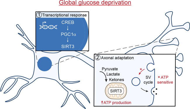
How does PGC1α affect synaptic ATP production? To uncover this mechanism, the authors focused on the PGC1α target gene Sirtuin 3 (Sirt3), a mitochondrially localized, NAD+-dependent protein deacetylase that they found to be upregulated by glucose depletion. Sirt3 is known to promote ETC efficiency, citric acid cycle intermediate generation, and the utilization of ketone bodies, pyruvate, and lactate (11). In other words, Sirt3 function could be the bridge between transcriptional changes induced by glucose deprivation and metabolic adaptations to maintain synaptic ATP levels. The authors nicely extend this finding in an in vivo model of nutrient deprivation across several time scales. While Sirt3 levels are unchanged in mice fasted overnight, Sirt3 expression increased in mice on a 6-mo alternative-day fasting cycle. Having established Sirt3 as a robust biomarker of glucose availability, the authors next tested whether Sirt3 drives metabolic reprograming at the synapse. They show that Sirt3 localizes to mitochondria and at the synapse and next question whether this localization equates to functional changes in synaptic ATP production (Fig. 1).
Figure 1.
Glucose deprivation induces an adaptive transcriptional response to maintain synapse function by upregulating mitochondrial Sirt3 to enhance oxidative metabolism. SV, synaptic vesicle.
For this the authors evaluate if Sirt3 knockdown (KD) affects the energetically sensitive process of synaptic vesicle recycling. If glucose-deprived neurons induce Sirt3 to accelerate or “upshift” mitochondrial ATP production in the absence of glycolysis, then Sirt3 KD should have no effect in the presence of glucose. Indeed, glucose supplemented Sirt3 KD neurons have no impairments in vesicle endocytosis. However, Sirt3 KD neurons, when supplied only with lactate and pyruvate (or ketone bodies), have slower vesicle endocytosis, indicating an insufficiency in mitochondrial ATP production. Direct measurement of ATP levels in presynaptic terminals using a synaptically localized luciferase reporter, Syn-ATP (2), revealed a deficit in basal mitochondrial ATP production, consistent with the effects of the respiration inhibitor oligomycin (7). However, these reduced ATP levels have no effect on other important measures of synaptic function including presynaptic calcium influx and the probability of vesicle release. Together these data support a model that sustained metabolic stress induces Sirt3 to enhance mitochondrial ATP production in the absence of glycolysis and invite future studies on the role of Sirt3 in neuronal ATP production when all fuel sources are available.
This work brings a molecular mechanism to a long-standing question of how neurons maintain stable synaptic function in the face of a changing metabolic environment. It also raises the question of how short and long time-scale adaptive mechanisms interact. Similar to other chronic manipulations such as activity deprivation that induces homeostatic synaptic plasticity (12), the metabolic plasticity described by Tiwari and colleagues (8) is transcriptional and therefore conceivably has a global effect on all synapses. These findings raise many exciting questions related to the interface between neuronal activity and metabolism. The authors show that glucose deprivation manifests as a loss of ATP that is integrated by AMP kinase activation; however, where in the cell this signal originates (e.g., in the dendrites, axons, or synaptic terminals) and its sensitivity to the amount and duration of glucose loss remain to be determined. Moreover, can signals in addition to glucose deprivation also drive Sirt3 expression and metabolic rewiring? Finally, it will be fascinating to define how long metabolic rewiring persists and whether reintroduction of glucose can restore “normal” metabolic function. This work sheds important light on an underappreciated aspect of neuronal cell biology, neuronal metabolism, and in so doing, generates important insights into the adaptations that keep our minds running.
References
- 1.Attwell, D., and Laughlin S.B.. 2001. J. Cereb. Blood Flow Metab. 10.1097/00004647-200110000-00001 [DOI] [PubMed] [Google Scholar]
- 2.Rangaraju, V., et al. 2014. Cell. 10.1016/j.cell.2013.12.042 [DOI] [Google Scholar]
- 3.Yellen, G. 2018. J. Cell Biol. 10.1083/jcb.201803152 [DOI] [PMC free article] [PubMed] [Google Scholar]
- 4.Magistretti, P.J., and Allaman I.. 2015. Neuron. 10.1016/j.neuron.2015.03.035 [DOI] [PubMed] [Google Scholar]
- 5.Divakaruni, A.S., et al. 2017. J. Cell Biol. 10.1083/jcb.201612067 [DOI] [Google Scholar]
- 6.Ashrafi, G., et al. 2017. Neuron. 10.1016/j.neuron.2016.12.020 [DOI] [Google Scholar]
- 7.Ashrafi, G., et al. 2020. Neuron. 10.1016/j.neuron.2019.11.020 [DOI] [Google Scholar]
- 8.Tiwari, A., et al. 2024. J. Cell Biol. 10.1083/jcb.202305048 [DOI] [Google Scholar]
- 9.Altarejos, J.Y., and Montminy M.. 2011. Nature Rev. Mol. Cell Biol. 10.1038/nrm3072 [DOI] [PMC free article] [PubMed] [Google Scholar]
- 10.Austin, S., and St-Pierre J.. 2012. J. Cell Sci. 10.1242/jcs.113662 [DOI] [PubMed] [Google Scholar]
- 11.Sidorova-Darmos, E., et al. 2018. Front. Cell. Neurosci. 10.3389/fncel.2018.00196 [DOI] [PMC free article] [PubMed] [Google Scholar]
- 12.Turrigiano, G.G. 2008. Cell. 10.1016/j.cell.2008.10.008 [DOI] [Google Scholar]


