1. PATIENT PRESENTATION
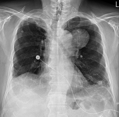
A 75‐year‐old man, with a medical history of colon cancer and lung metastasis who was treated with chemotherapy and lung tumor resection, presented to the emergency department with distressing symptoms of chest pain and profuse sweating. An initial chest x‐ray revealed a troubling finding– a protruding mass in the left mediastinum (Figure 1). Computed tomography (CT) was performed.
FIGURE 1.

Chest x‐ray showed a left mediastinal mass with loss of aortic silhouette.
2. DIAGNOSIS
Thoracic aortic aneurysm
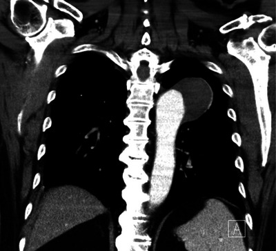
Subsequent chest CT confirmed the presence of a substantial saccular aneurysm involving the aortic arch (Figure 2). Prompt intervention was crucial, and the patient underwent thoracic endovascular aortic aneurysm repair. Fortunately, the procedure proceeded smoothly without any complications, and the patient's recovery was marked by its uneventfulness.
FIGURE 2.

Chest computed tomography showed a partial thrombosed aortic arch saccular aneurysm, about 6.4 cm in diameter.
Thoracic aortic aneurysms can manifest as distinctive radiographic features on chest x‐rays, including mediastinal widening, an enlarged aortic knob, or deviation of the trachea. However, it is often challenging to differentiate between an enlarged aortic silhouette due to a tortuous aorta, an aortic aneurysm, or a mediastinal tumor in direct contact with the aortic wall based solely on chest x‐ray findings. 1 Hence, it is crucial to maintain a low threshold for ordering tomographic imaging to precisely define the anatomy of the aorta in these cases.
CONFLICT OF INTEREST STATEMENT
Both of the authors have no conflicts of interest.
ACKNOWLEDGMENTS
There was no sponsor involved in any parts of this work.
Yap L‐H, Chen P‐W. Man with chest pain. JACEP Open. 2023;4:e13077. 10.1002/emp2.13077
REFERENCE
- 1. Isselbacher EM. Thoracic and abdominal aortic aneurysms. Circulation. 2005;111(6):816‐828. [DOI] [PubMed] [Google Scholar]


