Dear Editor:
Terra firma-forme dermatosis (TFFD) primarily affects children and young adults with normal hygiene. It is characterized by asymptomatic, benign, dirt-like dermatosis that is easily removed by scrubbing with 70% isopropyl alcohol (alcohol swab test)1,2. The most commonly involved areas are the face, neck, trunk, ankles, and umbilicus, although the genital area may also be affected. Few cases of TFFD have been reported3,4. Herein, we present two cases of penile TFFD.
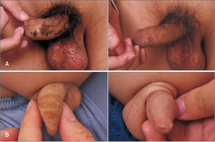
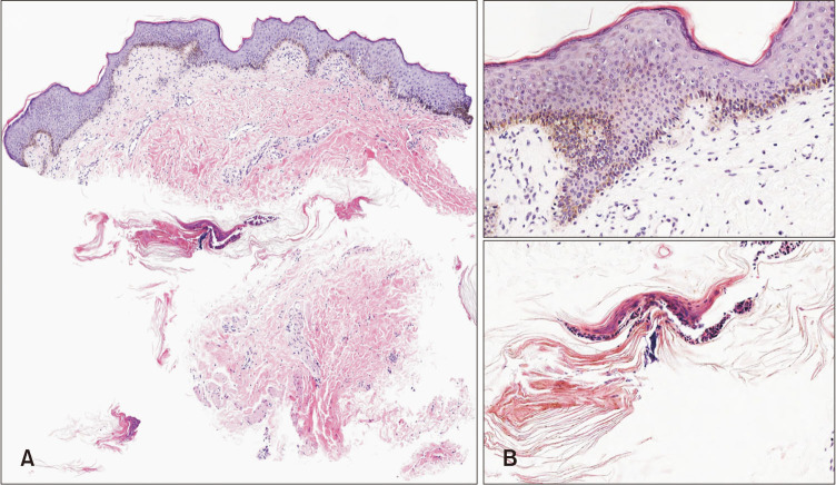
Case 1: A healthy 13-year-old boy presented with a three-month history of a brownish lesion on the skin of the penile shaft. He showered daily and had good hygiene habits. Physical examination revealed grouped, linear, brownish, flat-topped papuloplaques on the penile shaft (Fig. 1A). Punch biopsy revealed lamellar hyperkeratosis with keratin globules, mild acanthosis, papillomatosis, and increased melanin pigmentation along the basal layer (Fig. 2). Interestingly, the lesion almost completely disappeared when the area was gently scrubbed with an alcohol swab for conducting the biopsy (Fig. 1A), and there was no lesion recurrence.
Fig. 1. (A) Grouped, linear, brownish, flat-topped papuloplaques were seen on the penile shaft. (B) Most of the lesions disappeared after scrubbing with an alcohol swab.
Fig. 2. Findings from punch biopsy of the penile lesion. Mild acanthosis and papillomatosis (A: H&E, ×100), increased melanin pigmentation along the basal layer and lamellar hyperkeratosis with keratin globules (B: H&E, ×400) are noted.
Case 2: A healthy 8-year-old boy with normal hygiene habits presented to the clinic with a four-month history of a brownish lesion on the skin of the penis. His parents tried to remove the lesion by rubbing it with soap, but there was no improvement. Physical examination revealed brownish, flat-topped papuloplaques on the penile shaft (Fig. 1B). As the lesion could almost completely be scrubbed off with an alcohol swab (Fig. 1B), further biopsy was unnecessary. No recurrence was noted at the one-month follow-up.
TFFD is characterized by the occurrence of dirt-like plaques despite good hygiene1,2,4. It is important to distinguish TFFD from other papillomatous dermatoses such as dermatosis neglecta, acanthosis nigricans, and epidermal nevi because they are very similar in appearance to TFFD5. Especially, patients with dermatosis neglecta had poor hygiene, and the lesion was easily removed with detergents5. Dermoscopic findings of large, polygonal, plate-like, brown scales arranged in a mosaic pattern could be useful for diagnosing TFFD2. TFFD lesions characteristically disappear completely after wiping with 70% ethyl or isopropyl alcohol, although they are not removed when washed with soap. Alcohol swabbing is the gold standard treatment for TFFD. Stacked hyperkeratosis can also be removed with keratolytic agents such as 2% salicylic acid, lactic acid, and urea lotion2. Furthermore, fresh lemon juice can be used as an alternative to alcohol in patients with alcohol intolerance3.
Currently, the etiology of TFFD is considered to be hyperkeratosis due to impaired desquamation and delayed keratinization which leads to the progressive accumulation of sebum and dirt2. However, the dirt-like dermatosis could also be attributed to the creation of a favorable environment for bacterial overgrowth due to contact of the skin of the genital area with urine, which is slightly acidic2,3.
When treating patients with brownish hyperkeratotic lesions in whom TFFD is suspected, the patient’s hygiene habits should be checked and an alcohol swab test should be performed prior to biopsy, as this can help avoid unnecessary tests and treatments.
ACKNOWLEDGMENT
We received the patient’s consent form about publishing all photographic materials.
Footnotes
CONFLICTS OF INTEREST: The authors have nothing to disclose.
FUNDING SOURCE: None.
References
- 1.Berk DR. Terra firma-forme dermatosis: a retrospective review of 31 patients. Pediatr Dermatol. 2012;29:297–300. doi: 10.1111/j.1525-1470.2011.01422.x. [DOI] [PubMed] [Google Scholar]
- 2.Sechi A, Patrizi A, Savoia F, Leuzzi M, Guglielmo A, Neri I. Terra firma-forme dermatosis: a systematic review. Int J Dermatol. 2021;60:933–943. doi: 10.1111/ijd.15301. [DOI] [PubMed] [Google Scholar]
- 3.Chen HS, Li FG, Zhu Y, Wang T, Fan YM. Penile terra firma-forme dermatosis in a child: ultrastructural observation and topical ethacridine lactate and tacrolimus treatment. Eur J Dermatol. 2019;29:222–224. doi: 10.1684/ejd.2019.3505. [DOI] [PubMed] [Google Scholar]
- 4.Jung JW, Jung HJ, Jang YH, Shin DH, Kim SA. Terra firma-forme dermatosis: a report of four cases and review of the literature. Korean J Dermatol. 2018;56:276–279. [Google Scholar]
- 5.Moon J, Kim MW, Yoon HS, Cho S, Park HS. A case of terra firma-forme dermatosis: differentiation from other dirty-appearing diseases. Ann Dermatol. 2016;28:413–415. doi: 10.5021/ad.2016.28.3.413. [DOI] [PMC free article] [PubMed] [Google Scholar]


