

Abstract
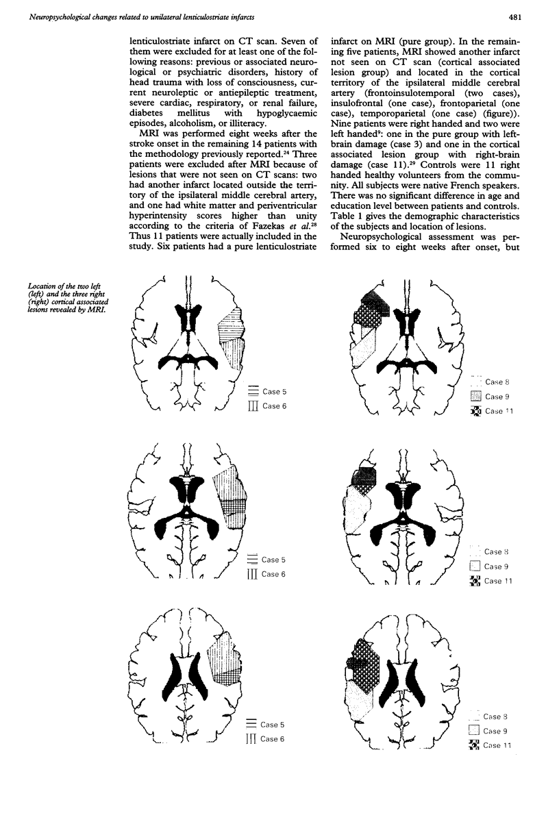
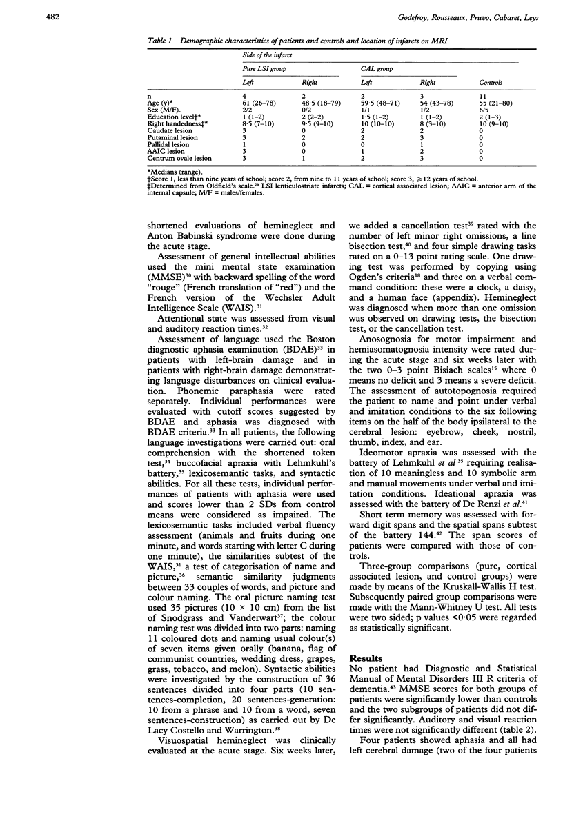
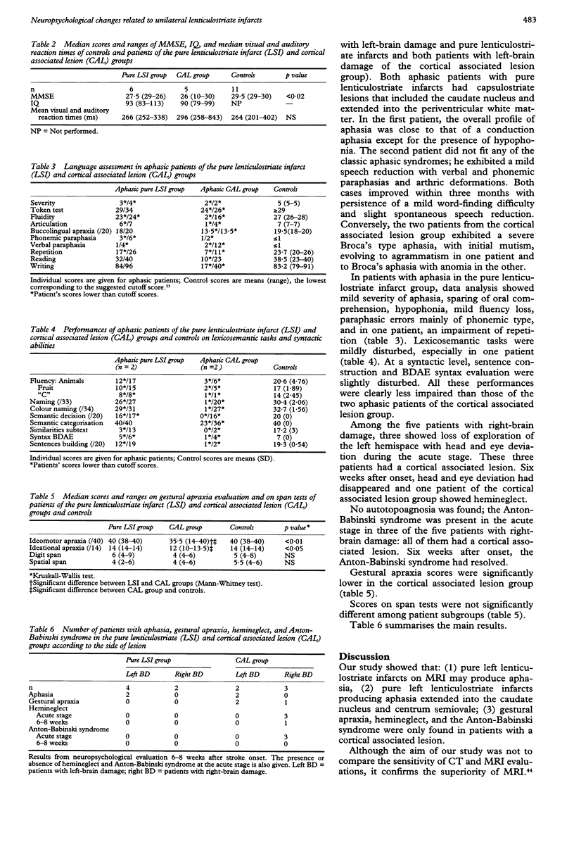
According to previous studies, focal capsulostriatal lesions may produce aphasia, hemineglect, gestural apraxia, frontal lobe dysfunction, and memory impairment. A few reports of capsulostriate infarcts secondary to involvement of lenticulostriate arteries have confirmed that aphasia and hemineglect may occur whereas gestural apraxia, anosognosia and frontal-lobe symptoms are rare. Most studies used CT scan assessment and did not exclude possible associated lesions. Neuropsychological changes in 11 patients with lenticulostriate infarcts diagnosed by CT scan were prospectively investigated. MRI in five of the 11 patients showed an associated cortical lesion not seen on CT scan. Patients with pure lenticulostriate infarcts on MRI may exhibit aphasia of mild severity whereas Broca's aphasia, hemineglect, gestural apraxia, and anosognosia were only seen in the subgroup with associated cortical lesions. Aphasia in patients with pure lenticulostriate infarcts was characterised by prominent expressive and lexicosemantic task impairments. The results strongly suggest that cortical involvement is critical to the extent and severity of neuropsychological changes in patients with lenticulostriate infarcts.
Full text
PDF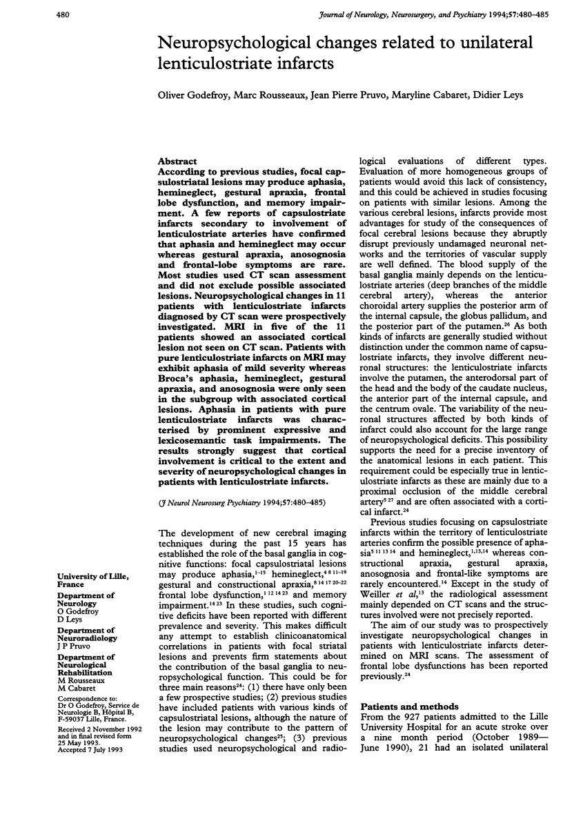
Images in this article
Selected References
These references are in PubMed. This may not be the complete list of references from this article.
- Adams H. P., Jr, Damasio H. C., Putman S. F., Damasio A. R. Middle cerebral artery occlusion as a cause of isolated subcortical infarction. Stroke. 1983 Nov-Dec;14(6):948–952. doi: 10.1161/01.str.14.6.948. [DOI] [PubMed] [Google Scholar]
- Agostoni E., Coletti A., Orlando G., Tredici G. Apraxia in deep cerebral lesions. J Neurol Neurosurg Psychiatry. 1983 Sep;46(9):804–808. doi: 10.1136/jnnp.46.9.804. [DOI] [PMC free article] [PubMed] [Google Scholar]
- Alexander G. E., DeLong M. R., Strick P. L. Parallel organization of functionally segregated circuits linking basal ganglia and cortex. Annu Rev Neurosci. 1986;9:357–381. doi: 10.1146/annurev.ne.09.030186.002041. [DOI] [PubMed] [Google Scholar]
- Alexander M. P., Naeser M. A., Palumbo C. L. Correlations of subcortical CT lesion sites and aphasia profiles. Brain. 1987 Aug;110(Pt 4):961–991. doi: 10.1093/brain/110.4.961. [DOI] [PubMed] [Google Scholar]
- Anderson S. W., Damasio H., Tranel D. Neuropsychological impairments associated with lesions caused by tumor or stroke. Arch Neurol. 1990 Apr;47(4):397–405. doi: 10.1001/archneur.1990.00530040039017. [DOI] [PubMed] [Google Scholar]
- Basso A., Della Sala S. Ideomotor apraxia arising from a purely deep lesion. J Neurol Neurosurg Psychiatry. 1986 Apr;49(4):458–458. doi: 10.1136/jnnp.49.4.458. [DOI] [PMC free article] [PubMed] [Google Scholar]
- Brunner R. J., Kornhuber H. H., Seemüller E., Suger G., Wallesch C. W. Basal ganglia participation in language pathology. Brain Lang. 1982 Jul;16(2):281–299. doi: 10.1016/0093-934x(82)90087-6. [DOI] [PubMed] [Google Scholar]
- Cambier J., Elghozi D., Strube E. Hémorragie de la tête du noyau caudé gauche. Désorganisation du discours et de l'expression graphique, perturbations des séries gestuelles. Rev Neurol (Paris) 1979;135(11):763–774. [PubMed] [Google Scholar]
- Caplan L. R., Schmahmann J. D., Kase C. S., Feldmann E., Baquis G., Greenberg J. P., Gorelick P. B., Helgason C., Hier D. B. Caudate infarcts. Arch Neurol. 1990 Feb;47(2):133–143. doi: 10.1001/archneur.1990.00530020029011. [DOI] [PubMed] [Google Scholar]
- Costello A. L., Warrington E. K. Dynamic aphasia: the selective impairment of verbal planning. Cortex. 1989 Mar;25(1):103–114. doi: 10.1016/s0010-9452(89)80010-3. [DOI] [PubMed] [Google Scholar]
- Damasio A. R., Damasio H., Chui H. C. Neglect following damage to frontal lobe or basal ganglia. Neuropsychologia. 1980;18(2):123–132. doi: 10.1016/0028-3932(80)90058-5. [DOI] [PubMed] [Google Scholar]
- Damasio A. R., Damasio H., Rizzo M., Varney N., Gersh F. Aphasia with nonhemorrhagic lesions in the basal ganglia and internal capsule. Arch Neurol. 1982 Jan;39(1):15–24. doi: 10.1001/archneur.1982.00510130017003. [DOI] [PubMed] [Google Scholar]
- De Renzi E., Faglioni P. Normative data and screening power of a shortened version of the Token Test. Cortex. 1978 Mar;14(1):41–49. doi: 10.1016/s0010-9452(78)80006-9. [DOI] [PubMed] [Google Scholar]
- DeWitt L. D., Grek A. J., Buonanno F. S., Levine D. N., Kistler J. P. MRI and the study of aphasia. Neurology. 1985 Jun;35(6):861–865. doi: 10.1212/wnl.35.6.861. [DOI] [PubMed] [Google Scholar]
- Decroix J. P., Graveleau P., Masson M., Cambier J. Infarction in the territory of the anterior choroidal artery. A clinical and computerized tomographic study of 16 cases. Brain. 1986 Dec;109(Pt 6):1071–1085. doi: 10.1093/brain/109.6.1071. [DOI] [PubMed] [Google Scholar]
- Donnan G. A., Bladin P. F., Berkovic S. F., Longley W. A., Saling M. M. The stroke syndrome of striatocapsular infarction. Brain. 1991 Feb;114(Pt 1A):51–70. [PubMed] [Google Scholar]
- Folstein M. F., Folstein S. E., McHugh P. R. "Mini-mental state". A practical method for grading the cognitive state of patients for the clinician. J Psychiatr Res. 1975 Nov;12(3):189–198. doi: 10.1016/0022-3956(75)90026-6. [DOI] [PubMed] [Google Scholar]
- Godefroy O., Rousseaux M., Leys D., Destée A., Scheltens P., Pruvo J. P. Frontal lobe dysfunction in unilateral lenticulostriate infarcts. Prominent role of cortical lesions. Arch Neurol. 1992 Dec;49(12):1285–1289. doi: 10.1001/archneur.1992.00530360087023. [DOI] [PubMed] [Google Scholar]
- Hier D. B., Mondlock J., Caplan L. R. Behavioral abnormalities after right hemisphere stroke. Neurology. 1983 Mar;33(3):337–344. doi: 10.1212/wnl.33.3.337. [DOI] [PubMed] [Google Scholar]
- Kertesz A., Ferro J. M. Lesion size and location in ideomotor apraxia. Brain. 1984 Sep;107(Pt 3):921–933. doi: 10.1093/brain/107.3.921. [DOI] [PubMed] [Google Scholar]
- Lehmkuhl G., Poeck K., Willmes K. Ideomotor apraxia and aphasia: an examination of types and manifestations of apraxic symptoms. Neuropsychologia. 1983;21(3):199–212. doi: 10.1016/0028-3932(83)90038-6. [DOI] [PubMed] [Google Scholar]
- Levine R. L., Lagreze H. L., Dobkin J. A., Turski P. A. Large subcortical hemispheric infarctions. Presentation and prognosis. Arch Neurol. 1988 Oct;45(10):1074–1077. doi: 10.1001/archneur.1988.00520340028006. [DOI] [PubMed] [Google Scholar]
- Mazaux J. M., Orgogozo J. M., Henry P., Loiseau P. Troubles du langage au cours des lésions thalamiques. Etude par le test de Goodglass et Kaplan. Rev Neurol (Paris) 1979 Jan;135(1):59–64. [PubMed] [Google Scholar]
- Mendez M. F., Adams N. L., Lewandowski K. S. Neurobehavioral changes associated with caudate lesions. Neurology. 1989 Mar;39(3):349–354. doi: 10.1212/wnl.39.3.349. [DOI] [PubMed] [Google Scholar]
- Mesulam M. M. Large-scale neurocognitive networks and distributed processing for attention, language, and memory. Ann Neurol. 1990 Nov;28(5):597–613. doi: 10.1002/ana.410280502. [DOI] [PubMed] [Google Scholar]
- Metter E. J., Riege W. H., Hanson W. R., Kuhl D. E., Phelps M. E., Squire L. R., Wasterlain C. G., Benson D. F. Comparison of metabolic rates, language, and memory in subcortical aphasias. Brain Lang. 1983 May;19(1):33–47. doi: 10.1016/0093-934x(83)90054-8. [DOI] [PubMed] [Google Scholar]
- Ogden J. A. Anterior-posterior interhemispheric differences in the loci of lesions producing visual hemineglect. Brain Cogn. 1985 Jan;4(1):59–75. doi: 10.1016/0278-2626(85)90054-5. [DOI] [PubMed] [Google Scholar]
- Oldfield R. C. The assessment and analysis of handedness: the Edinburgh inventory. Neuropsychologia. 1971 Mar;9(1):97–113. doi: 10.1016/0028-3932(71)90067-4. [DOI] [PubMed] [Google Scholar]
- Olsen T. S., Bruhn P., Oberg R. G. Cortical hypoperfusion as a possible cause of 'subcortical aphasia'. Brain. 1986 Jun;109(Pt 3):393–410. doi: 10.1093/brain/109.3.393. [DOI] [PubMed] [Google Scholar]
- Perani D., Vallar G., Cappa S., Messa C., Fazio F. Aphasia and neglect after subcortical stroke. A clinical/cerebral perfusion correlation study. Brain. 1987 Oct;110(Pt 5):1211–1229. doi: 10.1093/brain/110.5.1211. [DOI] [PubMed] [Google Scholar]
- Puel M., Demonet J. F., Cardebat D., Bonafé A., Gazounaud Y., Guiraud-Chaumeil B., Rascol A. Aphasies sous-corticales. Etude neurolinguistique avec scanner X de 25 cas. Rev Neurol (Paris) 1984;140(12):695–710. [PubMed] [Google Scholar]
- Ringelstein E. B., Zeumer H., Angelou D. The pathogenesis of strokes from internal carotid artery occlusion. Diagnostic and therapeutical implications. Stroke. 1983 Nov-Dec;14(6):867–875. doi: 10.1161/01.str.14.6.867. [DOI] [PubMed] [Google Scholar]
- Rousseaux M., Steinling M., Griffié G., Quint S., Cabaret M., Lesoin F., Mazingue M., Destée A. Corrélations de l'asphasie thalamique avec le débit sanguin cérébral. Rev Neurol (Paris) 1990;146(5):345–353. [PubMed] [Google Scholar]
- Schenkenberg T., Bradford D. C., Ajax E. T. Line bisection and unilateral visual neglect in patients with neurologic impairment. Neurology. 1980 May;30(5):509–517. doi: 10.1212/wnl.30.5.509. [DOI] [PubMed] [Google Scholar]
- Snodgrass J. G., Vanderwart M. A standardized set of 260 pictures: norms for name agreement, image agreement, familiarity, and visual complexity. J Exp Psychol Hum Learn. 1980 Mar;6(2):174–215. doi: 10.1037//0278-7393.6.2.174. [DOI] [PubMed] [Google Scholar]
- Vallar G., Perani D. The anatomy of unilateral neglect after right-hemisphere stroke lesions. A clinical/CT-scan correlation study in man. Neuropsychologia. 1986;24(5):609–622. doi: 10.1016/0028-3932(86)90001-1. [DOI] [PubMed] [Google Scholar]
- Wallesch C. W., Kornhuber H. H., Brunner R. J., Kunz T., Hollerbach B., Suger G. Lesions of the basal ganglia, thalamus, and deep white matter: differential effects on language functions. Brain Lang. 1983 Nov;20(2):286–304. doi: 10.1016/0093-934x(83)90046-9. [DOI] [PubMed] [Google Scholar]
- Weiller C., Ringelstein E. B., Reiche W., Thron A., Buell U. The large striatocapsular infarct. A clinical and pathophysiological entity. Arch Neurol. 1990 Oct;47(10):1085–1091. doi: 10.1001/archneur.1990.00530100051013. [DOI] [PubMed] [Google Scholar]
- Yeterian E. H., Van Hoesen G. W. Cortico-striate projections in the rhesus monkey: the organization of certain cortico-caudate connections. Brain Res. 1978 Jan 6;139(1):43–63. doi: 10.1016/0006-8993(78)90059-8. [DOI] [PubMed] [Google Scholar]