Abstract
Purpose
We have previously reported that a plasmid DNA vaccine encoding prostatic acid phosphatase (pTVG-HP) had greater clinical activity when given in combination with pembrolizumab to patients with metastatic, castration-resistant prostate cancer. The current trial was conducted to evaluate vaccination with PD-1 blockade, using nivolumab, in patients with early, recurrent (M0) prostate cancer.
Methods
Patients with M0 prostate cancer were treated with pTVG-HP (100 µg administered intradermally) and nivolumab (240 mg intravenous infusion) every 2 weeks for 3 months, and then every 4 weeks for 1 year of total treatment. Patients were then followed for an additional year off treatment. The primary objectives were safety and complete prostate-specific antigen (PSA) response (PSA<0.2 ng/mL).
Results
19 patients were enrolled. No patients met the primary endpoint of complete PSA response; however, 4/19 (21%) patients had a PSA decline >50%. Median PSA doubling times were 5.9 months pretreatment, 25.6 months on-treatment (p=0.001), and 9.0 months in the subsequent year off-treatment. The overall median radiographic progression-free survival was not reached. Grade 3 or 4 events included adrenal insufficiency, fatigue, lymphopenia, and increased amylase/lipase. 9/19 (47%) patients developed immune-related adverse effects (irAE). The development of irAE and increased CXCL9 were associated with increased PSA doubling time. Quantitative NaF PET/CT imaging showed the resolution of subclinical lesions along with the development of new lesions at each time point.
Conclusions
In this population, combining nivolumab with pTVG-HP vaccination was safe, and immunologically active, prolonged the time to disease progression, but did not eradicate disease. Quantitative imaging suggested that additional treatments targeting mechanisms of resistance may be required to eliminate tumors.
Trial registration number
Keywords: Nivolumab, Prostatic Neoplasms, Vaccination
WHAT IS ALREADY KNOWN ON THIS TOPIC
A DNA vaccine encoding prostatic acid phosphatase (pTVG-HP) has been previously evaluated in patients with prostate-specific antigen (PSA)-recurrent (stage M0) prostate cancer and did not prolong time to disease progression when used as a monotherapy. When this vaccine was given in combination with PD-1 blockade; however, PSA declines and objective tumor responses were observed in patients with metastatic castration-resistant prostate cancer.
WHAT THIS STUDY ADDS
In this trial, pTVG-HP was given in combination with nivolumab to patients with M0 prostate cancer, and, while no patients experienced a complete PSA response, most patients experienced stable disease as reflected by profound changes in PSA doubling time; few patients had progression over 1 year of treatment and 1 year of follow-up off treatment. Quantitative imaging demonstrated response in some lesions but the development of new lesions over time.
HOW THIS STUDY MIGHT AFFECT RESEARCH, PRACTICE OR POLICY
Future studies will evaluate whether targeting resistant lesions with radiation therapy, or by addition of agents that block additional mechanisms of resistance, can improve the therapeutic effect of this regimen.
Introduction
Surgery and/or radiation therapy can be curative for localized prostate cancer. Notwithstanding, approximately one-third of patients have a recurrence of prostate cancer after these local therapies.1 This is first detectable as a rise in serum prostate-specific antigen (PSA), which occurs with a median of 8 years prior to detection by standard CT or bone scintigraphy imaging, termed M0 prostate cancer.2 While androgen deprivation can be used in this stage of disease, many patients are keen to avoid the treatment-related adverse effects of androgen deprivation. There is currently much interest in using newer imaging methods, such as prostate-specific membrane antigen (PSMA) PET/CT, to identify metastases not visible by standard imaging, to enable directed radiotherapy to recurrent lesions. A randomized phase 2 trial evaluated patients in this population for stereotactic ablation of lesions detectable by PSMA PET, versus observation, and found that treatment could delay the progression of disease.3 However, therapies that might eradicate the disease at this early stage, or further delay disease progression and the need for androgen deprivation, are highly desirable.
We have previously evaluated a tumor vaccine, using a plasmid DNA encoding prostatic acid phosphatase (PAP, pTVG-HP, aka MVI-816) in patients with castration-sensitive and castration-resistant M0 prostate cancer. Phase I trials of dose or schedule demonstrated that this approach was safe, and suggested that treatment might slow the growth of prostate cancer as determined by changes in PSA doubling time.4 5 A randomized phase 2 trial, however, evaluating this vaccine as a monotherapy versus control, showed that while there was evidence of biological activity as measured by immune response to the target antigen and changes in individual lesions detected by NaF PET/CT imaging, there was no significant increase in time to metastatic progression.6 However, a trial using this vaccine in combination with pembrolizumab in patients with metastatic, castration-resistant prostate cancer demonstrated objective responses in terms of decreases in tumor volumes and serum PSA with 32% of patients remaining on trial beyond 6 months without progression.7 8 The combination of vaccine and PD-1 blockade was found to increase the infiltration of tumors by CD8+T cells and increase PD-L1 expression within tumor compared with vaccine alone.8 In addition, the combination was found to lead to increased serum cytokines and chemokines associated with T-cell activation (eg, IFNα, IFNγ, and IL-12) and T-cell recruitment (eg, CXCL9 and CXCL10).7
The findings from this latter trial suggested that the combination of antitumor vaccination and PD-1 blockade might have greater therapeutic efficacy if employed in an earlier stage of disease, prior to the development of large metastatic disease that might have multiple mechanisms of immune resistance. Consequently, the current trial was designed to evaluate pTVG-HP in combination with PD-1 blockade in patients with castration-sensitive M0 prostate cancer who had previously undergone prostatectomy. The primary endpoints were safety and PSA complete response rate (serum PSA<0.2 ng/mL). The rationale for restricting it to patients who underwent prostatectomy, rather than including patients who had undergone primary radiation therapy, was to be able to best interpret a complete PSA response since patients receiving primary radiation therapy can have a detectable PSA from remaining normal prostate tissue despite having eradication of metastatic prostate cancer. In addition, we reasoned that the native prostate could have other mechanisms of immune regulation that could potentially impair a systemic immune response to other prostate cancer cells. Patients with rising serum PSA were treated for up to 1 year with pTVG-HP and nivolumab, and then followed for 1-year off-treatment, to determine if treatment elicited prolonged disease control. The study was designed using a Simon-optimal two-stage design in which 21 patients were to be treated in the first stage, with expansion to 41 patients total if >1 PSA complete responses were observed.
Materials and methods
Study agent and regulatory information
pTVG-HP (a.k.a. MVI-816, Madison Vaccines, Madison, Wisconsin, USA) is a plasmid DNA encoding the full-length human PAP cDNA.9 The trial protocol was reviewed and approved by all local and federal regulatory entities. All patients gave written informed IRB-approved consent for participation.
Patient population
Eligible subjects were those with a histological diagnosis of prostate adenocarcinoma who had previously undergone prostatectomy and who then had subsequent castration-sensitive PSA recurrence (stage M0), and without evidence of metastatic disease by conventional imaging (CT scans and bone scintigraphy). Patients were initially required to have a PSA doubling time of <12 months. Patients were not excluded for having received prior androgen deprivation administered with radiation therapy or at the time of prostatectomy, but androgen deprivation treatment for more than 24 months was prohibited, and patients were required to have a normal serum testosterone level (>50 ng/dL) at screening. CT scans and bone scintigraphy were used to determine eligibility and progression; evidence of metastatic disease by PET imaging was not used for eligibility or treatment response assessment. Patients were required to have an ECOG performance score of <2, and normal bone marrow, liver, and renal function.
Study design and procedures
Enrolled patients were treated with 100 µg pTVG-HP (MVI-816) administered intradermally every 2 weeks for six vaccinations, and then every 4 weeks for nine vaccinations, to complete 1 year of treatment. Nivolumab (240 mg administered intravenously) was given on the same day after each vaccination (figure 1). Patients were evaluated every 4 weeks with serum PSA, serum PAP, and safety labs. These labs included CBC, creatinine, electrolytes, glucose, bilirubin, ALT, AST, alkaline phosphatase, amylase, LDH, and TSH. All toxicities were graded according to the NCI Common Terminology Criteria Grading System, V.5. CT scans of the abdomen and pelvis and bone scans were performed every 6 months or as clinically indicated. No adjuvant was given with the vaccine for the first two immunizations. For each individual subject, if the PSA at week 4 was higher than at baseline, 200 µg granulocyte-macrophage colony-stimulating factor (GM-CSF, Sargramostim, Berlex Laboratories, Montville, New Jersey, USA) was coadministered intradermally with the vaccine at that visit and for all subsequent visits. There were no dose reductions permitted, and treatment was held if a patient developed >grade 3 toxicity. If the toxicity was attributed to nivolumab and not a vaccine, the vaccine schedule was continued. Treatment with nivolumab was discontinued for grade 4 toxicity or recurrent grade 3 toxicity. Patients came off study at the time of radiographic progression (by CT or bone scan), if there was undue toxicity, or at the discretion of the patient or physician that other therapies were warranted. Patients who completed 1 year of treatment were monitored at quarterly intervals, with collection of serum PSA, for an additional year.
Figure 1.
Schema. Shown is the trial schema. pTVG-HP, plasmid DNA encoding prostatic acid phosphatase. PET/CT, positron emission tomography / computed tomography scan.
The protocol was modified within the first year to include all patients with evidence of PSA rise, not just those with a doubling time <12 months, to speed accrual. In addition, the protocol was modified to include exploratory sodium fluoride positron emission tomography / computed tomography (NaF PET/CT) scans at baseline, 3 months and 6 months after the first 10 patients were accrued. This was to permit exploratory monitoring of bone-metastatic disease not detectable by bone scan, using TRAQinform IQ (AIQ Solutions, Madison, Wisconsin, USA).6 The use of this methodology to identify small bone lesion, and changes and response characteristics over time, have been previously reported.10 11 Specifically, test–retest limits of agreement for the relative change in SUVtotal were used to classify lesion-level response as partially responding (PRL), stable disease (SL), and progressing disease (PL).10 The primary study endpoint was complete PSA response (PSA<0.2 ng/mL). Secondary objectives were to evaluate the 2-year metastasis-free survival rate (MFS), median radiographic progression-free survival (PFS), changes in PSA doubling time, PSA response (<50% of baseline), and to determine whether GM-CSF was required as an adjuvant. Exploratory objectives included immunological evaluations and quantitative bone imaging assessments.
The protocol used a Simon-optimal two-stage design, with the plan to enroll 21 patients for the first stage and expand the trial by an additional 20 patients if >1 patient experienced a complete PSA response. With this design, the trial had 90% power to detect an increase in the PSA complete rate from 5% (null hypothesis) to 20% (alternative hypothesis) at the one-sided 0.05 significance level. If the true PSA complete response rate was only 5%, the study would be terminated after the first stage with 66% probability. Due to slow accrual and the unlikelihood of reaching this endpoint, the trial was closed early to further accrual after 19 patients were enrolled.
Clinical response evaluation
Serum PSA and PAP values were collected every 4 weeks during the first year of treatment and then every 3 months during the second year off treatment. PSA rise was not used to define progression. A complete PSA response was defined as a PSA<0.2 ng/mL with a confirmatory PSA<0.2 ng/mL at least 4 weeks later. CT scans of the abdomen/pelvis and bone scans were obtained every 6 months. The appearance of lesions consistent with metastatic disease was used to define radiographic progression. Lesions detected by 18F-NaF-PET/CT or other advanced imaging (such as choline PET/CT, fluciclovine, or PSMA PET scans) were not used to define metastatic disease or disease progression.
Immunological response evaluation
Measures of antigen-specific immune response were performed by IFNγ and granzyme B fluorescent ELISPOT with fresh (not cryopreserved) peripheral blood mononuclear cells (PBMC) as previously described.6 7 Due to the COVID-19 pandemic, 11 blood samples could not be evaluated in real time and were assessed later with cryopreserved samples. Immune response resulting from immunization was defined as a PAP-specific response detectable post-treatment that was statistically significant (compared with media-only control by t-test), at least threefold higher than the pretreatment value, and with a frequency >1:100,000 PBMC, as we have previously reported.5 7 8 12
Plasma samples were obtained at baseline and various post-treatment time points and were evaluated by Luminex multiplex analysis for 35 cytokine and chemokine analytes, according to the manufacturer’s instructions (Cytokine 35-plex human panel, ThermoFisher), and as previously reported.7 These analytes included EGF, Eotaxin, FGF-basic, G-CSF, GM-CSF, HGF, IFN-alpha, IFN-gamma, IL-1 beta, IL-1 alpha, IL-1RA, IL-2, IL-2R, IL-3, IL-4, IL-5, IL-6, IL-7, IL-8, IL-9, IL-10, IL-12 (p40/p70) IL-13, IL-15, IL-17A, IL-17F, IL-22, IP-10, MCP-1, MIG, MIP-1 alpha, MIP-1 beta, RANTES, TNF-alpha, and VEGF.
Statistical analysis
The primary efficacy endpoint was a complete PSA response. Secondary efficacy endpoints included the 2-year MFS, radiographic PFS, and changes in PSA doubling time. The complete PSA response and 2-year MFS rates were reported along with the corresponding two-sided 95% CIs which were constructed using the Wilson score method. The PSA doubling time was calculated for each patient as the logarithm of 2 divided by the slope of a linear regression of the log(PSA) over time (months). Time to radiographic progression was defined as the number of months from study enrolment to the date of the recorded progression event. Time to PSA and time to PAP progression were defined as the number of months from treatment start date to the earliest time at which a 25% increase over nadir was observed, with a minimum increase of 2 ng/mL, and confirmed by a subsequent measurement, or a minimum of 12 weeks for patients without a decline in value. For patients without a progression event, time to progression was censored at the last date of disease assessment. Radiographic PFS was analyzed using the Kaplan-Meier method. The log-rank test was used to compare PFS between patients with less than a twofold change of PSA doubling time over the treatment period to patients with greater than a twofold change. Comparisons of PSA doubling time from the pretreatment to the on-treatment and post-treatment assessments were conducted using the nonparametric Wilcoxon rank sum test. Baseline characteristics were summarized in terms of medians and ranges or frequencies and percentages. The frequencies of adverse events with an attribution of at least possibly treatment-related during the treatment period were summarized by type and severity in tabular format. Changes in cytokine and chemokine levels from the pre-treatment to the week 4 and week 12 on-treatment assessments were conducted using a paired t-test. All reported p values are two sided and a p<0.05 was used to define statistical significance. Statistical analyses were conducted by using SAS software (SAS Institute), V.9.4.
Results
Trial conduct and adverse effects
Between October 2018 and January 2022, 19 patients with PSA-recurrent castration-sensitive prostate cancer, without evidence of metastatic disease by conventional imaging (stage M0), were accrued at a single academic center (University of Wisconsin Carbone Cancer Center). As shown in table 1, while patients had low initial serum PSA levels, this was a high-risk population with a median PSA doubling time of 5.9 months.
Table 1.
Demographics
| Number | 19 |
| Age (years) | |
| Median | 68 |
| Range | 63–75 |
| Race/ethnicity | |
| White (non-Hispanic) | 18 (95%) |
| American Indian or Alaska Native | 1 (5%) |
| Gleason Score at Prostatectomy | |
| 6 | 1 (5%) |
| 7 | 7 (37%) |
| 8 | 7 (37%) |
| 9 | 4 (21%) |
| T stage | |
| T1 | 1 (5%) |
| T2 | 7 (37%) |
| T3 | 10 (53%) |
| TX | 1 (5%) |
| N stage | |
| N0 | 13 (68%) |
| N1 | 2 (11%) |
| NX | 4 (21%) |
| Prior therapy | |
| Prostatectomy | 19 (100%) |
| Adjuvant/salvage radiation therapy | 16 (84%) |
| Androgen deprivation therapy (<24 months) | 6 (32%) |
| Baseline PSA | |
| Median (ng/mL) | 3.1 |
| Range (ng/mL) | 1.96–10.11 |
| Baseline PSA doubling time | |
| Median (months) | 5.9 |
| 0–3 months | 5 (26%) |
| 3–6 months | 5 (26%) |
| 6–12 months | 6 (32%) |
| >12 months | 3 (16%) |
| Range (months) | 1.1–45.3 |
PSA, prostate-specific antigen.
Enrolled patients were treated with pTVG-HP (MVI-816) vaccine and nivolumab every 2 weeks for six treatments, and then every 4 weeks for nine treatments, to complete 1 year total. Patients were then followed for an additional year off treatment (figure 1). GM-CSF adjuvant was added to each subsequent vaccine treatment beginning at week 4 if the serum PSA was not lower than baseline. Overall, 5/19 (26%) patients received no GM-CSF adjuvant due to PSA decline detectable at week 4. As shown in table 2, one patient developed a grade 4 event of increased lipase (and mildly symptomatic grade 2 pancreatitis). Two other patients had amylase and/or lipase elevations without symptoms. Grade 3 events included single events of adrenal insufficiency, fatigue, non-cardiac chest pain, lymphopenia, and increased amylase and lipase. Common grade 1 or 2 events included hyperthyroidism or hypothyroidism, fatigue, chills, back pain, and injection site reactions. Two patients had treatment held or delayed due to adverse events, but overall 17/19 (89%) completed 1 year of treatment. One patient discontinued due to disease progression at month 6 and one due to the development of adrenal insufficiency. Overall, 9/19 (47%) experienced an immune-related adverse reaction of any grade, including adrenal insufficiency, thyroid dysfunction, pancreatitis, and rash.
Table 2.
Adverse events
| Grade 1 | Grade 2 | Grade 3 | Grade 4 | |
| Cardiac | ||||
| Palpitations | 1 (5%) | |||
| Endocrine | ||||
| Adrenal insufficiency | 1 (5%) | |||
| Hyper/hypothyroidism | 1 (5%) | 4 (21%) | ||
| Gastrointestinal | ||||
| Abdominal pain | 1 (5%) | |||
| Bloating | 1 (5%) | |||
| Diarrhea | 3 (16%) | 1 (5%) | ||
| Dry mouth | 1 (5%) | |||
| Microcolitis | 1 (5%) | |||
| Pancreatitis | 1 (5%) | |||
| General/constitutional | ||||
| Chills | 5 (25%) | |||
| Fatigue | 8 (42%) | 1 (5%) | ||
| Fever | 2 (11%) | |||
| Infusion-related reaction | 1 (5%) | |||
| Injection site reaction | 4 (21%) | |||
| Non-cardiac chest pain | 1 (5%) | 1 (5%) | ||
| Lab Investigations | ||||
| Creatinine increased | 1 (5%) | |||
| Lipase increased | 1 (5%) | 1 (5%) | ||
| Lymphocyte count decreased | 1 (5%) | |||
| Amylase increased | 2 (11%) | 1 (5%) | ||
| Metabolism and nutrition | ||||
| Anorexia | 2 (11%) | |||
| Hypoalbuminemia | 1 (5%) | |||
| Musculoskeletal | ||||
| Arthralgia | 1 (5%) | |||
| Back pain | 1 (5%) | 2 (11%) | ||
| Generalized muscle weakness | 1 (5%) | |||
| Pain in extremity | 1 (5%) | |||
| Nervous system | ||||
| Dysgeusia | 1 (5%) | |||
| Headache | 1 (5%) | |||
| Psychiatric | ||||
| Insomnia | 1 (5%) | |||
| Renal system | ||||
| Hematuria | 1 (5%) | |||
| Respiratory system | ||||
| Cough | 1 (5%) | |||
| Dyspnea | 1 (5%) | |||
| Sore throat | 1 (5%) | |||
| Skin | ||||
| Pruritus | 2 (11%) | |||
| Rash | 2 (11%) | 2 (11%) | ||
| Urticaria | 1 (5%) |
Shown are all adverse events that were deemed to be at least possibly related to treatment. The numbers represent the number of patients experiencing a particular event at any point during the treatment period, with the highest grade reported for any single individual.
Immunological response
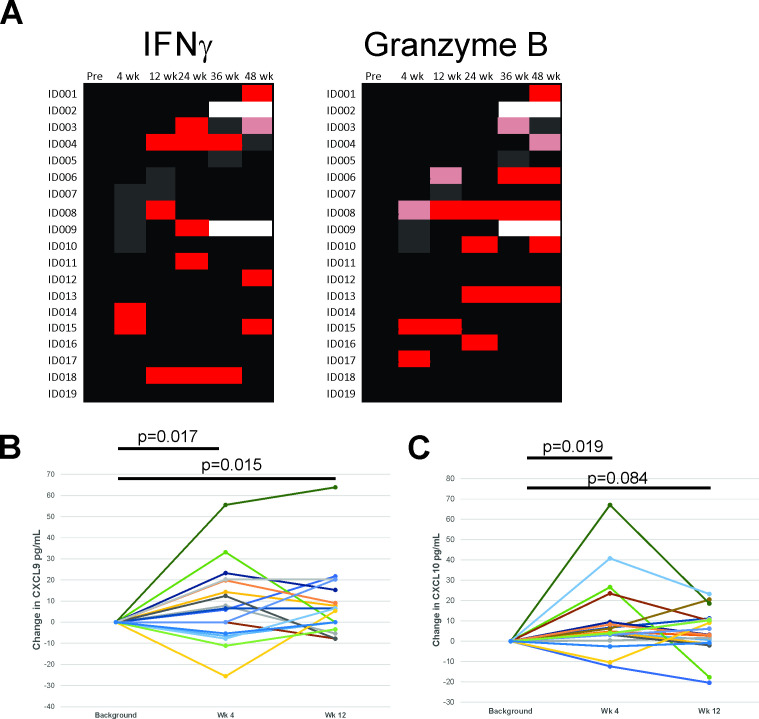
ELISPOT was used to evaluate T-cell immunity to the PAP vaccine target antigen. As shown in figure 2A, antigen-specific IFNγ-secreting and/or granzyme B-secreting T cells were detected in 15/19 (79%) of patients and detected at least twice following treatment in 8/19 (42%) of patients. Sera were evaluated for changes of 35 separate cytokines and chemokines from pretreatment to weeks 4 and 12. As shown in figure 2B,C, CXCL9 and CXCL10 were significantly increased in the serum after treatment. No other cytokines or chemokines were significantly increased or decreased after treatment.
Figure 2.
Immune evaluations. (A) Peripheral blood mononuclear cells (PBMC) were obtained from all patients and evaluated for PAP-specific T-cell immunity by fluorescent ELISPOT. A positive immune response (red, pink) was defined as a statistically significant response to the PAP peptide pool compared with media alone and that was at least threefold greater than pretreatment and >1:1 00 000 cells. Black/gray=no significant response, Blank=no data. Red/black=assay performed with fresh cells in real time. Pink/gray=assay performed with cryopreserved cells. Shown are responses for IFNγ release (left) or granzyme B release (right). Sera obtained pretreatment and at weeks 4 and 12 of treatment were evaluated for changes in cytokines and chemokines. Shown are the changes from pretreatment of CXCL9 (B) and CXCL10 (C). Statistical comparisons are by paired t-test, and p<0.05 was considered statistically significant.
Clinical effects
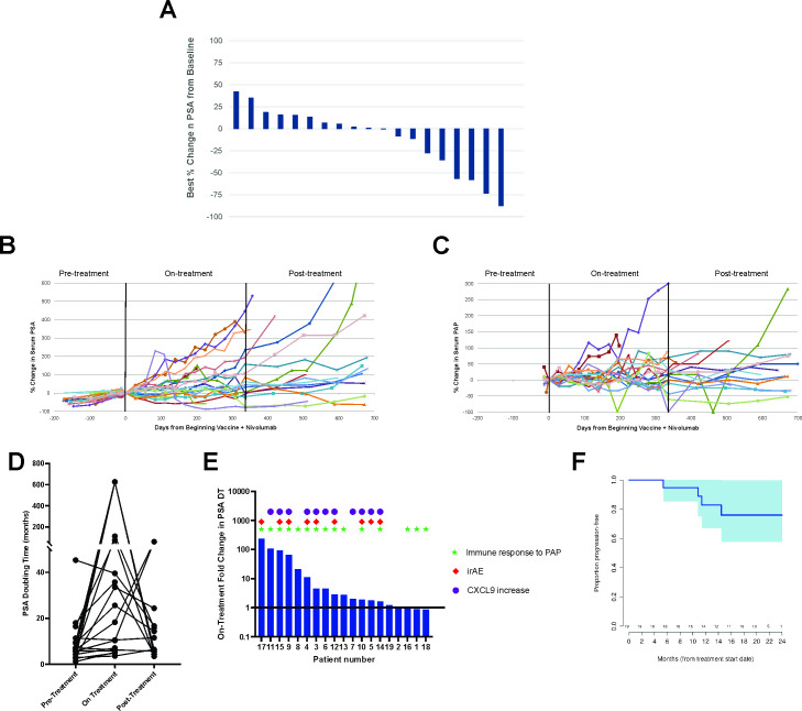
Serum PSA and PAP values were obtained monthly during the first year of treatment and at quarterly intervals in the second year. No patient (0%, 95% CI 0% to 17%) experienced a complete PSA response, the primary efficacy endpoint of the trial. 9/19 (47%) patients experienced any decrease in serum PSA from baseline, and 4/19 (21%) had a PSA decline >50%, as shown in figure 3A. The median time to PSA progression was 6.4 months (95% CI 2.8 to 11.0 months), and using the same metrics, the median time to PAP progression was 22.5 months (95% CI not estimated due to censoring, online supplemental figure 1). PSA and PAP levels remained stable over the period of treatment for most individuals. However, while two patients had stable or decreasing PSA values after completing treatment, PSA values tended to rise in the year following treatment (figure 3B,C). This is more clearly illustrated by the PSA doubling time over these pretreatment, on-treatment, and post-treatment time periods. The PSA doubling time increased from a median of 5.9 months pretreatment to 25.6 months on treatment (p=0.001). Median PSA doubling time in the post-treatment period was 9.0 months (figure 3D). Of three patients with PSA DT>12 months prior to treatment, two experienced at least a twofold increase in PSA DT on treatment. The development of post-treatment immune response to the PAP target antigen detected by ELISPOT was not highly associated with an increase in PSA doubling time. However, the development of immune-related adverse events, and increase in CXCL9 in the serum at week 4, were both associated with the on-treatment increase in PSA doubling time (figure 3E).
Figure 3.
Clinical evaluations. Serum PSA and PAP values were collected from all individuals prior to treatment (for PSA), over the course of treatment, and in the 1-year period off treatment. (A) Shown are the best percentage changes in serum PSA from day 1. Also shown are the individual plots over time of % change in serum PSA (B) or serum PAP (C) from the value obtained on day 1. (D) PSA doubling times for each patient were calculated from the values available up to 1 year prior to treatment, during the 1 year on treatment, and for the 1 year off treatment. (E) Fold change in PSA doubling time from pretreatment to on-treatment is displayed for each patient with symbols representing which patients developed an ELISPOT response at any post-treatment time point to PAP, which patients developed an immune-related adverse event (irAE), and which patients demonstrated an increase in serum CXCL9 at week 4 of treatment. (F) Shown is the time to radiographic progression. PAP, prostatic acid phosphatase; PSP, prostate-specific antigen.
jitc-2023-008067supp001.pdf (149.8KB, pdf)
CT scans and bone scans were performed at 6-month intervals or as clinically indicated. The median time to radiographic progression was not reached (figure 3F). 15/19 (79%, 95% CI 57% to 91%) remained metastasis-free at 2 years. When evaluating change in PSA doubling time for individual patients, there was no significant difference detected when comparing PFS between patients with less than a doubling of the PSA doubling time to those with at least twofold increase in PSA doubling time over the year of treatment (p=0.23).
Quantitative imaging
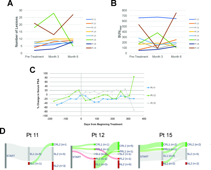
NaF PET/CT scans were acquired from 8 patients pretreatment and at months 3 and 6. Images were analyzed using TRAQinform IQ to permit automated identification and quantification of change in individual bone lesions detectable by NaF PET/CT that were not detectable by standard bone scintigraphy.10 11 Figure 4A shows the total number of lesions in bone detected at each time point for each individual. Figure 4B shows the total functional burden (sum of SUVtotal in all detected lesions) at each time point per individual. In general, there were not consistent trends in the number of lesions or SUVtotal of all detectable lesions for each individual. Three of the eight individuals (patients 11, 12 and 15) had relatively stable serum PSA values over the year of treatment (figure 4C). Sankey plots (figure 4D) of these three subjects depict how lesions changed (intralesional response classification based on change in SUVtotal) at each time point, as well as how new lesions appeared and changed with ongoing treatment. All patients were found to develop new lesions by month 6. These findings suggested that treatment elicited antitumor responses at individual tumor sites, but that new lesions continued to emerge over time.
Figure 4.
Quantitative bone imaging analysis. NaF PET/CT scans were obtained within 2 weeks prior to study start (baseline), and at months 3 and 6 of treatment for eight subjects. Images were analyzed by AIQ Solutions using TRAQinform IQ software to automatically quantify the number of lesions for each patient at each time point (A), and the SUVtotal of all lesions per patient at each time point (B). (C) Percentage change from baseline of serum PSA values for three of the eight subjects. (D) Sankey plots depict change in SUVtotal for each individual lesion over time. CRL, complete response of lesion; NL, new lesion (response classifications based on deviation beyond established limits of agreement); PL, progressive lesion; PRL, partial response of lesion; SL, stable lesion.
Discussion
We report the results of a clinical trial in which patients with non-castrate M0 prostate cancer were treated with a DNA vaccine encoding PAP (pTVG-HP) and nivolumab. The primary endpoint was to determine if treatment could eradicate the disease, leading to a complete PSA response. The trial was halted early after accrual of 19 patients due to slow accrual and because the primary endpoint was unlikely to be met even with the enrollment of two additional patients as initially planned. Treatment, however, did lead to antitumor effects as evidenced by quantitative imaging and a prolonged PSA doubling time on treatment. Treatment was also associated with the development of immune response to the target antigen and increases in serum CXCL9 and CXCL10 chemokines. Taken together, findings from this trial demonstrate that (1) treatment of patients with M0 prostate cancer with pTVG-HP and nivolumab elicited similar types of immune responses and immune-related adverse events as identified in patients with mCRPC, and this treatment was associated with an improved clinical course; (2) GM-CSF is not required as a vaccine adjuvant; (3) pTVG-HP and PD-1 blockade can elicit treatment responses, but not a protective durable immunity; and (4) treatment of patients in this population will likely require additional treatments, such as ablative radiation to resistant lesions, to eliminate prostate tumors and avoid the need for subsequent androgen deprivation.
In this trial, we observed that the majority of patients developed post-treatment immune response to the PAP target antigen. This was not significantly associated with prolonged PSA doubling time. Patients also developed increases in serum CXCL9 and CXCL10 that were detectable at 4 weeks and 12 weeks after the start of treatment. Increases in CXCL9 at 4 weeks were significantly associated with an increase in PSA doubling time. While the number of patients overall was low in this trial, we observed similar trends in patients with mCRPC treated with pTVG-HP and pembrolizumab.7 In that trial, we also observed a non-significant trend for increased time to progression in patients who developed immune response to the PAP target antigen and in those who developed increases in serum IFNγ, CXCL9, and CXCL10.7 Collectively, these findings suggest a similar immune-mediated treatment effect following vaccination and PD-1 blockade, and identify rational on-treatment biomarkers that could be employed for future larger clinical trials. In fact, these other measures may serve as better biomarkers of a clinical effect than an immune response to the vaccine antigen.
9/19 (47%) patients in the current trial developed immune-related adverse effects. This is similar to the frequency (28/66, 42%) of individuals with mCRPC who developed immune-related adverse effects following treatment with pTVG-HP and pembrolizumab.7 The same types of events were observed, notably adrenal insufficiency, thyroid dysfunction, pancreatitis, and rash, and there were no unusual new sites of toxicity. The most common immune-related adverse effect observed was thyroid dysfunction (hyperthyroidism and/or hypothyroidism), occurring in 5/19 (26%) of patients on this trial and 16/66 (24%) of patients in the mCRPC trial. Of note, this frequency is higher than what was observed with pembrolizumab alone used in the treatment of patients with mCRPC, in which 13/258 (5%) patients developed any grade of thyroid dysfunction.13 Similar rates of thyroid dysfunction have been reported in 3/35 (5.7%) patients with mCRPC treated with single-agent atezolizumab,14 and in 26/374 (7.0%) patients with mCRPC treated with atezolizumab and enzalutamide.15 While it is unclear why the thyroid would be a specific site of autoimmune toxicity, and the thyroid does not express the PAP target antigen, our findings suggest that the combination of PD-1 blockade with pTVG-HP may increase the risk of thyroid dysfunction. In any case, this was an easily managed toxicity.
A secondary endpoint of the current trial was to evaluate the need for GM-CSF as a vaccine adjuvant. In a phase I trial using a DNA vaccine encoding a different target antigen, we had identified that the development of T cell immune response to the target antigen was affected by the vaccine schedule but not by the inclusion of GM-CSF as an adjuvant, permitting us to conclude that GM-CSF was not necessary as an adjuvant for that vaccine.12 In the current trial, GM-CSF was added as an adjuvant only if a PSA decline was not observed at week 4. Five patients (26%) had PSA declines without any use of GM-CSF. Of the 14 patients in whom GM-CSF was added, three (21%) developed any subsequent PSA decline. From this, we conclude that while GM-CSF may have provided a modest improvement for a few patients, it was not absolutely required as an adjuvant.
In the current trial, we observed that treatment elicited changes in serum PSA levels, with most patients having a prolonged PSA doubling time over the year of treatment, and 3/19 (16%) patients having a PSA decline over that time period. The median time to radiographic progression was longer than expected, based on this high-risk cohort (median pretreatment PSA doubling time of 5.9 months) and based on our prior findings in a randomized phase 2 trial in this population.6 16–18 However, no patient experienced a complete PSA response, and in the 1-year period of follow-up off treatment, the majority of patients had a rise in serum PSA. By quantitative imaging, we observed that lesions detectable at baseline frequently resolved with treatment, however, some lesions persisted. New lesions that appeared at month 3 typically resolved at month 6, suggesting these might be due to flare phenomena. However, new lesions appeared again at 6 months. Collectively, these suggest that treatment elicited responses in individual lesions, but new lesions continued to appear and some stable lesions persisted. This could explain the observation of stable disease, suggesting that there is heterogeneity of response with some individual lesions responding and resolving with treatment, and perhaps persistent lesions giving rise to new metastatic sites. The evaluation of individual responding and non-responding lesions is an area of importance for future exploration.
The clinical findings from this trial demonstrate that there is a treatment effect with pTVG-HP and nivolumab, but suggest that treatment should either be continued indefinitely until disease progression or that other treatments should be added to this treatment combination to target mechanisms of resistance. The findings from quantitative imaging that some lesions persist and that new lesions develop over time, however, suggest that the latter approach would be preferable. At present, the majority of patients with non-castrate M0 prostate cancer receive advanced imaging by PSMA PET/CT and are then considered for targeted radiation therapy. In fact, we observed that the adoption of this approach slowed this trial’s accrual. A randomized trial conducted in patients with castration-sensitive M0 prostate cancer demonstrated that targeted radiation therapy of small metastatic lesions can delay the development of further metastatic disease, but did not suggest that disease was eliminated.3 Taken together, we propose that these approaches might be more optimally combined. That is, by using repetitive, quantitative imaging of individual lesions by PSMA PET/CT following treatment with pTVG-HP and nivolumab, it may be possible to identify resistant lesions amenable to targeted radiation therapy with the goal of eradicating recurrent prostate cancer. This is an approach we aim to evaluate in a future clinical trial.
Acknowledgments
We thank the referring physicians and participating patients.
Footnotes
Twitter: @HamidEmamekhoo
Contributors: DGM oversaw the experimental design and immunological analysis, and wrote the manuscript. He is responsible for the overall content as the guarantor. JE was the study biostatistician, designed the trial, analyzed all results, and edited the manuscript. TPT, EW and LJ conducted the laboratory analyses. HE and GL oversaw the conduct of the clinical trial.
Funding: Grant support was provided by NIH (P30 CA014520). Nivolumab was provided by Bristol Myers Squibb. pTVG-HP was provided by Madison Vaccines. The TRAQinform IQ bone imaging analysis was conducted by AIQ Solutions, and funded by Madison Vaccines.
Competing interests: DGM has ownership interest, has received research support, and serves as consultant to Madison Vaccines, which has licensed material described in this manuscript and supported this trial. None of the other authors have relevant potential conflicts of interest.
Provenance and peer review: Not commissioned; externally peer reviewed.
Supplemental material: This content has been supplied by the author(s). It has not been vetted by BMJ Publishing Group Limited (BMJ) and may not have been peer-reviewed. Any opinions or recommendations discussed are solely those of the author(s) and are not endorsed by BMJ. BMJ disclaims all liability and responsibility arising from any reliance placed on the content. Where the content includes any translated material, BMJ does not warrant the accuracy and reliability of the translations (including but not limited to local regulations, clinical guidelines, terminology, drug names and drug dosages), and is not responsible for any error and/or omissions arising from translation and adaptation or otherwise.
Data availability statement
Data are available on reasonable request. The data generated and/or analyzed during this study are available from the corresponding author on reasonable request.
Ethics statements
Patient consent for publication
Not applicable.
Ethics approval
This study involves human participants and was approved by Institutional Review Board of the University of WisconsinProtocol 2018-0418. Participants gave informed consent to participate in the study before taking part.
References
- 1. Oefelein MG, Smith ND, Grayhack JT, et al. Long-term results of radical retropubic prostatectomy in men with high grade carcinoma of the prostate. J Urol 1997;158:1460–5. [PubMed] [Google Scholar]
- 2. Pound CR, Partin AW, Eisenberger MA, et al. Natural history of progression after PSA elevation following radical prostatectomy. JAMA 1999;281:1591–7. 10.1001/jama.281.17.1591 [DOI] [PubMed] [Google Scholar]
- 3. Phillips R, Shi WY, Deek M, et al. Outcomes of observation vs stereotactic ablative radiation for oligometastatic prostate cancer: the ORIOLE phase 2 randomized clinical trial. JAMA Oncol 2020;6:650–9. 10.1001/jamaoncol.2020.0147 [DOI] [PMC free article] [PubMed] [Google Scholar]
- 4. McNeel DG, Dunphy EJ, Davies JG, et al. Safety and immunological efficacy of a DNA vaccine encoding prostatic acid phosphatase in patients with stage D0 prostate cancer. J Clin Oncol 2009;27:4047–54. 10.1200/JCO.2008.19.9968 [DOI] [PMC free article] [PubMed] [Google Scholar]
- 5. McNeel DG, Becker JT, Eickhoff JC, et al. Real-time immune monitoring to guide plasmid DNA vaccination schedule targeting prostatic acid phosphatase in patients with castration-resistant prostate cancer. Clin Cancer Res 2014;20:3692–704. 10.1158/1078-0432.CCR-14-0169 [DOI] [PMC free article] [PubMed] [Google Scholar]
- 6. McNeel DG, Eickhoff JC, Johnson LE, et al. Phase II trial of a DNA vaccine encoding prostatic acid phosphatase (pTVG-HP [MVI-816]) in patients with progressive, nonmetastatic, castration-sensitive prostate cancer. J Clin Oncol 2019;37:3507–17. 10.1200/JCO.19.01701 [DOI] [PMC free article] [PubMed] [Google Scholar]
- 7. McNeel DG, Eickhoff JC, Wargowski E, et al. Phase 2 trial of T-cell activation using MVI-816 and pembrolizumab in patients with metastatic, castration-resistant prostate cancer (mCRPC). J Immunother Cancer 2022;10:e004198. 10.1136/jitc-2021-004198 [DOI] [PMC free article] [PubMed] [Google Scholar]
- 8. McNeel DG, Eickhoff JC, Wargowski E, et al. Concurrent, but not sequential, PD-1 blockade with a DNA vaccine elicits anti-tumor responses in patients with metastatic, castration-resistant prostate cancer. Oncotarget 2018;9:25586–96. 10.18632/oncotarget.25387 [DOI] [PMC free article] [PubMed] [Google Scholar]
- 9. Johnson LE, Frye TP, Arnot AR, et al. Safety and immunological efficacy of a prostate cancer plasmid DNA vaccine encoding prostatic acid phosphatase (PAP). Vaccine 2006;24:293–303. 10.1016/j.vaccine.2005.07.074 [DOI] [PubMed] [Google Scholar]
- 10. Lin C, Bradshaw T, Perk T, et al. Repeatability of quantitative 18F-Naf PET: a multicenter study. J Nucl Med 2016;57:1872–9. 10.2967/jnumed.116.177295 [DOI] [PMC free article] [PubMed] [Google Scholar]
- 11. Kyriakopoulos CE, Heath EI, Ferrari A, et al. Exploring spatial-temporal changes in (18)F-sodium fluoride PET/CT and circulating tumor cells in metastatic castration-resistant prostate cancer treated with enzalutamide. J Clin Oncol 2020;38:3662–71. 10.1200/JCO.20.00348 [DOI] [PubMed] [Google Scholar]
- 12. Kyriakopoulos CE, Eickhoff JC, Ferrari AC, et al. Multicenter phase I trial of a DNA vaccine encoding the androgen receptor ligand-binding domain (pTVG-AR, MVI-118) in patients with metastatic prostate cancer. Clin Cancer Res 2020;26:5162–71. 10.1158/1078-0432.CCR-20-0945 [DOI] [PMC free article] [PubMed] [Google Scholar]
- 13. Antonarakis ES, Piulats JM, Gross-Goupil M, et al. Pembrolizumab for treatment-refractory metastatic castration-resistant prostate cancer: multicohort, open-label phase II KEYNOTE-199 study. J Clin Oncol 2020;38:395–405. 10.1200/JCO.19.01638 [DOI] [PMC free article] [PubMed] [Google Scholar]
- 14. Petrylak DP, Loriot Y, Shaffer DR, et al. Safety and clinical activity of atezolizumab in patients with metastatic castration-resistant prostate cancer: a phase I study. Clin Cancer Res 2021;27:3360–9. 10.1158/1078-0432.CCR-20-1981 [DOI] [PubMed] [Google Scholar]
- 15. Powles T, Yuen KC, Gillessen S, et al. Atezolizumab with enzalutamide versus enzalutamide alone in metastatic castration-resistant prostate cancer: a randomized phase 3 trial. Nat Med 2022;28:144–53. 10.1038/s41591-021-01600-6 [DOI] [PMC free article] [PubMed] [Google Scholar]
- 16. Smith MR, Kabbinavar F, Saad F, et al. Natural history of rising serum prostate-specific antigen in men with castrate nonmetastatic prostate cancer. J Clin Oncol 2005;23:2918–25. 10.1200/JCO.2005.01.529 [DOI] [PubMed] [Google Scholar]
- 17. Freedland SJ, Humphreys EB, Mangold LA, et al. Death in patients with recurrent prostate cancer after radical prostatectomy: prostate-specific antigen doubling time subgroups and their associated contributions to all-cause mortality. J Clin Oncol 2007;25:1765–71. 10.1200/JCO.2006.08.0572 [DOI] [PubMed] [Google Scholar]
- 18. Antonarakis ES, Zahurak ML, Lin J, et al. Changes in PSA kinetics predict metastasis- free survival in men with PSA-recurrent prostate cancer treated with nonhormonal agents: combined analysis of 4 phase II trials. Cancer 2012;118:1533–42. 10.1002/cncr.26437 [DOI] [PMC free article] [PubMed] [Google Scholar]
Associated Data
This section collects any data citations, data availability statements, or supplementary materials included in this article.
Supplementary Materials
jitc-2023-008067supp001.pdf (149.8KB, pdf)
Data Availability Statement
Data are available on reasonable request. The data generated and/or analyzed during this study are available from the corresponding author on reasonable request.


