Abstract
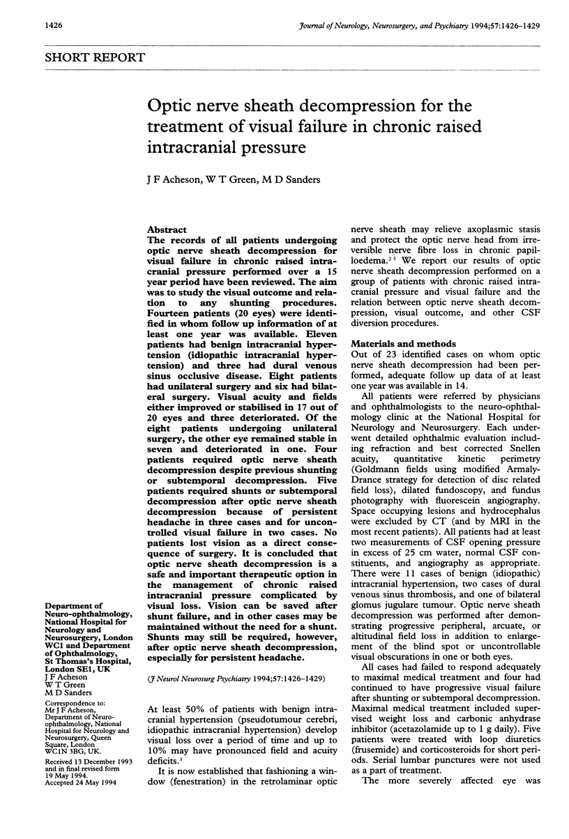
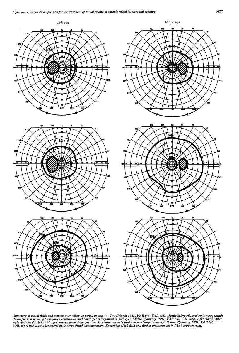
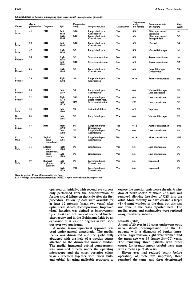
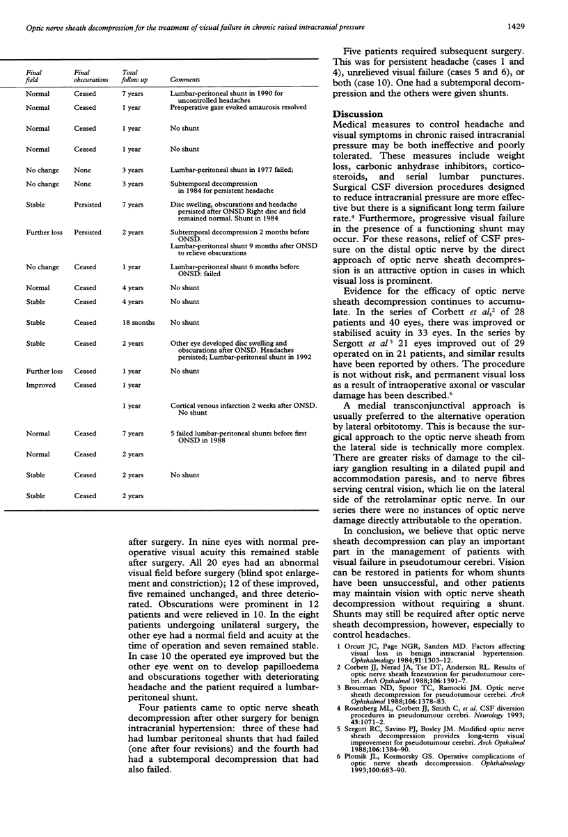
The records of all patients undergoing optic nerve sheath decompression for visual failure in chronic raised intracranial pressure performed over a 15 year period have been reviewed. The aim was to study the visual outcome and relation to any shunting procedures. Fourteen patients (20 eyes) were identified in whom follow up information of at least one year was available. Eleven patients had benign intracranial hypertension (idiopathic intracranial hypertension) and three had dural venous sinus occlusive disease. Eight patients had unilateral surgery and six had bilateral surgery. Visual acuity and fields either improved or stabilised in 17 out of 20 eyes and three deteriorated. Of the eight patients undergoing unilateral surgery, the other eye remained stable in seven and deteriorated in one. Four patients required optic nerve sheath decompression despite previous shunting or subtemporal decompression. Five patients required shunts or subtemporal decompression after optic nerve sheath decompression because of persistent headache in three cases and for uncontrolled visual failure in two cases. No patients lost vision as a direct consequence of surgery. It is concluded that optic nerve sheath decompression is a safe and important therapeutic option in the management of chronic raised intracranial pressure complicated by visual loss. Vision can be saved after shunt failure, and in other cases may be maintained without the need for a shunt. Shunts may still be required, however, after optic nerve sheath decompression, especially for persistent headache.
Full text
PDF


Selected References
These references are in PubMed. This may not be the complete list of references from this article.
- Brourman N. D., Spoor T. C., Ramocki J. M. Optic nerve sheath decompression for pseudotumor cerebri. Arch Ophthalmol. 1988 Oct;106(10):1378–1383. doi: 10.1001/archopht.1988.01060140542020. [DOI] [PubMed] [Google Scholar]
- Corbett J. J., Nerad J. A., Tse D. T., Anderson R. L. Results of optic nerve sheath fenestration for pseudotumor cerebri. The lateral orbitotomy approach. Arch Ophthalmol. 1988 Oct;106(10):1391–1397. doi: 10.1001/archopht.1988.01060140555022. [DOI] [PubMed] [Google Scholar]
- Orcutt J. C., Page N. G., Sanders M. D. Factors affecting visual loss in benign intracranial hypertension. Ophthalmology. 1984 Nov;91(11):1303–1312. doi: 10.1016/s0161-6420(84)34149-5. [DOI] [PubMed] [Google Scholar]
- Plotnik J. L., Kosmorsky G. S. Operative complications of optic nerve sheath decompression. Ophthalmology. 1993 May;100(5):683–690. doi: 10.1016/s0161-6420(93)31588-5. [DOI] [PubMed] [Google Scholar]
- Rosenberg M. L., Corbett J. J., Smith C., Goodwin J., Sergott R., Savino P., Schatz N. Cerebrospinal fluid diversion procedures in pseudotumor cerebri. Neurology. 1993 Jun;43(6):1071–1072. doi: 10.1212/wnl.43.6.1071. [DOI] [PubMed] [Google Scholar]
- Sergott R. C., Savino P. J., Bosley T. M. Modified optic nerve sheath decompression provides long-term visual improvement for pseudotumor cerebri. Arch Ophthalmol. 1988 Oct;106(10):1384–1390. doi: 10.1001/archopht.1988.01060140548021. [DOI] [PubMed] [Google Scholar]


