

Abstract
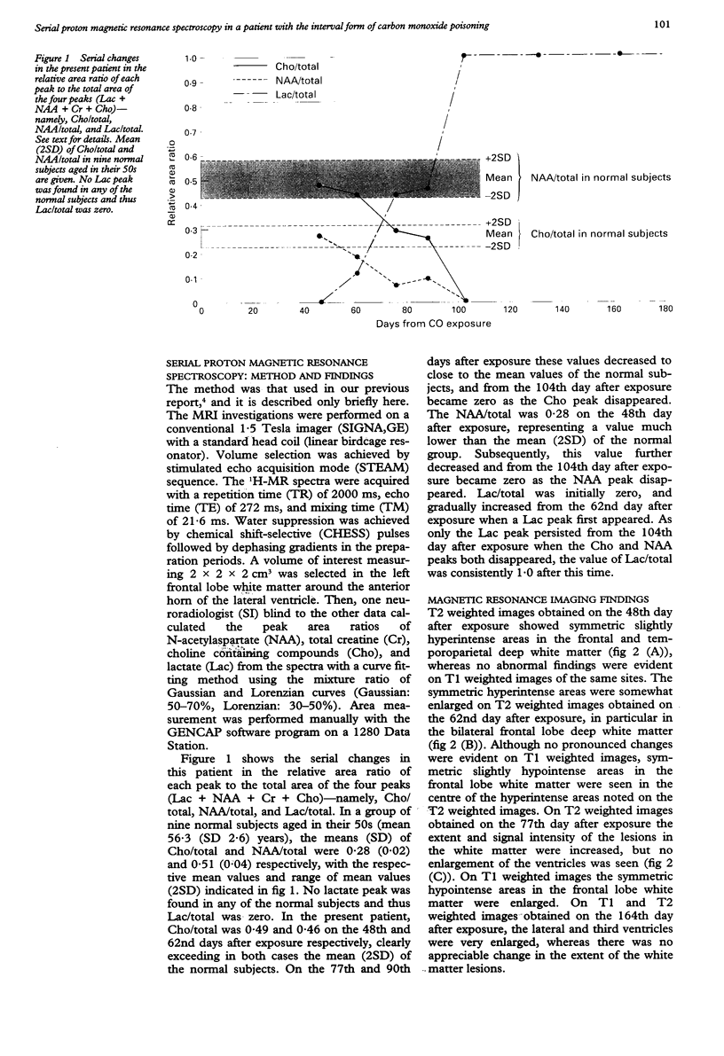
Serial proton magnetic resonance spectroscopy (1H-MRS) studies were performed from immediately after the appearance of sequelae in a patient with the interval form of carbon monoxide (CO) poisoning. The volume of interest was set over the frontal lobe white matter. In the early period a persistent increase in choline was found, which was thought to reflect the course of progressive demyelination. The appearance of lactate and decrease in N-acetylaspartate reflected the point at which neuron injury became irreversible. These were followed later by the finding of irreversible changes on MRI and single photon emission computed tomography. The findings suggest that 1H-MRS may be a useful modality to determine neuron viability and prognosis early in the course of the interval form of CO poisoning.
Full text
PDF
Images in this article
Selected References
These references are in PubMed. This may not be the complete list of references from this article.
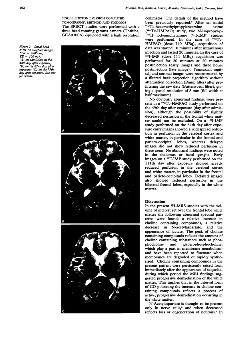
- Chang K. H., Han M. H., Kim H. S., Wie B. A., Han M. C. Delayed encephalopathy after acute carbon monoxide intoxication: MR imaging features and distribution of cerebral white matter lesions. Radiology. 1992 Jul;184(1):117–122. doi: 10.1148/radiology.184.1.1609067. [DOI] [PubMed] [Google Scholar]
- Choi I. S. Delayed neurologic sequelae in carbon monoxide intoxication. Arch Neurol. 1983 Jul;40(7):433–435. doi: 10.1001/archneur.1983.04050070063016. [DOI] [PubMed] [Google Scholar]
- Matsuda H., Oskoie S. D., Kinuya K., Tsuji S., Sumiya H., Tonami N., Hisada K. Tc-99m HMPAO brain perfusion tomography atlas using a high resolution SPECT system. Clin Nucl Med. 1990 Jun;15(6):428–431. doi: 10.1097/00003072-199006000-00014. [DOI] [PubMed] [Google Scholar]
- Menon D. K., Sargentoni J., Peden C. J., Bell J. D., Cox I. J., Coutts G. A., Baudouin C., Newman C. G. Proton MR spectroscopy in herpes simplex encephalitis: assessment of neuronal loss. J Comput Assist Tomogr. 1990 May-Jun;14(3):449–452. doi: 10.1097/00004728-199005000-00024. [DOI] [PubMed] [Google Scholar]
- Murata T., Koshino Y., Omori M., Murata I., Nishio M., Horie T., Umezawa Y., Isaki K., Kimura H., Itoh S. In vivo proton magnetic resonance spectroscopy study on premature aging in adult Down's syndrome. Biol Psychiatry. 1993 Sep 1;34(5):290–297. doi: 10.1016/0006-3223(93)90086-s. [DOI] [PubMed] [Google Scholar]
- Nadler J. V., Cooper J. R. N-acetyl-L-aspartic acid content of human neural tumours and bovine peripheral nervous tissues. J Neurochem. 1972 Feb;19(2):313–319. doi: 10.1111/j.1471-4159.1972.tb01341.x. [DOI] [PubMed] [Google Scholar]
- Vieregge P., Klostermann W., Blümm R. G., Borgis K. J. Carbon monoxide poisoning: clinical, neurophysiological, and brain imaging observations in acute disease and follow-up. J Neurol. 1989 Dec;236(8):478–481. doi: 10.1007/BF00328511. [DOI] [PubMed] [Google Scholar]
- van der Knaap M. S., van der Grond J., van Rijen P. C., Faber J. A., Valk J., Willemse K. Age-dependent changes in localized proton and phosphorus MR spectroscopy of the brain. Radiology. 1990 Aug;176(2):509–515. doi: 10.1148/radiology.176.2.2164237. [DOI] [PubMed] [Google Scholar]