

Abstract
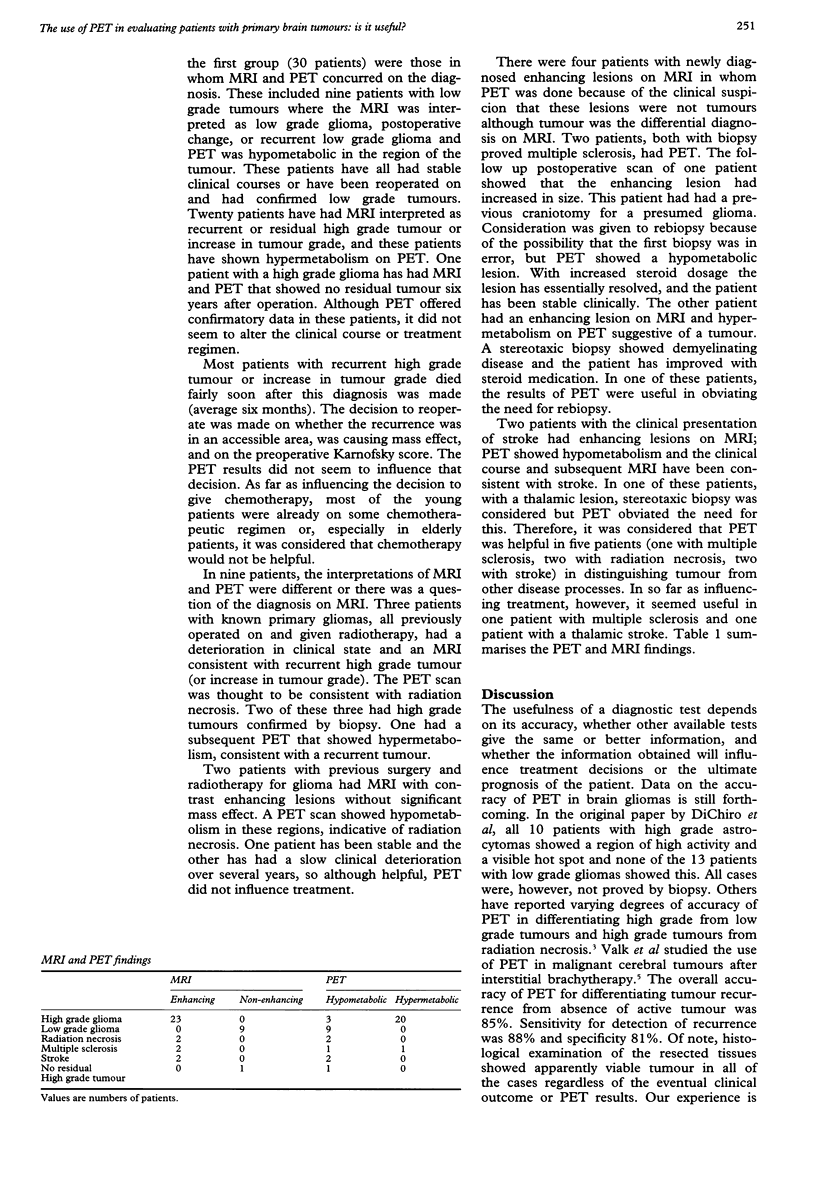
During an 18 month period 39 patients were evaluated with [18F] fluorodeoxyglucose-PET (FDG-PET) for primary brain tumours. These included patients with suspected newly diagnosed tumours and patients with known tumours who were being evaluated for possible recurrence or increasing tumour grade. Scans were performed on a 951-31 Siemen's PET scanner with 4 mm resolution. Scanning time was about 20 minutes per patient. All patients had undergone recent cerebral MRI. These patients were divided into two groups. In the first group (30) MRI and PET concurred on the diagnosis. The second group (nine) comprised those where the interpretation of MRI and PET was different or there was a question of the diagnosis on MRI. This group comprised three patients in whom MRI suggested recurrent tumour and PET inaccurately suggested radiation necrosis; two patients with newly diagnosed enhancing lesions on MRI in whom PET was useful in distinguishing strokes from tumour; two patients with prior gliomas with new enhancing isolated lesions on MRI in whom PET scan accurately depicted radiation necrosis; and two patients with newly diagnosed enhancing lesions on MRI in whom PET scan was helpful in distinguishing multiple sclerosis from tumour in one but not in the other. Therefore, of the 39 patients, PET was helpful in five in distinguishing tumour from other disease processes; but, in so far as influencing treatment, it seemed helpful in only two. Thus PET seems to be of limited value as an aid to evaluating and treating patients with suspected or known primary brain tumours.
Full text
PDF
Selected References
These references are in PubMed. This may not be the complete list of references from this article.
- Davis W. K., Boyko O. B., Hoffman J. M., Hanson M. W., Schold S. C., Jr, Burger P. C., Friedman A. H., Coleman R. E. [18F]2-fluoro-2-deoxyglucose-positron emission tomography correlation of gadolinium-enhanced MR imaging of central nervous system neoplasia. AJNR Am J Neuroradiol. 1993 May-Jun;14(3):515–523. [PMC free article] [PubMed] [Google Scholar]
- Di Chiro G., DeLaPaz R. L., Brooks R. A., Sokoloff L., Kornblith P. L., Smith B. H., Patronas N. J., Kufta C. V., Kessler R. M., Johnston G. S. Glucose utilization of cerebral gliomas measured by [18F] fluorodeoxyglucose and positron emission tomography. Neurology. 1982 Dec;32(12):1323–1329. doi: 10.1212/wnl.32.12.1323. [DOI] [PubMed] [Google Scholar]
- Di Chiro G., Oldfield E., Wright D. C., De Michele D., Katz D. A., Patronas N. J., Doppman J. L., Larson S. M., Ito M., Kufta C. V. Cerebral necrosis after radiotherapy and/or intraarterial chemotherapy for brain tumors: PET and neuropathologic studies. AJR Am J Roentgenol. 1988 Jan;150(1):189–197. doi: 10.2214/ajr.150.1.189. [DOI] [PubMed] [Google Scholar]
- Gutin P. H., Leibel S. A., Wara W. M., Choucair A., Levin V. A., Philips T. L., Silver P., Da Silva V., Edwards M. S., Davis R. L. Recurrent malignant gliomas: survival following interstitial brachytherapy with high-activity iodine-125 sources. J Neurosurg. 1987 Dec;67(6):864–873. doi: 10.3171/jns.1987.67.6.0864. [DOI] [PubMed] [Google Scholar]
- Janus T. J., Kim E. E., Tilbury R., Bruner J. M., Yung W. K. Use of [18F]fluorodeoxyglucose positron emission tomography in patients with primary malignant brain tumors. Ann Neurol. 1993 May;33(5):540–548. doi: 10.1002/ana.410330520. [DOI] [PubMed] [Google Scholar]
- Kornblith P. L., Walker M. Chemotherapy for malignant gliomas. J Neurosurg. 1988 Jan;68(1):1–17. doi: 10.3171/jns.1988.68.1.0001. [DOI] [PubMed] [Google Scholar]